Hemolytic anemia - what it is, clinical recommendations and treatment methods
Hemolytic anemia (HA) is a series of rare, but diagnosable and treatable blood diseases, during which increased destruction of red blood cells begins. HA, which is caused by intravascular destruction, appears as a result of the action of toxins, severe burns, blood poisoning, and transfusion of blood that is inappropriate for the rhesus group and blood type. Immunopathological processes also influence.
HA with extravascular (intracellular) destruction is hereditary in nature. The destruction of red blood cells occurs more often in the macrophages of the spleen, less often in the bone marrow, liver and lymph nodes. Probably a trio: anemia, inflammation of the spleen and jaundice. HA are divided into erythrocytopathies, erythrocytoenzymopathies and hemoglobinopathies.
A little about red blood cells
Erythrocytes, or red blood cells, are blood cells whose main function is to transport oxygen to organs and tissues.
- Red blood cells are formed in the red bone marrow, from where their mature forms enter the bloodstream and circulate throughout the body. The lifespan of red blood cells is 100-120 days. Every day, about 1% of them die and are replaced by the same number of new cells.
- If the lifespan of red blood cells is shortened, more of them are destroyed in the peripheral blood or spleen than have time to mature in the bone marrow - the balance is disrupted. The body responds to a decrease in the content of red blood cells in the blood by increasing their synthesis in the bone marrow, the activity of the latter increases significantly - 6-8 times.
As a result, an increased number of young red blood cell precursor cells - reticulocytes - is determined in the blood. The destruction of red blood cells with the release of hemoglobin into the blood plasma is called hemolysis.
HEMOLYSIS
HEMOLYSIS
(Greek, haima blood + lysis destruction, dissolution; synonym:
hematolysis, erythrocytolysis
) - the process of destruction of red blood cells, in which hemoglobin is released from them into the plasma. However, there is evidence [T. A. Prankerd, 1961] that disruption of the integrity of erythrocytes during hemolysis is not necessary and that the process may be limited by functional changes in erythrocytes with stretching of the cell membrane and changes in its permeability.
Blood after hemolysis of red blood cells (Hemolyzed blood) is a clear red liquid (lacquered blood).
It is necessary to distinguish between hemolysis in the body and in vitro.
Under the body's conditions, hemolysis occurs normally. This is the so-called physiological hemolysis occurring due to the natural aging of red blood cells. Hemolysis as a pathological phenomenon can occur under the influence of a number of factors: transfusion of incompatible blood, infusion of hypotonic solutions, the action of hemolytic poisons (see), hemotoxins (see); due to hereditary deficiency of enzyme systems in erythrocytes (see Enzymopenic anemia); in the presence of abnormal hemoglobins in erythrocytes (see Hemoglobinopathies), caused by both an anomaly in the primary structure of the hemoglobin molecule (see Sickle cell anemia) and a violation of the synthesis of polypeptide chains of hemoglobins (see Thalassemia); when antibodies to red blood cells appear (see Hemolytic anemia, immune hemolytic anemia; Hemolytic disease of newborns; Blood transfusion, post-transfusion anemia); under the influence of certain medications. In some diseases, “autohemolysins” are found in the blood serum, formed in response to the immunizing effect of denaturation and tissue breakdown products (in case of radiation sickness or tumor processes).
G. as a biophysic, the process is studied in vitro, since in vivo it is impossible to trace in detail the destruction of the erythrocyte. In addition, the study of the mechanism of G. is also necessary in order to prevent it in studies of the life expectancy of erythrocytes, in conditions of storage of canned blood and erythrocyte mass, when staging the complement fixation reaction and other tests carried out with blood or erythrocytes.
The red blood cell membrane consists of a bimolecular layer of lipids with monolayers of protein on both sides. Lipid molecules lie parallel to each other, but perpendicular to the plane of the membrane, with the polar heads of the phospholipids pointing outward and the long hydrocarbon chains pointing toward the center of the membrane. Protein chains are adsorbed on the polar heads. It is assumed that the interaction of protein and phospholipids is ensured by electrostatic forces and van der Waals forces (see Molecule). The length of lipid molecules is approximately 30 A, or 3 nm [H. Fricke, 1935], the thickness of the protein monolayer does not exceed 1 nm, the thickness of the cell membrane is approx. 8 nm [Danielli and Davson (JF Danielli, H. Davson), 1952]. According to O'Brien (JS O'Brien, 1967), the membrane of human erythrocytes contains cholesterol, phosphatidylserine, sphingomyelin, cerebrosides and other lipids. Water and small ions diffuse through the membrane at high speed through special pores of diameter. 0.3-0.4 nm. These pores are impenetrable for calcium and magnesium ions, for sugars, and even more so for large-molecular colloids. The half-time of water exchange through the membrane is 0.004 seconds, for the chlorine anion - 0.2 seconds, and for cations it is much longer: for the potassium cation approx. 30 hours, for sodium cation approx. 20 o'clock [Jandl (J. Jandl), 1965].
G. in vitro can be caused by physical. effects on red blood cells, chemical agents, hemolytic poisons of plant, animal and bacterial origin, the addition of blood serum from animals not immunized to red blood cells.
Physical influences include heating or repeated freezing and thawing of a suspension of erythrocytes or blood (thermal G.), placing erythrocytes in a hypotonic solution or in another environment that promotes an increase in the osmotic pressure of the internal environment of erythrocytes (osmotic G.), radiant energy, ultrasound, electrical energy. In this case, G. may be complete or incomplete, with greater or lesser changes and damage to erythrocytes.
Heating a suspension of erythrocytes to a temperature of 49° leads to their swelling, and at a temperature of 62-63° to their disintegration with the release of hemoglobin; Some erythrocyte fragments retain hemoglobin. G. during repeated freezing and thawing of erythrocytes occurs due to mechanical injury to them by pieces of ice and an increase in the concentration of substances inside the erythrocyte.
The mechanism of osmotic blood pressure is the penetration of water into the red blood cell (its volume increases and the membrane stretches). When the erythrocyte membrane is stretched by water, the pores expand through which hemoglobin molecules exit. According to Dawson (1940), the release of hemoglobin is preceded by an increase in the permeability of the erythrocyte membrane for potassium ions. With complete hemoglobin, erythrocyte hemoglobin is almost completely released into the plasma. In this case, free hemoglobin is first released, and then hemoglobin, labilely bound to phosphatides, is broken down; part of the hemoglobin remains firmly associated with the stroma (S. I. Afonsky, 1947); the stroma of erythrocytes has the appearance of the so-called. shadows
The volume of an erythrocyte, at which G. begins, is called the critical volume of an erythrocyte; It varies among different animal species. For human erythrocytes, the critical volume is 146% of the initial volume, for sheep erythrocytes - 126%, for rabbit erythrocytes - 137%.
The mechanism of osmotic digestion is similar when erythrocytes are placed in isotonic solutions of urea, glucose, glycerin, etc. When using urethane and alcohol anesthesia, the permeability of the erythrocyte membrane to water, potassium and hemoglobin decreases and osmotic digestion slows down.
Osmotic G. of erythrocytes is studied in a wedge, in practice for various diseases in the form of a test for the stability (resistance) of erythrocytes to hypotonic solutions of sodium chloride. The concentration of sodium chloride, at which osmotic G. begins, is taken as an indicator of the minimum osmotic resistance of erythrocytes. The concentration at which complete G. occurs is considered an indicator of the maximum resistance of erythrocytes. Red blood cells of a healthy person begin to hemolyze in 0.44-0.48% sodium chloride solution and are completely hemolyzed in 0.28-0.32% solution.
The intensity of gas under the influence of radiant energy depends on the wavelength, and the curve of the action of light on the gas process is parallel to the curve of hemoglobin adsorption. In the presence of small amounts of photosensitizers (eosin, fluorescein, erythrosin, hematoporphyrin, etc.), the hemolytic effect of radiant energy is enhanced. It is believed that photosensitizer dyes are adsorbed only on certain areas of the surface of the red blood cell, where, under the influence of radiant energy, pores appear for the release of hemoglobin into the plasma.
Ultrasound damages red blood cells as a result of pressure differences in the sound field. With low ultrasound energy, red blood cells are deformed, their shell becomes porous; with stronger energy, the structure of the red blood cell is destroyed.
Under the influence of direct electric current, hemoglobin is released from the erythrocyte, and the erythrocyte stroma is significantly destroyed (stromatolysis). Alternating current does not destroy red blood cells.
Among the chemical agents, nitrites, nitrobenzene, nitroglycerin, ether, benzene, sodium oleic acid, sodium cholic and deoxycholic acid, aniline compounds, saponin, l isolecithin, etc. have a hemolytic effect. hemolytic agents cause direct damage to the structure of erythrocyte membranes, disrupting the arrangement of lipid molecules in it, with the formation of pores. Humphrey et al. (1969) described hexagonally arranged pores with a diameter of 8-10 nm when erythrocytes are exposed to saponin; after G. streptolysin O, phospholipase C, defects (pores) with a diameter of up to 40-50 nm were discovered. D. L. Rubinstein and R. A. Rutberg (1948) established that under the influence of chemicals. hemolytic agents first break down the hemoglobin compounds with the lipoprotein complexes of the erythrocyte. Sodium oleic acid or bile acids cause G., damaging the membranes of the erythrocyte, dissolving its lecithin. The deeper parts of the stroma are also damaged, releasing the hemoglobin associated with it. The hemolytic effect of saponin is caused by damage to the erythrocyte by combining it with erythrocyte cholesterol: the addition of cholesterol to the medium with saponin inhibits the hemolytic effect of the latter.
Poisons of worms, insects (bees, karakurt spider, scorpion), and snakes have a hemolytic effect. The mechanism of the hemolytic action of animal poisons is apparently associated with a change in the structure of the lipoid component of the erythrocyte membrane and is similar to the action of the enzyme lecithinase. Hemolysins of many bacterial toxins (tetanolysin, staphylolysin, streptolysin O and S, etc.) have lecithinase activity.
G. under the influence of normal hemolysins contained in the blood serum of non-immunized mammals of one species in relation to the erythrocytes of animals of another species is associated with the primary toxicity of these serums for the body of the corresponding animal species. However, complete correspondence between the hemolytic activity and toxicity of foreign sera has not been identified. The processes of enzymatic destruction of lipoprotein complexes of the erythrocyte membrane occupy a significant place in the mechanism of hyperemia from the influence of foreign serums. As a result of the dissolution of some of the lipids, the surface of the erythrocyte is primarily damaged, colloid-osmotic forces lead to swelling of the erythrocyte, then similar to placing the erythrocyte in a hypotonic environment.
Under the influence of hemolysins, specific antibodies that can combine only with the red blood cells of an immunized animal, immune hemolysis occurs. Immune hemolysin (see Amboceptor) is an antibody found in the gamma globulin fraction of blood serum, in the IgG and IgM fractions. Hemolysins attach complement to the erythrocyte: to dissolve one erythrocyte, 30-50 molecules of hemolysin and 25,000 molecules of complement are needed. The active agent in the mechanism of specific G. is complement (see), and hemolysin plays only the role of a link between complement and erythrocytes.
The relatively small amount of hemolysin consumed during specific immune G. is apparently explained by local damage to the erythrocyte by complement. Brunius (F. Brunius, 1936) calculated that with specific G. hemolysins cover only 0.001% of the surface of the erythrocyte.
When complement acts on a sensitized red blood cell, potassium ions, phosphates and ribonucleotides pass from the cell through the damaged cell membrane. A disturbance in the ion balance is accompanied by the entry of water into the cell and its swelling, as well as a decrease in the cell surface and the achievement of a critical hemolytic volume. Further stretching of the membrane and an increase in its pores contribute to the release of hemoglobin from the erythrocyte. In order for protein molecules to pass through the membrane, the pore diameter must be at least 6 nm. Multiple pores often form, which can then coalesce and contribute to membrane rupture and destruction of the red blood cell. The formation of numerous pores, according to Humphrey, depends on an excess of complement.
When an animal is immunized with blood serum containing immune hemolysin, antibodies are formed in the body - antihemolysins, which specifically combine with hemolysins and, therefore, prevent their connection with the erythrocyte, as a result of which specific hemolysins are inhibited.
The following stages of hemolysis are distinguished: prehemolytic (increased permeability of the erythrocyte membrane), hemoglobinolysis (decomposition of hemoglobin), hemolysis proper (release of hemoglobin), stromatolysis (destruction of the stroma). In immune hepatitis, three more stages are distinguished: sensitization, the damaging effects of complement, and diffusion of hemoglobin from the erythrocyte.
See also Red blood cells.
Bibliography:
Afonsky S.I. On the issue of the chemical composition and properties of the stroma of horse erythrocytes, Uchen. zap. Kazansk vet. in-ta, t. 55, p. 20, 1947; Idelson L. I., Didkovsky N. A. and Ermilchenko G. V. Hemolytic anemias, M., 1975, bibliogr.;
Cabot E. and Meyer M. Experimental immunochemistry, trans. from English, M., 1968;
Lorie Yu. I. Autoimmune hemopathy, Ter. arkh., vol. 39, no. 2, p. 10, 1967, bibliogr.; Polikar A. Molecular cytology of animal cell membranes and its microenvironment, trans. from French, Novosibirsk, 1975; Rubinstein D. L. and Rutberg R. A. Cleavage of hemoglobin during chemical hemolysis, Biochemistry, v. 13, no. 2, p. 147, 1948; Blood and its disorders, ed. by R. J. Hardisty a. DJ Weatherall, Oxford, 1974; O'Brien JS Cell membrane-nes-composition, structure function, J. theor. Biol., v. 15, p. 307, 1967, bibliogr.; The cell surface, ed. by BD Kahan a. R. A. Reisfeld, NY-L., 1974; Cooper RA a. Shallil SY The red cell mebrane in hemolytic anemia, in the book: Modern treatment, ed. by L.S. Lessin a. W. F. Rosse, y. 8, p. 329, NY, 1971, bibliogr.; Davson H. a. Daniel li JF Permeability or natural membranes, L., 1952; Erythrocytes, thrombocytes, leukocytes, ed. by E. Gerlach ao, Stuttgart, 1973; Humhrey JH a. Dourmashkin RR The lesions in cell membranes caused by complement, in the book: Advanc, in immunol., ed. by F. J. Dixon, a. HG Kunkel, v, 11, p. 75, NY-L., 1969, bibliogr.; Ponder E. Red cell structure and its breakdown, Wien, 1955, bibliogr.; Red cell shape, physiology, pathology, ultrastructure, ed. by M. Bessis ao, NY ao, 1973; Shohet SB Hemolysis and changes in erythrocyte membrane lipids, New Engl. J. Med., v. 286, p. 577, p. 638, 1972, bibliogr.
L. M. Ishimova.
Causes
Modern science does not provide for the division of hemolytic anemia depending on the site of destruction of red blood cells. Paying more attention to the etiology and pathogenesis of the disease and, based on these principles, the disease is divided into 2 main classes:
- Acquired forms of HA, which are classified according to the factor that destroys red blood cells and causes this anemia (antibodies, hemolytic poisons, mechanical damage).
- Hereditary hemolytic anemias are classified according to the principle of localization of a genetic defect in red blood cells, due to which red blood cells become defective, functionally unstable and unable to live their intended time. Hereditary HAs include: membranopathies (microspherocytosis, ovalocytosis), enzyme defects (G-6-PDS deficiency), hemoglobinopathies (sickle cell anemia, thalassemia).
A large number of all GAs fall into acquired forms, but among them there are a number of variants, which, in turn, also have varieties due to individual causes of occurrence:
- The development of hemolytic anemia can be triggered by vitamin E deficiency;
- HA develops due to somatic mutations that change the membrane structure of red blood cells (paroxysmal cold nocturnal hemoglobinuria);
- The development of the disease is due to the influence of anti-erythrocyte antibodies produced on the own antigenic structures of erythrocytes (autoimmune - autoimmune hemolytic anemia) or isoantibodies that enter the blood from the outside (isoimmune - hemolytic disease of the newborn);
- Various chemicals that are foreign to the human body (organic acids, hemolytic poisons, salts of heavy metals, etc.) often have a negative effect on membrane structures;
- Damage to the membrane of red blood cells can be caused by the mechanical impact of artificial heart valves or injury to red blood cells in the capillary vessels of the feet when walking and running (marching hemoglobinuria);
- A parasite such as malarial plasmodium, which enters the human blood through the bite of a mosquito (female) of the genus Anopheles (malarial mosquito), is dangerous in terms of the occurrence of hemolytic anemia, as a symptom of “swamp fever”.
The most common form of acquired HA is autoimmune hemolytic anemia (AIHA).
Types of hemolysis
{banner_banstat0}
As mentioned above, an erythrocyte has its own life cycle, which usually reaches 125 days, after which its membrane is destroyed and the cell dies. This entire process occurs in the human spleen under the influence of the immune system. Depending on where hemolysis occurs, it is divided into several types.
Most experts in the field of studying blood diseases divide hemolysis of red blood cells into the following subtypes:
- hemolysis occurring inside the human body is called biological. It develops in the spleen, “old” red blood cells disintegrate under the influence of macrophages of the immune system, and hemoglobin practically does not enter the plasma. In special cases, the death of red blood cells is observed in the vascular bed (during an attack by poisonous snakes, or insects whose bite poses a danger to humans, or during a blood transfusion procedure from a donor who has a different blood type than the recipient), this process is accompanied by the release of hemoglobin in the blood plasma and its release with urine;
- chemical hemolysis of red blood cells is the breakdown of blood cells under the influence of any chemicals (acids, alkalis, toxins, as well as poisons of snakes and insects);
- the thermal type occurs when the blood is exposed to high temperatures, or, conversely, excessively low temperatures;
- the mechanical type is caused by injury to the membrane of red blood cells as it passes through the machine during the hemodialysis procedure, or by strong shaking of the test tube with the material for analysis;
- osmotic hemolysis is observed in laboratory conditions, injury to the erythrocyte membrane is observed in hypotonic liquids. Water enters the blood cell, it stretches, causing the membrane to burst.
Classification
All hemolytic anemias are classified according to this principle.
Acquired forms - develop under the certain influence of any external factors that have a destructive effect on red blood cells.
- Autoimmune hemolytic anemia;
- Anemia caused by mechanical erythrocyte damage or provoking parasitic, toxic effects of substances.
Hereditary or congenital forms are the result of the impact of genetically determined abnormalities on the vital activity of red blood cells.
- Membranopathies;
- Hemoglobinopathies (sickle cell anemia, thalassemia, etc.);
- Erythrocytopathies (Minkowski-Choffard anemia);
- Enzymopathies.
Symptoms
Hemolytic anemia manifests itself more prominently during crises. With sluggish processes, the symptoms are blurred. The specific type of pathology can only be determined with the help of additional diagnostics.
The main symptoms include:
| Anemia syndrome | manifests itself in pallor of the skin and mucous membranes. Accompanied by symptoms of lack of oxygen in the form of shortness of breath, dizziness, weakness, and increased heart rate. |
| Jaundice syndrome | An increase in the concentration of bilirubin as a breakdown product of red blood cells is expressed by yellowness of the skin and a change in the color of urine. |
| Hepatosplenomegaly | the enlargement of the spleen occurs due to intense hemolysis and can reach significant sizes. The liver is less susceptible to changes. But in some cases there is an increase in it, accompanied by heaviness in the hypochondrium. |
Hemolytic anemia may manifest itself with additional symptoms such as:
- abdominal pain;
- loose stools;
- bone soreness;
- elevated temperature;
- pain in the chest and kidney area.
Even if there are only 2 or 3 of the listed symptoms, this should be a reason for a medical examination. After all, toxic bilirubin with prolonged exposure to tissues and organs can disrupt their functions.
Symptoms
{banner_banstat2}
Depending on the severity of the disease, the symptoms of hemolysis may vary, for example, with a mild form, patients experience weakness, sometimes chills, a feeling of nausea, and yellowness of the eyeballs is often noted.
In the case of extensive hemolysis, after 8-9 hours from the onset of development, patients are concerned about:
- increasing headache and fatigue;
- sometimes vomiting occurs;
- in the area of the right hypochondrium there is severe pain, which can radiate to the lower back;
- The main symptoms of hemolysis include a change in urine color to brown or dark burgundy;
- a blood test shows a low level of red blood cells;
- there is a high content of “young” red blood cells;
- often the patient has a high temperature of up to 39 degrees;
- due to the breakdown of blood cells in the liver, the organ enlarges, resulting in liver failure;
- a few days after the onset of hemolysis, patients are diagnosed with jaundice;
- there is a high level of bilirubin in the blood;
- Due to the breakdown of hemoglobin in the kidneys, the tubules are blocked, which leads to the development of renal failure, up to complete urinary retention.
It is worth noting that hemolysis of erythrocytes is observed not only in the human body, for example, in laboratory conditions when studying a human blood test.
If blood cells break down during laboratory tests, the test results may be unreliable.
The main reasons for rapid blood clotting include:
- absence of preservatives in the test tube or their insufficient quantity;
- methodological instructions were violated when collecting material for research;
- violation of sterility during blood collection;
- the patient eating too fatty foods before a blood test;
- violation of methodological recommendations before the start of the study;
- violations of the rules for transporting blood tests;
- violation of the rules for storing blood tubes;
- exposure to high temperatures on blood tubes.
Unfortunately, the above reasons lead to repeated blood sampling and testing, which is undesirable, especially for young children, so medical personnel must strictly follow the instructions during the procedure.
Clinical forms
In clinical manifestations, several common forms of hemolytic anemia can be distinguished:
- Autoimmune anemia. This acquired disease is accompanied by extensive damage to blood cells. The main cause of the pathology is the formation of antibodies in red blood cells, which provoke hemolysis. Autoimmune hemolytic anemia occurs when immune cells perceive the body's red blood cells as foreign agents and strive to destroy them. The prognosis for this disease is unfavorable and requires a blood transfusion or bone marrow transplant.
- Minkowski-Choffard anemia (hereditary microspherocytosis) is characterized by abnormal permeability of the erythrocyte membrane through which sodium ions pass. The disease has an autosomal dominant hereditary nature. Development is wavy: alternating stable periods and hemolytic crises. Main signs: decrease in osmotic resistance of erythrocytes, predominance of altered erythrocytes - microspherocytes, reticulocytosis. In case of a complex course of the disease, surgical intervention (removal of the spleen) is necessary.
- Thalassemia. If the attending physician determines the congenital blood disease thalassemia, this is a consequence of impaired hemoglobin production in the chemical composition of the blood. In the absence of timely treatment, anemia only progresses, symptoms increase, turning the patient into a disabled person. This disease is congenital in origin and requires a set of tests, a detailed examination of the clinical patient, and treatment to maintain a period of remission.
- Porphyrias are a hereditary form of the disease and are caused by impaired formation of porphyrins, components of hemoglobin. The first sign is hypochromia, iron deposition gradually appears, the shape of red blood cells changes, and sideroblasts appear in the bone marrow. Porphyria can also be acquired due to toxic poisoning. Treatment is carried out by administering glucose and hematite.
- Sickle cell anemia is the most common type of hemoglobinopathy. A characteristic sign: red blood cells take on a sickle shape, which leads to them getting stuck in the capillaries, causing thrombosis. Hemolytic crises are accompanied by the release of black urine with traces of blood, a significant decrease in hemoglobin in the blood, and fever. The bone marrow contains a high content of erythrokaryocytes. During treatment, the patient is given an increased amount of fluid, oxygen therapy is administered and antibiotics are prescribed.
Three enemies of biochemistry
06.05.2021
1 - normal and icteric 2 - normal and chylosis 3 - normal and hemolysis
1) Hemolysis
Hemolysis can occur:
— Due to a violation of the preanalytical stage of blood sampling.
— In certain pathologies (diseases), pathological hemolysis occurs. That is, cells are destroyed much faster than necessary. Such processes occur in infectious diseases, poisoning with plant or synthetic poisons, hemolytic anemia, autoimmune diseases, after transfusion of inappropriate donor blood.
The hallmark of hemolysis is a clear, red serum after centrifugation of the blood. This coloring of the serum leads to a distortion of the true values of biochemical parameters. Therefore, it is necessary to repeat the selection of material to verify true pathological hemolysis.
2) Chylosis/lipemia
Chylesis is a physical clouding of the serum due to the presence of large amounts of fats (triglycerides and chylomicrons).
You may encounter this situation:
If the blood was not taken on an empty stomach. In this case, chyle is caused by “gastronomic” reasons: eating fatty, spicy, smoked foods, dairy (fermented milk) products, as well as alcoholic beverages, eggs, chocolate, bananas, etc. on the eve of blood (plasma) donation.
In all other cases, chylosis indicates a metabolic disorder. If the diet contains a lot of fatty foods and animal proteins, a disorder of lipid metabolism occurs, therefore, the blood serum will be fatty.
First of all, when chyle is detected, it is necessary to re-take a blood test, adhering to the above recommendations before this procedure, and if chyle is confirmed, consult a doctor.
3) Ictericity
Icterus is due to:
Most often, various liver diseases, in which the level of bilirubin in the blood rises sharply.
Sometimes an increase in the level of bilirubin in the blood can be associated with prolonged fasting of the patient on the eve of the test, although even a very long absence of food intake in a completely healthy person rarely leads to icterus in the resulting blood serum.
A high concentration of bilirubin in the blood can distort the value of a laboratory indicator. However, it should be borne in mind that it is not always possible to correct the increased level of bilirubin in the blood; in this case, you need to notify the laboratory about the characteristics of the patient’s health condition and this will be taken into account when performing research.
Laboratory diagnostics doctor (head)
Clinical diagnostic laboratory of the city clinic No. 4, Grodno Nefedova L.I.
Diagnostics
In the diagnosis of hemolytic anemia, a general clinical blood test is used (anemia and changes in the size and shape of red blood cells are detected), a biochemical blood test (including serum bilirubin, ALT, LDH), serum haptoglobin, hemosiderin and urine hemoglobin. To confirm the diagnosis, bone marrow puncture can be used (active processes of erythropoiesis are determined in the biopsy specimen).
The most characteristic of active intravascular hemolysis is spherocytosis of erythrocytes (with transfusion reactions, hereditary spherocytosis, with hemolytic anemia with warm antibodies). Schistocytosis (with intravascular prosthetics, microangiopathies), sickle erythrocytes (with sickle cell anemia), target erythrocytes (with liver pathologies, hemoglobinopathies), nucleated erythrocytes and basophilia (with beta thallasemia major) may also be observed.
Heinz bodies are found in unstable hemoglobin, activation of peroxidation, acanthocytes in anemia with spur-shaped red blood cells, agglutinated cells in cold agglutinin disease.
Changes in blood tests due to anemia
To diagnose anemia and determine its type, blood is taken from a finger and a vein. For some types of anemia, a urine test is performed for bile pigments, which are formed as a result of the breakdown of red blood cells and concomitant liver damage.
Determine in blood:
- Red blood cell content and hematocrit;
- Reticulocyte concentration;
- The presence of red blood cells of irregular shape , composition and size;
- Color index (CI) , based on this criterion, anemia is divided into three types: hypochromic - CP is reduced; normochromic – CP is not changed; hyperchromic - the CPU is increased.
- Serum iron concentration . Based on this criterion, anemia is divided into three types: normosideremic - the indicator is normal; hyposideremic – iron concentration is less than normal; hypersideremic – iron content is increased.
- Saturation of blood serum with transferrin , a protein that transports iron, for the formation of red blood cells;
- Leukocyte content . With anemia, both the general indicator and the content of leukocytes of any type (lymphocytes, monocytes, eosinophils, basophils) may change;
- Concentration of bilirubin and haptoglobin;
- Mean erythrocyte diameter (EDD) . Anemia is: normocytic, red cells are normal; microcytic – the size of red blood cells is reduced; macrocytic and megaloblastic - unnaturally large red blood cells are found.
Treatment of hemolytic anemia
With the development of hemolytic anemia, effective treatment is much more difficult than for any other types. This is due to the fact that it is not always possible to influence the mechanism that triggers hemolysis. The general treatment plan may include:
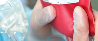
- Blood transfusions of washed red blood cells. Indicated when red blood counts drop to critical levels;
- The use of folic acid and vitamin B12 preparations;
- Transfusion of fresh frozen plasma and human immunoglobulin;
- Administration of glucocorticoid hormones (dexamethasone, prednisolone, methylprednisolone, cortinef);
- Use of cytostatics. Indicated exclusively for autoimmune hemolytic anemia;
- Prevention of infectious complications and exacerbations of existing chronic pathology;
- Surgical treatment in the form of splenectomy (removal of the spleen). The method provides a relatively good prognosis for recovery. Not effective for Minkowski-Choffard anemia and other types of hereditary hemolysis.
Any hemolytic anemia, the fight against which is started untimely, is a complex problem. It is unacceptable to try to deal with it on your own. Its treatment must be comprehensive and prescribed exclusively by a qualified specialist based on a thorough examination of the patient.
Acquired hemolytic anemia
The main group of drugs successfully used in the treatment of acquired anemia are glucocorticoids (prednisolone), which can completely block the hemolysis process. But treatment with such hormones is quite lengthy. For its full effectiveness, an accurately calculated dosage of the drug is required. If steroid hormones are ineffective, cytostatics are used.
If the cause of hemolytic anemia cannot be identified, then doctors begin symptomatic treatment. Usually it consists of transfusion of red blood cell suspension to the patient. This method is especially effective for low hemoglobin levels (less than 50) and during periods of hemolytic progression.
Congenital or hereditary anemia
Typically, drug treatment of such anemia provides only temporary improvements in the patient’s general condition, but, unfortunately, does not prevent relapses of hemolytic crises. Here, surgical intervention will be more effective.
It involves performing a splenectomy - removal of the spleen. This operation has shown 100% effectiveness; patients are almost completely cured, although the pathological properties of red blood cells are preserved. Typically, indications for surgery are attacks of renal colic, splenic infarction, severe anemia, or frequently recurring crises.


