Liver fibrosis is the proliferation of connective tissue in the liver, which occurs when hepatocytes (liver cells) are damaged by hepatitis B, C, and D viruses, alcohol, toxic substances and other factors. Progression of liver fibrosis leads to cirrhosis, liver failure and portal hypertension.
What is liver fibrosis?
Fibrosis is an increase in the amount of connective tissue in the liver. It is not an independent disease, but represents a universal response of an organ to damage, regardless of the cause. The process of its formation is called fibrogenesis and can be observed during the progressive course of drug-induced, non-alcoholic and alcoholic fatty liver disease, viral hepatitis and other pathologies.
Traditional medicine
If liver fibrosis is treated comprehensively, then in this case, along with drug treatment, therapy using traditional medicine is indicated. Non-drug treatment is also indicated during the period of remission to reduce the likelihood of relapses. The following treatment methods are used:
To improve the flow of bile and strengthen the immune system, use a tincture made from ground milk thistle seeds. This drug has a hepatoprotective effect. To inhibit the growth of fibrous tissues, a tincture of white cinquefoil is used. To do this, ten grams of dry grass roots are mixed with one hundred milliliters of vodka and allowed to brew for thirty days. After this, the product is used twice a day, twenty to thirty drops, washed down with water.
Important! It is not recommended to use alcohol-containing tinctures in the treatment of liver fibrosis when the disease is caused by alcoholic hepatitis.
A choleretic and immunomodulatory effect can be achieved using a decoction of rose hips.
To prepare the decoction, the fruits are poured with hot water and infused in a thermos overnight. Use a couple of tablespoons three times a day. To obtain a choleretic effect, use a decoction of corn silk. This remedy has an anti-inflammatory effect and is indicated for pathological stagnation of bile. The decoction helps dissolve stones. Olive oil also has a choleretic effect. It is especially effective to use oil in combination with honey, mixing the components in equal parts. It is worth understanding that this kind of cleansing does not affect the process of scarring in the liver, but rather helps eliminate the accompanying symptoms.
Mechanism of damage
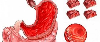
Stellate cells or Ito cells play a key role in the development of fibrosis. When the liver is damaged by various factors, such as excess fat accumulation, alcohol, viruses, drugs, toxins, stellate cells become active. Activated Ito cells begin to produce collagen, a protein that forms the basis of connective tissue. As a result, connective tissue accumulates in the liver. Progressive fibrosis can lead to irreversible damage to its structure - cirrhosis.
Symptoms
The difficulty of timely diagnosis is due to the fact that there are no specific symptoms and signs of the disease. The disease can be asymptomatic for a long time or be accompanied by signs that characterize the liver as a whole - an increase in its size, heaviness and pain in the right hypochondrium, an increase in the level of liver enzymes - alanine aminotransferase and aspartate aminotransferase in the blood. In later stages, fibrosis symptoms may include:
- enlarged spleen;
- dilation of the veins of the esophagus and bleeding from them;
- decrease in the level of hemoglobin, platelets, leukocytes in the blood.
Diagnostic methods
During a medical examination, the disease can be detected only in the last stages of the development of the process. Clinical studies are much more informative:
- CBC is a general blood test, which shows a decreased level of hemoglobin and red blood cells with an increased ESR;
- OAM is a general analysis that detects the presence of protein, casts and bilirubin;
- blood - biochemical analysis that detects an increase in all indicators related to the liver;
- Ultrasound of the liver - reveals an increase in the size of the organ and the appearance of fibrotic formations;
- elastometry - to assess the structure of the liver and the elasticity of its tissues;
- tomography - to determine the number of fibrous foci, from localization and quality indicators.
To accurately diagnose liver fibrosis, a biopsy may be necessary. It is performed under the control of ultrasound equipment by inserting a special trepanation needle. If there are contraindications to the procedure, a diagnosis can be made based on elastometry data.
Diagnosis of the disease
Biopsy
The gold standard for diagnosis is biopsy. A biopsy is the removal of a small area of liver tissue with a special needle for further study. Although the procedure is quite safe, it can nevertheless be accompanied by unpleasant pain. In this regard, non-invasive diagnostic methods, including laboratory and instrumental ones, are becoming increasingly relevant.
Laboratory methods
Laboratory methods are based on the determination of compounds in the blood that are indirect or direct markers of fibrosis. Indirect markers include indicators such as platelet count, prothrombin time, alanine aminotransferase and aspartate aminotransferase levels. Direct markers are transforming growth factor, type IV collagen, aminoterminal propeptide of type III procollagen, hyaluronic acid, matrix metalloproteinases, tissue inhibitors of matrix metalloproteinases.
The use of both direct and indirect markers individually has limited diagnostic value. The diagnostic accuracy increases when they are used in combination – as part of special panels. The most famous of them are “FibroTest” (France), “Fibrometer” (France), “FibroSpect” (USA), “ELF” (Great Britain), “Hepascor” (Australia). At the same time, diagnostic results obtained using laboratory panels, as a rule, require confirmation by instrumental methods.
Instrumental methods
Instrumental methods of non-invasive diagnostics include ultrasound and computed tomography. However, in the early stages of fibrosis, their accuracy is low.
Today, the most promising non-invasive diagnostic method is elastometry. It is based on assessing the elasticity of liver tissue using an ultrasound sensor. The sensor generates low-frequency vibrations that are transmitted to the liver tissue and create elastic waves. The speed of propagation of these waves is determined by the elasticity of the liver, a decrease in which indicates compaction of the organ due to an increase in the content of connective tissue in it. The lower the elasticity, the higher the stage of the disease. Studies have shown a high degree of agreement between elastometry and biopsy results.
Types of disease
Depending on the location of fibrotic growths, the following forms are distinguished:
- Venular. Scar fibers are noted in the central sections of the organ lobules.
- Pericellular. Connective tissue constrictions are detected around cellular structures.
- Septal. An unfavorable option, which is characterized by necrosis of large areas of liver tissue. The septa (septa) formed at the sites of necrosis change the structure of the organ.
- Periductal. Conical scar structures grow around the bile ducts.
Stages
The disease is usually classified into five stages:
- Stage 0
– no disease; - Stage 1
– mild fibrosis (deposition of connective tissue around liver cells - hepatocytes and intrahepatic bile ducts); - Stage 2
– moderate fibrosis (formation of connective tissue septa – fibrous septa between the portal tracts located at the junctions of the hepatic lobules); - Stage 3
– pronounced fibrosis (formation of fibrous septa between the portal tracts and the central veins located in the center of the hepatic lobules); - Stage 4
– cirrhosis (disruption of the normal lobular structure of the liver – the formation of false lobules due to the formation of fibrous septa).
Forms
Based on the degree of prevalence and localization of the pathological process, several main forms of the disease can be distinguished:
- venular – the focus is located in the central part of the organ;
- pericellular – inflammation is mainly concentrated around hepatocytes;
- septal – the presence of large necrotic areas, the formation of a large number of fibrous septa;
- periductal - characterized by constantly growing connective tissue located around the bile canaliculi;
- mixed form is the most common type of fibrosis, which includes all the signs described above.
The rate of development of fibrosis from the initial to the last stage is determined mainly by the type of pathology.
- The non-cirrhotic form can develop in various severe infectious diseases. In this case, sclerotic changes and thrombosis in the hepatic vessels may develop. This pathology is often the result of excessive alcohol consumption, the presence of hepatitis of various natures, exposure to toxic substances, and uncontrolled use of medications.
- The periportal form is complemented by potal hypertension and is characterized by an increased severity of the course. The disease begins with parasites, which occurs through contaminated water. This form is dangerous due to severe complications when the helminth enters the human body.
Prevention and treatment of the disease
In liver diseases accompanied by the formation of fibrosis, its treatment is an extremely important task. But much more relevant and justified is the prevention of the development and progression of this condition. Despite the large number of studies in this direction, there are currently no specific drugs for the treatment and prevention of fibrosis. Thus, the most rational approach is to use drugs in the treatment of liver pathology that can prevent the development and progression of the disease.
At the present stage, a large number of drugs used for liver diseases are available. However, not many of them have a mechanism of action that allows us to discuss the possibility of their use for the prevention of the development and progression of fibrosis. And practically none of these drugs have demonstrated this capability in clinical studies.
Causes
Today, experts identify many different causes of fibrosis. Often the disease occurs due to poor heredity or the presence of congenital pathologies, as well as after prolonged or uncontrolled use of certain medications, alcohol, or as a result of poisoning with toxic substances.
The main reasons for the appearance of fibrosis of the second stage:
- viral hepatitis (B, C, D), accompanied by acute inflammation;
- various viral infections in acute and chronic forms (infectious mononucleosis);
- cytomegalovirus infection (a type of herpes);
- long-term (more than eight years) alcohol consumption;
- a sharp weakening of the immune system;
- autoimmune hepatitis, when the body independently destroys healthy liver cells;
- deviation in the functioning of the biliary tract;
- presence of cholelithiasis;
- development of toxic hepatitis, accompanied by liver damage from various poisons, substances, etc.;
- use of certain medications (used in the treatment of tumors, rheumatism, etc.);
- venous congestion in the hepatic vessels.
Important! Excess weight, diabetes, and lack of essential nutrients can cause fibrosis.
Phosphogliv* – reliable protection
Domestic drug Phosphogliv *
represents an exception to this series.
Its active component - glycyrrhizic acid - in numerous experimental studies has demonstrated the ability to influence the key link in fibrogenesis - to suppress the production of connective tissue by stellate cells. This ability was confirmed in clinical studies of the drug Phosphogliv *
, which showed its beneficial effect on fibrosis in various liver diseases.
The drug has long been successfully used by doctors in the complex treatment of liver diseases. In addition, in patients with viral hepatitis who cannot, for one reason or another, receive antiviral treatment and are at particular risk for the development of fibrosis and cirrhosis, Phosphogliv *
used as a means of pathogenetic action.
The accumulated experience of medical use and the results of clinical studies have made it possible to include Phosphogliv *
in clinical recommendations and standards for the treatment of liver diseases.
Remember that only a doctor can diagnose a disease and prescribe appropriate treatment!