To the reference book The middle ear is an intermediate link between the outer and inner ear and is involved in the processes of converting sound vibrations.
Author:
- Sokolova Alla Vasilievna
otorhinolaryngologist, otosurgeon, doctor of the highest category
(Voted by: )
- Outer ear
- Middle ear
- Inner ear
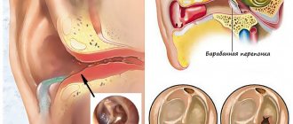
The middle ear is a part of the human auditory system, which is located between the outer and inner ear and is formed by the tympanic cavity with the auditory ossicles (hammer, incus, stapes). It is entrusted with the function of converting air vibrations into liquid vibrations, which are perceived by the hearing aid in the inner ear.
Review
Acute otitis media is most often a bacterial or viral infection that affects the middle ear, the air-filled space behind the eardrum that contains tiny vibrating bones. Children are more likely than adults to suffer from this disease.
Acute otitis media is often painful due to inflammation and fluid buildup in the middle ear.
Since ear infections often clear up on their own, treatment may begin with pain management and monitoring the problem. Otitis media in infants, and in severe cases in general, often requires antibacterial drugs. Long-term inflammation—persistent fluid in the middle ear and frequent infections—can cause hearing problems and other serious complications.
About the nature of sound
Sound is a wave that propagates through a medium and creates mechanical vibrations. Actually, we perceive these vibrations with the organ of hearing. Like any wave, sound is characterized by frequency and amplitude. Amplitude is the loudness of a sound and is measured in decibels (dB). Frequency is defined as the number of vibrations per second and is expressed in hertz (Hz). The higher the frequency, the higher the sound, and vice versa. A person is able to hear sound in a wide frequency range, but only sounds from 125 to 8000 Hz are important for life. For example, sound waves in the range of 500-4000 Hz correspond to the human voice.
The sound of a piccolo flute, a child's voice, birdsong, whispers are high frequencies, the sound of a double bass, the growling of animals, and thunder are low frequencies.
A decrease in the range of audible frequencies can change for various reasons; this is associated with changes in the inner ear (cochlea) and is called hearing loss.
Symptoms
Symptoms of inflammation usually develop quickly.
Signs and symptoms common in children include:
- pain in the ears, especially when lying down;
- poor sleep;
- crying more than usual;
- difficulty listening or responding to sounds;
- loss of balance;
- fever (38 C or higher);
- discharge of fluid from the ear;
- headache;
- loss of appetite.
Common symptoms in adults include:
- earache;
- release of fluid;
- hearing loss.
Causes
Inflammation in the middle ear is caused by bacteria or viruses. Often caused by a cold, flu, or allergies, it causes fluid to accumulate and swell in the nasal passages, throat, and auditory tubes.
The role of the auditory tube
The auditory tubes are a pair of narrow tubes that extend from the middle ear to the nasopharynx, behind the nasal passages. The laryngeal end of the tubes opens and closes to regulate air pressure in the middle ear and its drainage function.
Swelling, inflammation, and mucus in the eustachian tubes from an upper respiratory infection or allergies can block them, causing fluid to accumulate in the middle ear.
Ear infections are more common in children, in part because their eustachian tubes are narrower and more horizontal—factors that make it difficult for fluid to drain out of them, causing them to become clogged.
Role of adenoids
Adenoids are two small pads of tissue high at the back of the nose that play a role in the activity of the immune system. This feature makes them especially vulnerable to infection, inflammation and swelling.
Because the adenoids are located near the opening of the eustachian tubes, inflammation or enlargement of the adenoids can block the eustachian tubes, thereby facilitating middle ear infection. Inflammation of the adenoids will play a role in the development of otitis media in children because children have relatively large adenoids.
Other conditions that may be associated with an ear infection or the result of similar middle ear problems include the following:
- Otitis with effusion is inflammation and fluid formation (effusion) in the middle ear without bacterial or viral infection. This can happen because fluid accumulation persists after the ear infection has cleared. It may also occur due to some dysfunction or non-infectious blockage of the eustachian tubes.
- Chronic otitis media with effusion occurs when fluid remains in the middle ear and continues to return without bacterial or viral infection. This makes children susceptible to new ear infections and can affect their hearing.
- Chronic suppurative otitis is a persistent ear infection that often leads to rupture or perforation of the eardrum.
Embryology
At the beginning of the 2nd month of the intrauterine period, a tubal-tympanic recess is formed in the first pharyngeal pouch (I gill slit). Subsequently, its distal part expands and gives rise to the tympanic cavity, and the narrow proximal part turns into the Eustachian tube. The cartilaginous anlages of the auditory ossicles are formed in the embryonic connective tissue above the primary tympanic cavity; starting from the 7th month. During the prenatal period, the tissue surrounding them undergoes resorption, as a result of which the tympanic cavity expands and the auditory ossicles are suspended in a cay on a thin plate of epithelium. The tympanic cavity and auditory ossicles reach their final size at the end of the prenatal period.
Risk factors
- Age. Children ages 6 months to 2 years are more susceptible to ear infections because of the size and shape of their eustachian tubes and because their immune systems are underdeveloped.
- Children's group. Children in group settings are more likely to get colds and ear infections than children who stay at home because they are exposed to more infections, such as colds.
- Baby food. Babies who drink from a bottle, especially while lying down, are more likely to develop ear infections than babies who are breastfed.
- Seasonal factors. Ear infections are most common during the fall and winter, when colds and flu are common. People with seasonal allergies are at greater risk of ear infections during seasonal allergies.
- Contaminated air. Exposure to tobacco smoke or high levels of air pollution can increase the risk of ear infection.
Complications
Frequent infections and constant build-up of fluid can lead to serious complications:
- Hearing impairment. An ear infection is often accompanied by mild hearing loss. Persistent infection or fluid in the middle ear can lead to more significant hearing loss. If there is some permanent damage to the eardrum or other structures of the middle ear, permanent hearing loss may occur.
- Delayed speech development. When hearing is temporarily or permanently impaired in infants and young children, they may experience delays in speech, social and mental development.
- Spread of infection. Infections that do not respond well to treatment may spread to nearby tissue. Infection of the mastoid process, the bony protuberance behind the ear, is called mastoiditis. This infection can cause bone damage and pus-filled cavities. Rarely, serious middle ear infections spread to other tissues in the skull, including the brain or meninges surrounding the brain (meningitis).
- Rupture of the eardrum. Most tears heal within 72 hours. In some cases, surgery is necessary.
Prevention
The following tips may reduce your risk of developing otitis media:
- Prevent colds and other illnesses. Teach your children to wash their hands frequently and thoroughly, and not to share food or drinks. Teach your children to cough or sneeze into their hand. If possible, limit the time your child spends in a group setting. Try to keep your child at home when he is sick.
- Avoid secondhand smoke. Try to stay in non-smoking areas.
- Breastfeeding your baby. If possible, breastfeed your baby for at least six months. Breast milk contains antibodies that may provide protection against ear infections.
- If you bottle feed, keep your baby upright. Avoid propping bottles in your baby's mouth while he is lying down. Do not put the bottle in the crib with your baby.
- Talk to your doctor about getting vaccinated. Ask your doctor about which vaccines are appropriate for your child. Seasonal flu shots, pneumococcal and other bacterial vaccines can help prevent ear infections.
Diagnostics
Your doctor may diagnose an ear infection or other condition based on the symptoms you describe. To clarify the diagnosis and identify the cause of the disease, the doctor will use a special light-up instrument (otoscope) to look at the ears, throat and nasal passages.
Otoscopy
An otoscope is a specialized instrument that allows the doctor to examine the ear cavity and judge the presence of fluid behind the eardrum. Using an otoscope, the doctor can gently blow air against the eardrum. Typically, this air causes the eardrum to move. If the middle ear is filled with fluid, the doctor will hardly notice any movement of the eardrum.
Additional tests
Your doctor may perform other diagnostic tests if there is any doubt about the diagnosis.
- Tympanometry. This test measures the movement of the eardrum. The device that closes the ear canal regulates the air pressure in the canal, thereby causing the eardrum to move. The device quantifies how well the eardrum moves and a measure of the pressure inside the middle ear.
- Acoustic reflexometry. This test shows how the mobility of the eardrum changes in response to sound stimulation.
- Paracentesis. Sometimes the doctor may use a thin needle to pierce the eardrum to drain fluid from the middle ear, a procedure called paracentesis. If the infection does not respond well to previous treatment, then taking a smear of this fluid to determine the type of bacteria helps to choose the right antibiotic.
- Other tests. If your child has had persistent ear infections or persistent fluid build-up in the middle ear, your doctor may refer you to an audiologist, speech therapist, or therapist for testing of hearing, speech skills, language comprehension, or developmental abilities.
Anatomy
Rice.
1. Medial (labyrinthine) wall of the right tympanic cavity, stapes and mastoid cave: 1 - tensor tympani muscle; 2 - hemicanal of the tensor tympani muscle (partially opened); 3 3 hemicanal of the Eustachian tube; 4 - cape groove; 5 - cape; 6 — drum cells; 7 - dimple of the cochlear window; 8 - head of the stirrup; 9 - tendon of the stapedius muscle; 10 - mastoid cells; 11—tympanic sinus; 12 - pyramidal elevation; 13 — protrusion of the facial canal; 14 - protrusion of the lateral semicircular canal; 15 - mastoid cave; 16 — rear leg of the stirrup; 17 — stirrup membrane; 18 - tendon of the tensor tympani muscle (cut off); 19 - supratympanic recess. S. u. is located in the temporal bone and its formations are in close proximity to large vessels and nerves passing through the base of the skull (tsvetn. Fig. 1). S. u. consists of a tympanic cavity filled with air with auditory ossicles located in it, the eustachian tube, air cavities and the mastoid process.
The tympanic cavity (cavitas tympanica) is a space with a volume of approximately 0.8 - 1 cm3; located between the eardrum (see) and the bony labyrinth of the inner ear (see). It communicates through the eustachian tube (see Auditory tube) with the nasal part of the pharynx (see), and through the mastoid cave (see Mastoid process) with the mastoid cells. In a newborn, the tympanic cavity occupies a more horizontal position in the skull than in an adult.
There are 6 walls in the tympanic cavity. The upper, tegmental wall (paries tegmentaiis) is formed by a thin bone plate (roof of the tympanic cavity, T.; tegmen tympani) of the pyramid of the temporal bone. The lower, jugular wall (paries jugularis) borders the jugular fossa (fossa jugularis) of the pyramid of the temporal bone, in which lies the upper bulb of the internal jugular vein (bulbus v. jugularis sup.). In the posterior mastoid wall (paries mastoi-deus) there is an entrance to the mastoid cave (aditus ad antrum). On the medial wall in the cave there is a protrusion of the lateral semicircular canal (prominentia canalis semicircularis lat.), below which there is a protrusion of the facial nerve canal (prominence of the facial canal, T.; prominentia canalis facialis). In the superomedial part of the mastoid wall of the tympanic cavity, a pyramidal eminence (eminentia pyramidalis) is formed, inside which lies the stapedius muscle (m. stapedius). The tendon of this muscle emerges from the hole in the pyramidal eminence. The anterior wall of the tympanic cavity is called carotid (paries caroticus), because it forms the wall of the carotid canal, into which the internal carotid artery (a. carotis int.) passes. The carotid wall contains tympanic cells (cellulae tympanicae); the tympanic opening of the eustachian tube (ostium tympanicum tubae auditivae) is located on it. The lateral membranous wall (paries membranaceus) is mostly occupied by the tympanic membrane (membrana tympani).
Rice. 2. Lateral (membranous) wall of the right tympanic cavity, auditory ossicles, their ligaments and recesses of the tympanic cavity: 1 - roof of the tympanic cavity; 2 - superior incus ligament; 3 — anvil body; 4 - posterior malleus fold; 5 - supratympanic recess; 6 - short leg of the anvil; 7 - posterior ligament of the incus; 8 - mastoid cave; 9 - posterior recess of the eardrum; 10 — long leg of the anvil; 11—lenticular process; 12 - facial nerve (cut off); 13 - mastoid wall; 14 - eardrum (tense part); 15 - superior bulb of the internal jugular vein; 16 - internal carotid artery; 17 — drum cells; 18 — carotid-tympanic artery (cut off); 19 - carotid-tympanic canaliculus; 20 - fibrocartilaginous ring; 21 - carotid wall; 22 — hammer handle; 23 - anterior recess of the eardrum; 24 - bony part of the Eustachian tube; 25 - place of attachment of the tensor tympani muscle; 26 - muscle that strains the tympanic membrane (cut off); 27 - anterior malleus fold; .28 - drum string; 29 — neck of the malleus; 30 - incus-hammer joint; 31 — head of the malleus; 32 - superior ligament of the malleus
The medial, or labyrinthine, wall (paries labyrinthicus) is the outer wall of the bony labyrinth (color book fig. 2). It forms a rounded promontory (promontorium), which corresponds to the base of the cochlea. Above the promontory there is a dimple for the window of the vestibule (fossula fenestrae vestibuli), forming a niche with the window of the vestibule (fenestra vestibuli), or oval window, which includes the base of the stapes. Below the promontory, in the niche-shaped dimple of the cochlea window (fossula fenestrae cochleae), there is a cochlea window (fenestra cochleae), or round window. It is closed by the secondary tympanic membrane (membrana tympani secundaria) and leads to the scala tympani (scala tympani). The shape of the dimple of the cochlear window and the window itself is individually variable.
The tympanic cavity is conventionally divided into three sections: lower, middle and upper. The lower section, hypotympanum, is a part of the tympanic cavity located between the jugular wall and the lower edge of the eardrum. The middle section, mesotympanum, is limited by two horizontal planes, conditionally drawn through the lower and upper edges of the eardrum. The upper section, epitym panum, is located between the upper edge of the tympanic membrane and the roof of the tympanic cavity.
Rice. 1. Right auditory ossicles (top and inside view): I - malleus, II - incus, III - stirrup; 1 - short leg of the anvil; 2 — anvil body; 3 - long leg of the anvil; 4 - incus-stapedial joint; 5 - posterior leg of the stirrup; 6 — base of the stirrup; 7 - front leg of the stirrup; 8 — hammer handle; 9 - anterior process of the malleus; 10 — head of the hammer; 11 - incus-hammer joint.
The auditory ossicles (ossicula auditus) lie in the upper part of the tympanic cavity. They are strengthened by ligaments and connected to each other by joints, forming a movable three-membered chain between the tympanic membrane and the window of the vestibule (Fig. 1). In newborns, some connective tissue remains around the auditory ossicles, so their mobility is limited until about 6 months. postnatal period.
The shape of the bones varies individually. Auditory ossicles of the right and left S. often have different shapes and sizes.
The malleus (malleus) represents the outer link of this chain. It consists of a head (caput mallei), a handle (manubrium mallei) and two processes - a thin, long anterior process (processus ant.) and a short lateral process (processus lat.). The lower end of the handle of the malleus is fused with the tympanic membrane, the anterior process enters the petrotympanic fissure (fissura petrotympanica), the lateral process is adjacent to the tympanic membrane. The malleus is fixed by the anterior, superior and lateral ligaments (ligg. mallei ant., sup. et lat.).
The anvil (incus) is the middle link in the chain of auditory ossicles. It consists of a body (corpus incudis) and two legs (long and short). The body of the incus has an articular surface, the edges with the articular surface of the malleus form the incudomallearis joint (art. incudomallearis). The long leg (crus longum) has a curved end, on which there is a lenticular process (processus lenticularis), articulated with the stirrup. The short leg (crus breve) is connected through a ligament to the posterior wall of the tympanic cavity.
The stapes forms the internal link of the chain of auditory ossicles. It consists of a head (caput stapedis), anterior and posterior legs (crura ant. et post.) and a base (basis stapedis). The head of the stapes forms an incudostapedia joint (art. incudostapedia) with the lenticular process of the incus. Both legs, together with the base of the stirrup, limit the opening in which the membrane of the stirrup (membrana stapedis) is located. The base of the stapes is fixed in the window of the vestibule by the annular ligament (lig. annulare stapedis). This ligament is divided into anterior, posterior and lateral parts, differing in the direction of the fibers.
The movements of the auditory ossicles are regulated by the muscle that strains the eardrum (m. tensor tympani) and the stapedius muscle (m. stapedius).
Rice. 3. Vessels and nerves of the medial (labyrinthine) wall of the right tympanic cavity (the facial and carotid canals are opened): 1 - stylomastoid artery; 2 and 15 - tympanic nerve; 3 — knot-elbow; 4 - connecting branch of the facial nerve; 5 - greater petrosal nerve; 6 - superior tympanic artery; 7 - lesser petrosal nerve; 8 - semi-canal of the tensor tympani muscle; 9 - muscle that strains the tympanic membrane (cut off); 10 - carotid-tympanic nerve; 11—tubal branch of the tympanic plexus; 12 - internal carotid artery; 13 - carotid tympanic artery; 14 - semi-canal of the Eustachian tube; 16 - internal carotid plexus; 17 - inferior tympanic artery; 18 - glossopharyngeal nerve (lower node); 19 - tympanic plexus; 20 - jugular wall; 21 - internal jugular vein; 22 - cape; 23 — dimple of the cochlear window; 24 - posterior tympanic artery; 25 - drum string; 26 - stapedius nerve; 27 - stapedius muscle; 28 - stirrup; 29 — protrusion of the lateral semicircular canal; 30 - mastoid cave.
The mucous membrane of the tympanic cavity covers all formations located in it and continues into the mucous membrane of the Eustachian tube and mastoid cells (tsvetn. Fig. 3). Moving onto the auditory ossicles, the mucous membrane forms folds - the anterior and posterior hammer (plicae malleolares ant. et post.), the incus fold (plica incudis) and the stapes fold (plica stapedis). At the point where the tympanic string passes, a fold of the tympanic string (plica chordae tympani) is formed.
The supratympanic recess, or attic (recessus epitympanicus, s. at-ticus), has the shape of a dome and extends from the roof of the tympanic cavity to the tendon of the tensor tympani muscle. It contains the head of the malleus and the body of the incus. The anterior recess of the tympanic membrane (recessus membranae tympani ant.) is located between the tympanic membrane and the anterior ligament of the malleus. The posterior recess (recessus membranae tympani post.) is located between the tympanic membrane and the posterior ligament of the malleus. The superior recess (recessus membranae tympani sup.), or Prussian space, lies between the head of the malleus, the body of the incus and the loose part (pars flaccida) of the tympanic membrane.
Rice. 4. Schematic representation of the relationship of the right middle ear with the inner ear and adjacent vessels and nerves (external view). Orange indicates the projection of the formations of the middle ear, green - the inner ear, blue and blue - venous vessels, pink and red - arteries: 1 - anterior semicircular canal; 2 - vestibule; 3 - snail; 4 - trigeminal nerve ganglion; 5 - Eustachian tube; 6 - medial plate of the pterygoid process; 7 - tympanic cavity; 8 - internal carotid artery; 9 - styloid process; 10 - internal jugular vein; 11 - facial nerve; 12 - mastoid process; 13—external auditory opening; 14 - lateral semicircular canal; 15 - sigmoid sinus; 16 - mastoid cave; 17 - posterior semicircular canal; 18 - pyramid of the temporal bone
Innervation
. The mucous membrane of the tympanic cavity, eustachian tube and mastoid cells is innervated by the tympanic plexus (plexus tympanicus), located on the labyrinthine wall of the tympanic cavity (color. Fig. 4). This plexus contains small groups of multipolar neurons; formed by the tympanic nerve (n. tympanicus), which is a branch of the glossopharyngeal nerve, the connecting branch of the facial nerve (r. communicans) and the carotid-tympanic nerves (nn. caroticotympanici), extending from the internal carotid plexus (plexus caroticus int.). The tensor tympani muscle is innervated by a branch of the mandibular nerve (n. musculi tensoris tympani), the stapedius muscle by a branch of the facial nerve - by the stapedius nerve (n. stapedius).
Blood supply
S. u. arteries carry out: the inferior tympanic (a. tympanica inf.), branching from the ascending pharyngeal (a. pharyngea ascendens), stylomastoid (a. stylomastoidea) and posterior tympanic (a. tympanica post.) - from the posterior auricular (a. auricularis post.), anterior tympanic (a. tympanica ant.) - from the maxillary (a. maxillaris), superior tympanic (a. tympanica sup.) - from the middle meningeal (a. meningea media) and carotid-tympanic arteries (aa. caroticotympanicae ) - from the internal carotid artery. Venous outflow occurs through the tympanic veins (vv. tympanicae) into the pterygovenous plexus.
Lymphatic drainage
carried out through lymphatic vessels approaching the mastoid, retropharyngeal and deep parotid lymph nodes (nodi lymphatici mastoidei, retropha-ryngeales et parotidei profundi).
What does the diagnosis mean?
Acute otitis media. The doctor makes this diagnosis if he observes signs of fluid in the middle ear, if there are signs or symptoms of infection, and if the onset of symptoms was relatively unexpected.
Otitis with effusion. If the diagnosis is otitis media with effusion, the doctor has detected fluid in the middle ear, but there are currently no signs or symptoms of infection.
Chronic purulent otitis media. If the doctor makes this diagnosis, it means that he has discovered that a persistent ear infection is leading to a rupture or perforation of the eardrum.
Physiology
As the sound-conducting part of the hearing organ (see), S. at. carries out the transmission of sound vibrations from the environment to the auditory analyzer (see). The sound wave causes vibrations of the eardrum, which are transformed through the chain of auditory ossicles and transmitted to the base of the stapes; synchronous movement of the stapes causes vibrations in the perilymph, transmitted to the organ of Corti (see) and converted into nerve impulses. Ligamentous-muscular system S. at. also plays a protective role, protecting the inner ear from subthreshold and suprathreshold sound vibrations. Pneumatic system of cells of S. u. plays the role of a resonator of sound waves.
The Eustachian tube equalizes the pressure in the tympanic cavity with atmospheric pressure. The ciliated epithelium covering it has a protective function, protecting the middle ear from the penetration of infectious agents from the nasopharynx.
Treatment
Some ear infections resolve without antibiotic treatment. What's best for your child depends on many factors, including the child's age and the severity of symptoms.
Waiting tactics
Ear infections usually improve within the first two days, and most infections clear up on their own within one to two weeks without any treatment. The American Academy of Pediatrics and the American Academy of Family Physicians recommend waiting and seeing the outcome in the following cases:
- Children 6 to 23 months of age with mild internal pain in one ear for less than 48 hours and a temperature of less than 39°C.
- Children aged 24 months and older with mild internal ear pain on one or both sides for less than 48 hours and a temperature less than 39°C.
Some evidence suggests that antibiotic treatment may be effective for children with ear infections. Talk to your doctor about the benefits of antibiotics, their side effects, and the emergence of bacteria that are resistant to antibiotics when you overuse them.
Pain management
To reduce pain from an ear infection, your doctor may recommend using paracetamol (Calpol, Panadol, and others) or ibuprofen (Nurofen, Ibuclin, and others) to relieve the pain. Use medications as directed on the label. Use caution if giving aspirin to children or teenagers. Children and teenagers recovering from chickenpox or flu-like symptoms should never take aspirin because aspirin is associated with Reye's syndrome.
Antibiotic therapy
Your doctor may recommend treating otitis media with antibiotics in the following situations:
- Children 6 months of age and older with moderate/severe ear pain for more than 48 hours or a temperature of 39°C or higher.
- Children 6 to 23 months of age with mild internal ear pain lasting less than 48 hours and a temperature less than 39°C.
- Children aged 24 months and older with mild internal ear pain lasting less than 48 hours and a temperature less than 39°C.
In children under 6 months of age with confirmed acute otitis media, treatment should be started with antibiotics without an initial wait and observation period. Even after symptoms have improved, be sure to complete the course of antibiotics as recommended. Failure to follow this recommendation may result in recurrent infections and bacterial resistance to antibiotic therapy. Talk to your doctor about what to do if you accidentally miss an antibiotic dose.
Bypass surgery
If your child has recurrent otitis media or otitis media with effusion, the doctor may recommend a procedure to remove fluid from the middle ear. Recurrent otitis media can be said to occur if there have been three episodes of infection in six months or four episodes in a year, or at least one episode in the last six months. Recurrent otitis with effusion is a constant build-up of fluid in the ear after recovery or in the absence of any infection.
During an outpatient surgical procedure called a shunt, the surgeon creates a tiny hole in the eardrum that drains fluid from the middle ear. A tiny tube (shunt) is placed in the hole to help ventilate the middle ear and continue to drain fluid for the required amount of time. The shunt is installed for 3-6 months and removed surgically. The eardrum usually closes again after the shunt is removed.
Treatment of chronic suppurative otitis media
Chronic suppurative otitis media leads to perforation of the eardrum and is difficult to treat. It is often treated with antibiotics given as drops.
ACUTE OTITIS MEDIUM
What pathogenetic factor is leading in the development of otitis media?
What are the stages of acute otitis?
Is there a drug of choice in the treatment of acute otitis media?
What can be considered a criterion for recovery in acute otitis media?
Inflammatory diseases of the middle ear occur in all age groups. Among the total number of patients with various diseases of the ENT organs, acute otitis media is diagnosed in 20-30% of cases. This disease develops especially often in children, with the peak incidence occurring between 6 and 18 months; Before the age of three, 90% of children experience acute inflammation of the middle ear at least once [1]. Timely diagnosis and adequate treatment of otitis are often carried out with the participation of a general practitioner, so knowledge of the features of diagnosis and treatment of these diseases is extremely important to prevent possible adverse consequences of otitis.
Based on the nature of inflammation, catarrhal, serous and purulent otitis media are distinguished. Fibrinous, hemorrhagic inflammation and its mixed forms are also possible [4].
More often than other types of inflammation of the middle ear, catarrhal otitis media is observed, also called eustachitis, tubootitis, salpingootitis, etc. and developing as a result of dysfunction of the auditory tube. The cause of catarrhal otitis media is a more or less pronounced dysfunction of the auditory tube, leading to impaired ventilation of the tympanic cavity. This is possible for acute respiratory, acute infectious diseases, as well as influenza. The spread of infection from the upper respiratory tract to the mucous membrane of the auditory tube can lead to disruption of its patency, primarily in the area of the pharyngeal mouth. Tubootitis can also be caused by sudden changes in atmospheric pressure during the ascent and descent of an aircraft (aerootitis), or during the diving and ascent of divers and submariners (mareotitis).
Impaired ventilation of the tympanic cavity leads to the fact that the air contained in it is absorbed by the mucous membrane, and its replenishment is difficult due to the obstruction of the patency of the auditory tube. As a result, the pressure in the tympanic cavity decreases.
The main complaints with tubo-otitis are ear congestion, decreased hearing, sometimes tinnitus, autophony (the resonance of one’s own voice in the affected ear). Ear pain is usually absent or mild, the general condition remains satisfactory. Otoscopy data are very important for making a diagnosis. In this case, a retraction of the tympanic membrane is noted, accompanied by the following characteristic signs: apparent shortening of the handle of the malleus, a sharp protrusion of the short process towards the ear canal; disappearance or deformation of the light cone. Sometimes a radial injection of the vessels of the tympanic membrane along the handle of the malleus or a circular injection in the area of the annulus tympanicus is determined.
Hearing in acute tubo-otitis is slightly reduced, due to the type of sound conduction disturbance, mainly at low frequencies. Sometimes patients note improved hearing after yawning or swallowing saliva, accompanied by the opening of the lumen of the auditory tube.
Acute inflammatory processes of the upper respiratory tract are the causes of the development of temporary dysfunctions of the auditory tube. These disorders are more persistent in adenoid vegetations, various chronic diseases of the nasal cavity and paranasal sinuses (chronic purulent or polypous rhinosinusitis, especially with choanal polyps, deviated nasal septum, hypertrophy of the posterior ends of the inferior turbinates, etc.), and tumors of the nasopharynx.
Against the background of dysfunction of the auditory tube, exudative otitis media can develop, characterized by the presence of serous-mucosal effusion in the tympanic cavity. Today, various designations for the disease are accepted: “secretory otitis media,” “serous otitis media,” “mucous,” etc. The leading pathogenetic factor of exudative otitis media is also a persistent violation of the ventilation and drainage functions of the auditory tube. The very name of this form of otitis indicates increased secretion of mucus and a protracted course of the disease. Its characteristic signs are the appearance of a thick viscous secretion in the tympanic cavity, slowly increasing hearing loss and the absence of perforation of the eardrum. In the development of the disease, along with persistent tuberculous dysfunction, a change in the immunobiological properties of the body, a decrease in general and local resistance, also plays an important role.
Against the background of rarefaction in the non-ventilated tympanic cavity, transudation occurs, the migration of a small number of neutrophilic leukocytes and lymphocytes; transudate leaks into the tympanic cavity.
During this period, the patient notes ear congestion; sometimes mild autophony and hearing loss are observed. During otoscopy, the eardrum is retracted, gray in color, with injected vessels along the handle of the malleus; sometimes air bubbles are visible in the tympanic cavity.
The appearance of fluid in the tympanic cavity is subjectively manifested by a feeling of fullness and pressure in the ear, sometimes noise in the ear and more severe conductive hearing loss. There is often a sensation of fluid transfusion (splashing) when the head position changes, accompanied by improved hearing. This can be explained by the fact that when the head is tilted, the fluid in the tympanic cavity moves, thereby opening up the niches of the windows of the labyrinth, which leads to improved hearing. During otoscopy during this period, fluid is often visible through the eardrum, the level of which is determined in the form of an arcuate line that moves when the position of the head changes.
Restoring ventilation of the tympanic cavity can lead to recovery. However, with continued disruption of the tubular function, secretory otitis media takes a chronic course, turning into fibrosing otitis media, characterized by the appearance of a scar process in the tympanic cavity - the so-called adhesive otitis media develops, leading to severe persistent hearing loss.
Diagnosis of exudative otitis media is complex and not always timely. This is due to the asymptomatic course of the disease, which does not cause any significant pain and does not lead to disruption of the general condition of the patient. The patient gets used to a moderate decrease in hearing in one ear, gradually increasing, and stops paying attention to it, especially if the other ear hears normally. The doctor should take into account that the asymptomatic course of exudative otitis media is now becoming more common. Otoscopy, preferably with magnification, is of great importance in diagnosis. To clarify the diagnosis, a study of the function of the auditory tube is performed using publicly available tests; Impedance measurement is also performed, which reveals a flattened curve. Hearing is examined using tuning forks and audiometry.
Acute purulent otitis media is an acute inflammation of the mucous membrane of the tympanic cavity, and all parts of the middle ear are involved to one degree or another in the process.
This is a widespread disease of the middle ear, which can be either mild or, rapidly developing, cause a severe general inflammatory reaction of the body. However, in both cases, it often leaves behind an adhesive process, accompanied by difficult-to-treat hearing loss, or becomes chronic, often progressive, which also leads to hearing loss and often to severe complications. A distinctive feature of this disease at present is a less acute onset and sluggish course, and in childhood - a tendency to relapse.
The cause of the disease is infection in the tympanic cavity with reduced local and general resistance. Most often (up to 80%) the causative agents of acute purulent otitis media in adults and children are S. pneumoniae and H. influenzae, somewhat less often - M. catarrhalis, S. pyogenes, S. aureus or associations of microorganisms. Viral otitis is observed mainly during epidemics of viral diseases.
The most common route of infection is tubogenic - through the auditory tube. With various general infectious diseases, local inflammatory processes in the upper respiratory tract, the protective function of the epithelium of the auditory tube is disrupted, and the microflora penetrates into the tympanic cavity. Less commonly, the infection enters the middle ear through a damaged eardrum due to injury or through a wound to the mastoid process. In this case, they talk about traumatic otitis media. The third route of infection into the middle ear, hematogenous, is relatively rarely diagnosed; it is possible with infectious diseases such as influenza, scarlet fever, measles, typhoid, tuberculosis.
The inflammatory reaction in acute purulent otitis media from the very beginning affects not only the mucous membrane of the middle ear, but also the periosteum closely adjacent to it. The middle ear is filled with inflammatory exudate, which may initially be serous and then becomes purulent. The mucous membrane becomes sharply thickened, erosions and ulcerations appear on its surface. At the height of inflammation, the tympanic cavity is filled with exudate, granulations and thickened mucous membrane. If the drainage function of the auditory tube is impaired, this leads to bulging of the eardrum; as a result of strong pressure of purulent exudate and circulatory disorders, melting of some area and perforation of the eardrum often occurs, followed by otorrhea.
As the inflammatory changes subside, the amount of discharge decreases and suppuration stops completely. After the discharge from the ear stops, the perforation of the eardrum may heal, but ear congestion persists for some time. The criterion for recovery is normalization of the otoscopic picture and complete restoration of hearing.
In its course, acute purulent otitis media can be mild and quickly resolving, sluggish and protracted, acute and violent; as a rule, it ends with complete recovery; if this does not happen, it can cause chronic otitis media. In some cases, acute purulent otitis media is complicated by mastoiditis or even the development of intracranial complications or sepsis, although the latter conditions more often occur with chronic purulent inflammation of the middle ear.
The clinical picture of a typical acute purulent otitis media is characterized by a staged course. Local and general symptoms of the disease are expressed differently, depending on the stage and severity of the process. It is customary to distinguish pre-perforation, perforation and reparative stages of acute purulent otitis media. The process does not always go through all three stages. Thanks to the mobilization of the body's natural defenses, as well as during intensive therapy, the disease can already acquire an abortive course at the first stage.
The initial, pre-perforative stage of the disease is characterized by pronounced local and general symptoms. The leading complaint is pain in the ear, often very sharp, radiating to the temple and crown. Steadily growing, it sometimes becomes painful and unbearable. Pain occurs as a result of inflammatory infiltration of the mucous membrane of the tympanic cavity and the accumulation of exudate in it; in this case, irritation of the receptor endings of the branches of the trigeminal and glossopharyngeal nerves occurs. Sometimes there is pain on palpation and percussion of the mastoid process, which is caused by inflammation of its mucous membrane. At the same time, congestion and noise in the ear occur, and conductive hearing loss is detected with a slight deterioration in bone conduction of sound. With influenza, as well as measles and scarlet fever, the inner ear is sometimes involved in the process, which is manifested by a more significant impairment of sound perception. During this period, the general condition of the patient is often disturbed - signs of intoxication appear, body temperature rises to 38-39°C, changes characteristic of the inflammatory process are detected in the peripheral blood.
During otoscopy, the injection of blood vessels along the handle of the malleus and the radial vessels of the membrane is first visible, accompanied by shortening of the light cone. Then the hyperemia of the eardrum increases, becomes diffuse, its identifying points disappear, the membrane protrudes, becomes infiltrated, and sometimes becomes covered with a whitish coating. The duration of the initial stage of acute otitis media ranges from several hours to two to three days.
The perforated stage is characterized by perforation of the eardrum and the appearance of suppuration. In this case, the pain in the ear quickly subsides, the patient’s well-being improves, and the body temperature decreases. Discharge from the ear is initially profuse, mucopurulent, sometimes mixed with blood. During otoscopy, a so-called “pulsatile reflex” can be observed, when pus enters through the perforation in portions, synchronously with the pulse.
Gradually, the amount of discharge decreases, it becomes thick and acquires a purulent character. Suppuration usually lasts five to seven days. Perforation in acute otitis media is usually small; more extensive perforations occur with scarlet fever, measles, and tuberculosis.
The reparative stage is characterized not only by the cessation of suppuration and, in most cases, spontaneous scarring of the perforation, but also by the gradual restoration of hearing. Along with the cessation of discharge, hyperemia and infiltration of the eardrum disappear, its shine appears, and identifying contours become visible. Small perforations (up to 1 mm) close quite quickly, leaving no marks. With a large perforation, the middle fibrous layer at the site of the defect usually does not regenerate; in this case, if the perforation does close, this area looks atrophic, sometimes there are deposits of lime salts. Fibrous adhesive changes after otitis media often remain in the tympanic cavity itself, limiting the mobility of the auditory ossicles.
The typical course of acute purulent otitis media can be disrupted at any stage of the process. In some cases, the disease immediately takes on a sluggish, protracted nature, accompanied by mild general symptoms. Perforation of the eardrum does not occur, but a viscous, thick secretion accumulates in the tympanic cavity, which is difficult to evacuate. Following this, an adhesive (adhesive) process often develops in the tympanic cavity. Sometimes, on the contrary, from the very beginning the course of the disease can be extremely severe, with high fever, severe headache, vomiting, dizziness and a sharp deterioration in general condition. In some cases, even before perforation, the infection can quickly spread from the middle ear into the cranial cavity and lead to severe intracranial complications.
If, despite the perforation of the eardrum, the temperature does not decrease and the patient’s condition does not improve, this is usually associated with the transition of inflammation to the mastoid process, i.e., with the development of mastoiditis. Suppuration that does not stop for a long time (three to four weeks), when after cleaning the ear pus fills the ear canal again, indicates empyema of the mastoid process, which, as a rule, causes melting of its bone bridges.
In the normal course of otitis, changes in the peripheral blood are manifested by moderate leukocytosis without a pronounced shift of the formula to the left, and a mild increase in ESR. With a severe disease, pronounced leukocytosis is observed, sometimes up to 20.0•109/l and higher, with a noticeable shift to the left.
Treatment of acute otitis media is carried out differentially, depending on the specific nosological form, the severity of clinical symptoms and the characteristics of the patient’s somatic status. In all cases, the earlier it is started, the higher the effectiveness of treatment.
Considering the important role of tubular dysfunction in the pathogenesis of various forms of acute otitis media, first of all, measures are taken to restore the aeration of the tympanic cavity through the lumen of the auditory tube. In order to reduce swelling of the mucous membrane in the area of its pharyngeal mouth, the patient is prescribed vasoconstrictor nasal drops: naphthyzin, sanorin, tizin, nazivin, a combination drug - polydex with phenylephrine, xylometazoline [3], etc. Sometimes antihistamines (diphenhydramine) help reduce swelling of the mucous membrane , Suprastin, Tavegil, Claritin, Telfast, etc.). To prevent infected mucus from entering the nasopharynx through the auditory tube into the tympanic cavity, the patient should be warned against blowing his nose too vigorously. The nose should be cleaned one nostril at a time, without straining too much. For the same purpose, in the presence of inflammatory changes in the nasopharynx, it is not recommended to blow the auditory tubes according to Politzer; preference is given to catheterization of the auditory tube, performed after thorough anemization of its pharyngeal mouth. Through a catheter, a few drops of a 0.1% solution of adrenaline or dexamethasone can be injected into the lumen of the auditory tube. In case of acute eustachitis, the complex of treatment measures includes various physiotherapeutic procedures: ultraviolet radiation, UHF on the nose, laser therapy on the area of the mouth of the auditory tube, pneumomassage of the eardrum.
With adequate treatment, eustachitis usually goes away within a few days. The effectiveness of treatment of this disease depends on the timely elimination of pathology of the nasal cavity, paranasal sinuses and nasopharynx, which provoke the occurrence and further course of tubo-otitis.
Treatment of exudative otitis media should be comprehensive. First of all, one should also strive to restore the function of the auditory tube using the above methods. To improve tubular function, the ears are blown using the Politzer method or through an ear catheter, with simultaneous massage of the eardrum using a Siegle funnel. Dexamethasone, antibiotics, and chymotrypsin are injected into the lumen of the auditory tube through a catheter. The introduction of proteolytic enzymes and lidase through endaural electrophoresis is quite effective. Vasoconstrictor drugs are used in the nose in the form of drops, but their long-term use is undesirable, since the substances they contain reduce the mucociliary activity of the ciliated epithelium of the nasal cavity and auditory tube.
The prescription of antihistamines is recommended only in cases where serous otitis media develops against the background of allergies. General strengthening agents and vitamins are also indicated. The complex of therapeutic measures has recently increasingly included the anti-inflammatory drug fenspiride (erespal) [2], as well as immunocorrectors (for example, polyoxidonium 0.006 g intramuscularly every other day - a total of six to ten injections; derinat intramuscularly 5.0 ml every other day - five injections).
In cases where the function of the auditory tube is not restored, the exudate does not resolve and hearing does not improve, surgical methods are used to evacuate secretions from the tympanic cavity. The most widely used is tympanic cavity bypass.
For acute purulent otitis media, treatment is complex, and it is prescribed depending on the stage of the disease. An outpatient regimen is recommended, and in case of a pronounced increase in temperature or general malaise, bed rest. If there is a suspicion of an incipient complication, the patient should be urgently hospitalized.
The measures discussed above are carried out in order to restore or improve the ventilation and drainage functions of the auditory tube.
An important place in the treatment of acute purulent otitis media is occupied by catheterization of the auditory tube. Blowing of the auditory tube in acute otitis media using a catheter is performed with the aim of draining the middle ear, eliminating the vacuum in the tympanic cavity that always occurs with this disease, as well as introducing medications into it. A solution of amoxicillin clavulanate and dexamethasone in a 3:1 ratio with the addition of one or two drops of a 0.1% adrenaline solution is injected through an ear catheter. Catheterization helps normalize the function of the auditory tube and eliminates inflammation. Catheterization is carried out from the very beginning of the disease, which often makes it possible to achieve an abortive course of the process; at stages II-III of acute inflammation of the middle ear, blowing with a catheter also gives a good therapeutic effect.
The basis of drug treatment for acute suppurative otitis media is antibiotic therapy.
The prescription of antibiotics is, of course, indicated already in the pre-perforation stage. The drug of choice for the treatment of uncomplicated forms of otitis in adults is amoxicillin orally, 0.25–0.5 g three times a day for 10 days. If there is no effect after three days of amoxicillin therapy, the drug should be changed to augmentin (0.375 or 0.625 g orally two to three times a day) or cefuroxime axetil (0.25 or 0.5 g orally twice a day) [5]. In case of intolerance to b-lactam antibiotics, modern macrolides are prescribed (Rulid 0.15 orally twice a day; spiramycin 1.5 million IU orally twice a day). For complicated forms of otitis, fluoroquinolone drugs of III-IV generations are prescribed: Sparflo 400 mg orally on the first day, then 200 mg per day; avelox 400 mg orally once a day, the duration of treatment depends on the severity of the disease.
Even with a sharp improvement in the patient’s general condition and mitigation of local symptoms, the course of antibiotic therapy should not be stopped prematurely; its duration should be at least 8-10 days. Premature discontinuation of drugs can lead to relapse of the disease and the formation of adhesions in the tympanic cavity, which, in turn, can lead to persistent hearing loss [6].
For pain relief in the initial (preperforative) stage of the disease, paracetamol 1 g is prescribed four times a day. A good analgesic effect at this stage is provided by an endaural microcompress according to Tsytovich with an alcohol-glycerin mixture (equal parts of a 3% alcohol solution of boric acid and glycerin). A gauze or cotton wool moistened with this mixture is inserted into the external auditory canal until it comes into contact with the eardrum; the opening of the external auditory canal is obstructed with cotton wool moistened with Vaseline or greasy cream. Such a compress can be left in the ear for 4–6 hours. Otipax ear drops, which contain lidocaine hydrochloride, phenazone, sodium thiosulfate, ethyl alcohol and glycerin, have a pronounced analgesic, anti-inflammatory and anti-exudative effect.
A warming semi-alcohol compress is also applied topically to the ear, which accelerates the resolution of the inflammatory process. However, when, after applying a compress, the patient notices increased pain in the ear, the compress should be removed immediately so as not to provoke the development of complications.
If, despite the treatment, the patient’s condition does not improve, he is still bothered by severe pain in the ear, a high temperature persists, pain is detected when pressing on the mastoid process, and a protrusion of the eardrum is observed during otoscopy, then paracentesis is performed - an incision of the eardrum. Paracentesis should be performed according to emergency indications when signs of irritation of the inner ear or meninges appear (dizziness, vomiting, severe headache, etc.).
In the presence of perforation of the eardrum, the main attention is to ensure the free outflow of pus. Turundas should be changed frequently, while clearing the ear canal of pus. If the pus thickens, it can be removed by infusing a 3% solution of hydrogen peroxide, which, when combined with pus, forms foam. Foam and pus are removed from the depths of the ear canal using a probe with cotton wool wound around it. The patient should be instructed how to independently remove purulent secretions from the depths of the external auditory canal two to three times a day.
Medications can be administered into the middle ear using transtympanic injection. The above mixture of antibiotic and dexamethasone (and subsequently enzymes that prevent the formation of scars in the tympanic cavity - trypsin, chymopsin, lidase, etc.) is poured into the external auditory canal in an amount of 1 ml and injected by gently pressing the tragus into the external opening of the auditory canal . In this case, the medicinal substance passes through the tympanic cavity, the auditory tube and can enter the nasal cavity and mouth. Catheterization and transtympanic drug injection are effective treatment methods.
For thick purulent discharge, mucolytics are prescribed internally (fluimucil, ACC, fluifort, sinupret), erespal is an anti-inflammatory drug that reduces hypersecretion and swelling of the mucous membrane and stimulates the function of the ciliated epithelium of the auditory tube. At home, physiotherapeutic procedures (UV irradiation, UHF or microwave therapy, laser therapy) and warm compresses on the ear also contribute to a faster recovery.
After removing the purulent secretion, a medicinal solution prescribed by a doctor, heated to 37°C, is poured into the ear. This can be a 0.5-1% dioxidine solution, cypromed ear drops, which contain the antibacterial drug ciprofloxacin with a wide spectrum of antibacterial action; Otofa drops containing the active substance rifamycin, etc. It is not recommended to prescribe alcohol drops in the second stage of otitis, since alcohol often causes irritation of the mucous membrane of the tympanic cavity and severe pain.
Suppuration usually stops after a few days, which marks the transition of the disease to the final reparative stage. The perforation of the eardrum most often closes, forming an inconspicuous scar. During this period, it is important to achieve as complete a hearing restoration as possible. Antibiotic therapy is canceled, ear toilet is stopped, thermal procedures are also completed. After the disappearance of perforation, the main attention should be paid to restoring the ventilation function of the auditory tube and increasing the body's resistance. The auditory tube is blown through the Politzer method or through a catheter, and it is possible to introduce enzyme preparations into the tympanic cavity that prevent the formation of adhesions. For the same purpose, pneumomassage of the eardrum is performed using a pneumatic Siegle funnel, and endaural iontophoresis with lidase. It is recommended to continue vitamin therapy; biostimulants are prescribed - apilak, actovegin, cigapan, etc.
To ensure the restoration of hearing function, control audiometry is performed. In a typical favorable course, recovery occurs with the elimination of the inflammatory process and complete restoration of hearing.
Literature.
- Bogomilsky M. R., Chistyakova V. R. Pediatric otorhinolaryngology // Textbook for universities. - M.: GEOTAR-MED, 2001. - P. 78-95.
- Levina Yu. V., Luchikhin L. A., Krasyuk A. A. The use of erespal in the treatment of exudative otitis media // Vestn. otorinolar., 2003. - 4. - pp. 35-37.
- Palchun V. T., Polyakova T. S., Luchikhin L. A. New dosage form of xylometazoline // Proceedings of the Russian Conference of Otolaryngologists November 19-20, 2002 - pp. 371-373.
- Palchun V. T., Magomedov M. M., Luchikhin L. A. Otorhinolaryngology. — M.: Medicine. - 2002. - P. 382-408.
- Strachunsky L. S., Kozlov S. N. Modern antimicrobial therapy // Guide for doctors. - M.: Borges, 2002. - 436 p.
- Butler CC, Williams RG The etiology, Pathophysiology and Management of Otitis Media with Effusion. — Curr. Infect. Dis. Rep., 2003, 3: 205-213.
L. A. Luchikhin, Doctor of Medical Sciences, Professor of Russian State Medical University, Moscow