To the reference book Sensorineural hearing loss is a lesion of one or more parts of the auditory analyzer.
Symptoms: hearing loss, tinnitus, dizziness and imbalance. Author:
- Sokolova Alla Vasilievna
otorhinolaryngologist, otosurgeon, doctor of the highest category
4.17 (Votes: 6)
Sensorineural hearing loss is a hearing loss that affects any of the sound-perceiving structures of the auditory analyzer. The conversion of sound wave energy in the organ of Corti of the cochlea (in some sources defined as sensorineural hearing loss) or the conduction of nerve impulses to the cerebral cortex may be impaired. Timely correction of pathological changes allows you to slow down or prevent the progression of the disease and avoid the development of complete deafness.
What is sensorineural hearing loss?
The perception of sounds is ensured by the coordinated work of all parts of the hearing organ. The outer ear (concha and ear canal) conduct sound waves to the middle ear. The eardrum converts the wave into mechanical vibrations. The auditory ossicles transmit these vibrations to the inner ear. The movement of fluid in the cochlea irritates the hair cells, which convert mechanical impulses into nerve impulses. The auditory pathways transmit signals to the temporal lobes of the brain, where the central part of the auditory analyzer is located. After processing impulses by cerebral structures, a person understands what he heard.
Sensorineural hearing loss (SNHL) is one of the common diseases and an urgent problem in otorhinolaryngology. It has been shown that 1–6% of the world's population suffers from severe hearing loss, which impedes social communication [2, 9]. At the same time, NHL dominates among all forms of hearing loss, accounting for 74% [6, 10, 18, 16]. It should be noted that every year the number of patients with various variants of NCT is steadily increasing [2, 9, 15]. This may be due to improved diagnostics (the widespread introduction of audiometric equipment into outpatient practice), but a real increase in the number of sensorineural hearing impairments cannot be ruled out.
Depending on the course, chronic, acute and sudden NST are distinguished. In chronic NHL, in most cases there is bilateral persistent hearing loss observed over many years with slow or rapid progression. In acute and sudden NTS, the process is one-sided and most often reversible.
The division of acute hearing loss into acute and sudden hearing loss is based mainly on the time factor: sudden hearing loss occurs within a few minutes to 12 hours. If this period is increased to a day, NST is considered acute. However, the causes of these forms of hearing loss are almost the same - they are predominantly vascular and inflammatory pathologies. That is, from a clinical point of view, there is no difference in the approaches to their treatment, which makes, in our opinion, inappropriate to distinguish between these two forms of NST.
Regardless of the course of NTS, it is not a nosological form, since it occurs against the background of many diseases (vertebrobasilar insufficiency; various infectious diseases - influenza, mumps, measles, herpes, meningococcal meningitis, syphilis, scarlet fever, etc.; blood diseases ; Meniere's disease; neuroma of the VIII pair of cranial nerves, otosclerosis, etc.); intoxication with various ototoxic drugs (aminoglycoside antibiotics, streptomycin, quinine preparations, cytostatics, loop diuretics, analgesics, etc.), household (nicotine, alcohol) and industrial (gasoline, aniline, fluorine, mercury, etc.) toxic substances; various types of traumatic effects (mechanical, acoustic and vibration injuries, barotrauma and air concussion); genetic abnormalities (hereditary hearing loss); age-related changes (presbycusis), etc. [12, 17–19, 25].
Unfortunately, there is still no consensus on both the pathogenesis of NTS and the treatment strategy. This is apparently due to the fact that NST is a functional response of the inner ear and other parts of the auditory analyzer to multifactorial pathological influences [3, 8, 9, 11].
The results of many years of research we obtained using modern methods of otoneurological, audiological (tone threshold, suprathreshold, speech and ultrasound [US] audiometry, impedance measurement, registration of various classes of otoacoustic emission and auditory evoked potentials, including electrocochleography), vestibulological (coordination of movements of the upper limbs, static and dynamic balance, video-oculography, oculomotor reactions, bithermal caloric and rotational tests), neurovisual (computer and magnetic resonance imaging of the brain, cervical spine and temporal bone), biochemical (in-depth study of the lipid profile, indicators of rheological properties of blood, lipid peroxidation and antioxidant blood system), hemodynamic (extra- and intracranial ultrasound, Dopplerography, duplex scanning of the brain, 24-hour blood pressure monitoring) and angiographic methods (digital subtraction angiography) indicate that NST is a lesion not only of the organ of Corti and root VIII nerve, but also the peripheral and central parts of the vestibular analyzer, in most cases caused by disturbances in the venous phase of blood circulation in the vertebrobasilar region, vascular and intravascular factors of microcirculation with the development of hydrops of the labyrinth. Changes in vascular and intravascular microcirculation factors during NCT lead to disruption of the metabolism of the neuroepithelium and nerve fibers, primarily changing lipid metabolism. We identified statistically significant manifestations of dyslipidemia in the majority of patients with NCT: an increase in triglyceride concentration by 1.3; low-density lipoprotein cholesterol – 1.4; lipoprotein (a) – 4.0; atherogenic coefficient – 2.4 times.
Therefore, therapy for NST should be aimed at stabilizing cell membranes, dehydration, improving the rheological properties of blood, venous outflow from the cranial cavity, metabolism of brain cells and conductivity along nerve fibers, as well as enhancing regenerative processes.
The polymorphism of clinical manifestations and the polyetiology of acute and sudden NTS pose a difficult task for the ENT doctor: to determine the optimal treatment in the shortest possible time, taking into account the depth of the process, the etiological and pathogenetic mechanisms of sudden damage to the auditory and vestibular analyzers. Treatment of patients with these forms of NTS should be immediate, intensive and complex, prescribed depending on the prevailing etiological and pathogenetic factors. However, some approaches to NCT therapy are common to all patients.
Firstly, this is the earliest possible emergency hospitalization and initiation of treatment for patients with acute NCT. Our studies have shown that, along with other factors, the time of initiation of therapy affects the prognosis of restoration or improvement of hearing in this pathology.
Secondly, treatment of patients with acute NCT is carried out in three stages.
The first stage is emergency therapy using detoxification and drugs that improve intravascular hemodynamics (lasts 3–5 days). During this period of time, a comprehensive examination of the patient is carried out, helping to establish the etiology and pathogenesis of the lesion. The second stage is etiological and pathogenetic treatment (lasts 10–15 days). Note that both of these stages of treatment are carried out in a hospital setting and drugs are mainly administered parenterally (intravenously or intramuscularly).
The third stage is follow-up treatment of the patient on an outpatient basis in order to further improve or stabilize the effect obtained (lasts 1–3 months). This stage involves the administration of drugs orally.
Thirdly, as our studies have shown, in many cases the psycho-emotional status of patients with acute NTS changes. A comprehensive randomized study showed that the use of drugs aimed at reducing anxiety and depression leads to better results in terms of restoration or improvement of hearing.
We consider the etiological and pathogenetic approach to the treatment of acute NTS to be appropriate and most adequate, ensuring the best treatment results.
Based on anamnestic and clinical data, as well as the results of studies, all patients with acute NCT, depending on the etiology of the lesion, can be divided into three main groups and several subgroups.
1. Acute NCT of vascular origin, i.e. caused by vertebrobasilar insufficiency caused by arterial hypertension, cerebral atherosclerosis, diabetes mellitus, pathology of the cervical spine.
2. Acute NST of viral etiology.
In this group, three subgroups can be distinguished depending on the involvement in the pathological process and the level of damage to the vestibular analyzer - viral ganglionitis (damage to the spiral ganglion - the 1st nucleus of the auditory analyzer), acute post-influenza cochleovestibular neuritis (damage to the auditory and vestibular receptors) and arachnoiditis of the posterior cranial fossa with predominant damage to the cerebellopontine angle of the corresponding side (damage to the auditory and vestibular receptors, spiral and vestibular nuclei, cochleovestibular nerve and stem nuclei of the auditory and vestibular analyzers).
3. Acute NST of traumatic etiology. In this group, we distinguish sudden hearing loss that arose against the background of a closed craniocerebral injury, a transverse fracture of the temporal bone, and a shock wave.
We have developed and applied treatment complexes for acute NCT depending on its etiology and pathogenesis. Thus, in the group of patients with acute NCT of vascular origin, the main attention is paid to drugs that improve blood circulation in the brain, vascular and intravascular microcirculation factors, dehydration and stimulant drugs, as well as normalization of blood pressure, lipid and carbohydrate metabolism.
The main etiopathogenetic direction of treatment of patients with sudden NCT of viral etiology is the use of anti-inflammatory, detoxifying agents and glucocorticosteroid drugs.
In the treatment of patients with acute NTS of traumatic etiology, the correction of vascular disorders resulting from injury comes to the fore. Treatment of patients in this group should also be aimed at preventing inflammatory brain damage as a complication of traumatic brain injury and improving the energy metabolism of brain cells.
In case of chronic NCT, the basis of treatment is clinical observation with audiological monitoring and courses of appropriate therapy aimed at slowing the development of neuropathy and, consequently, the progression of hearing loss. In other words, if with acute NST we try to restore or at least improve hearing, then with chronic NST our task is to stabilize auditory thresholds.
When treating NST, it is necessary to remember the presence of blood-brain and blood-labyrinthine barriers, which prevent the entry of drugs directly into the labyrinth.
Complex therapy for any form of NST should include drugs that can improve metabolism, enhance regenerative processes in the neuroepithelium and slow down the development of neuropathy. For this purpose, B vitamins are traditionally used, primarily B1, B6 and B12, which have been used for many years in the complex treatment of diseases of the peripheral nervous system. It is difficult to name a neurological disease whose treatment would not include these vitamins, which are most in demand in modern clinical practice. Their versatile influence is due to a wide range of pharmacodynamic properties and participation as coenzyme forms in most metabolic processes.
Vitamin B1 (thiamine) is involved in energy processes in nerve cells, in particular in the Krebs cycle, and the regeneration of damaged nerve fibers [23]. In addition to participating in carbohydrate metabolism, thiamine is a modulator of neuromuscular transmission [21] and has antioxidant activity [20].
Vitamin B6 (pyridoxine) is a cofactor for more than 100 enzymes, and due to its ability to regulate amino acid metabolism, it normalizes protein metabolism [13, 22]. In addition, in recent years it has been proven that vitamin B6 has an antioxidant effect [24], participates in the synthesis of catecholamines, histamine and γ-aminobutyric acid, and increases intracellular reserves of magnesium, which plays an important role in the metabolic processes of the nervous system.
Vitamin B12 (cyanocobalamin) plays an important role in cell division, hematopoiesis, regulation of lipid and amino acid metabolism. It is involved in the most important biochemical processes of myelination of nerve fibers.
Currently, otorhinolaryngologists rarely use water-soluble preparations of vitamins B1, B6 and B12 for monotherapy, since their complex use, which provides a synergistic effect, is considered the most effective. One of the most effective complexes of these vitamins is considered to be the drug Milgamma® (Worwag Pharma GmbH & Co. KG, Germany). One ampoule of the drug contains 100 mg of thiamine hydrochloride and pyridoxine hydrochloride, 1000 mg of cyanocobalamin. It also contains the local anesthetic lidocaine (20 mg), which makes injections virtually painless. It should be noted that Milgamma® has a small ampoule volume - only 2 ml, which increases patient adherence to therapy. The effectiveness of this drug has been proven for neuropathies of various origins [1, 4]. In particular, the effectiveness of the use of Milgamma for NCT has been confirmed by studies conducted in recent years [7, 14].
Despite their undeniable effectiveness, the use of water-soluble B vitamins for treatment has its limitations. This primarily concerns thiamine, which is destroyed by intestinal thiaminases, and increasing its dose does not contribute to the entry of the vitamin into the blood. The fat-soluble analogue of thiamine, benfotiamine, is characterized by significantly greater bioavailability and the absence of a “saturation” effect. It is stable in an acidic environment and is not destroyed by intestinal thiaminases, which makes it possible to achieve the maximum effect when using it. Benfotiamine penetrates well through the blood-brain barrier, as well as through the lipophilic membranes of nerve cells, having better pharmacokinetic properties compared to thiamine [20, 22]. As a result of taking benfotiamine, the content of thiamine in red blood cells is 3 times higher than when taking water-soluble thiamine. Benfotiamine is part of the drug Milgamma ® compositum (tablet form). The standard treatment course of this drug is to take 3 tablets per day for 2–3 months.
Consecutive administration of Milgamma and Milgamma compositum for various diseases of the nervous system makes it possible to stimulate natural mechanisms for restoring the function of nervous tissue in neuropathies of various origins and help reduce pain syndromes.
Our many years of experience in the step-by-step use of Milgamma and Milgamma compositum (Milgamma® - intramuscularly No. 10 in a hospital setting → Milgamma® compositum 1 tablet 3 times a day for 1–3 months on an outpatient basis) both in the complex treatment of NST, and in the form monotherapy (for chronic NST) indicates good tolerability of such therapy, improvement (for acute NST) and stabilization (chronic NST) of hearing under its influence, as well as a decrease in the severity of subjective tinnitus.
In addition to drug therapy for chronic NTS, we use a number of physical treatment methods: endaural phonoelectrophoresis (PEF), intra- and supravascular laser irradiation of blood, fluctuating currents.
FEF is the combined use of ultrasound with medicinal electrophoresis. The effectiveness of FEF is many times higher than the effectiveness of ultrasound and electrophoresis separately, since when using this method, a faster and deeper delivery of drugs into the tissue is achieved. With endaural FEF, 1.5–2.0 ml of a medicinal substance heated to 36–37 °C is poured into the ear canal. The ultrasound emitter is lowered into the scaphoid fossa, the active electrode is placed on the mastoid process of the opposite ear. The duration of the procedure is 5–15 minutes at an ultrasound frequency of 880 kHz and a power of up to 0.2 W/cm2 with a galvanic current of no more than 0.1 mA. An ultrasound generator is used for the procedure (Ultrasound - T-5, ENT-8). Any physiotherapy unit (Potok-1) can serve as a source of direct current.
Our clinical and experimental studies have shown:
With endaural FEF, the drug enters the fluid of the inner ear without filling the tympanic cavity with a solution.
The administration of drugs using endaural FEF affects the protein metabolism of the inner ear; the creation of a drug depot in the tissues surrounding the external auditory canal ensures a prolonged supply of drugs to the inner ear.
Endaural FEF has a stimulating effect on biologically active points of the outer ear.
Systemic helium-neon laser irradiation of the blood normalizes the lipid peroxidation system and the antioxidant system, suppressing the first and activating the second. We use two ways to apply this method: intravascular and supravascular. Intravascular irradiation of blood is carried out using the ALOC-1 device after puncture of the ulnar vein, while the output power at the end of the light guide is 1.0 mW. Supravascular helium-neon laser irradiation is carried out using the LTN-117 apparatus. The light guide is directed strictly perpendicular to the ulnar vein. The output power at the end of the light guide is 20 mW, while a power of at least 1 mW is achieved in the vein with an exposure of 25–30 minutes.
Fluctuarization is a method of exposure for therapeutic purposes to a sinusoidal current of low strength and low voltage, which randomly varies in amplitude and frequency within the range of 100–2000 Hz. Under the influence of these currents, a vascular reaction occurs, tissue trophism and enzymatic activity are activated.
Currently, for fluctuarization we use the device SLUKH-OTO-1 (developed by the laboratory of pathology of the inner ear of the Moscow Research Institute of Ear, Throat and Nose). The device has electrodes of positive and negative polarity, which can be used on one or both sides. During fluctuarization, a positive electrode is inserted into the external auditory canal, and a negative electrode is inserted into the nasopharynx, to the mouth of the auditory tube. The electrodes have a thread for attaching cotton swabs soaked in saline solution. The voltage regulator of the device gradually increases the current until sensations of painless vibration, tingling or phosphenes appear in the extreme lateral abduction of the eye. The current value on the device scale is individual for each patient, but does not exceed 1.5 mA. The device operates in the so-called. bipolar symmetrical electrical noise. The course of treatment usually consists of 10 procedures lasting 10–15 minutes. Comparison of tonal threshold audiograms before and after treatment revealed an improvement in bone conduction hearing sensitivity within 10–20 dB in 80% of patients in the low and high frequency range. At the same time, patients note an increase in speech intelligibility.
Types of sensorineural hearing loss
The International Classification of Diseases (ICD-10) identifies the following forms of sensorineural hearing loss:
- H90.3 - double-sided;
- H90.4 - unilateral with preserved function of the second ear;
- H90.5 - unspecified.
And also H91:
- H91.0 - ototoxic hearing loss;
- H91.1—presbycusis;
- H93.3—damage to the auditory nerve.
In clinical practice, congenital, or early, form of sensorineural hearing loss is distinguished and acquired (all other cases). Based on the duration of the disease, hearing loss can be:
- sudden (hearing decreases within 12 hours);
- acute (hearing loss occurs within 1–3 days, the disorder persists for about a month);
- subacute (hearing loss occurs over 1–3 months);
- chronic (the process drags on for 3 months or more).
Taking into account the nature of its course, hearing loss can be reversible, stable and progressive.
2. Sudden hearing loss
Acute sensorineural hearing loss can have such a rapid course that it is classified as a separate group, the so-called “sudden hearing loss.”
. In this case, significant or even complete deafness develops within several hours (up to half a day). This situation has a number of specific features. Sudden hearing loss often ends with an equally rapid recovery. If sudden sensorineural hearing loss occurs during sleep, the patient is awakened by a sensation that is described as a broken telephone wire, that is, sudden silence wakes the patient in the same way as a loud sound. Most often, sudden hearing loss is unilateral.
Visit our Therapy page
Degrees of sensorineural hearing loss
Based on hearing thresholds, there are 4 degrees of sensorineural hearing loss:
- First. The problem of sound perception is noticeable only in certain situations (noisy room, conversation with several interlocutors at once, etc.) The threshold is increased to 25–40 dB.
- Second. The perception of quiet speech and whispers is seriously difficult. The threshold is increased to 41–55 dB.
- Third. Problems arise with speaking. The hearing threshold is 56–70 dB.
- Fourth. The patient hears only loud sounds from sources located in the immediate vicinity. The threshold is increased to 71–90 dB.
Deafness is diagnosed if a person cannot hear even very loud sounds, more than 91 dB.
How is the diagnosis carried out?
To suggest the presence of a disease, a patient’s complaints about poor hearing are sufficient. At the initial examination, a complete medical history is collected from an ENT doctor to identify the suspected cause of hearing loss. Based on the primary data, an additional examination is prescribed using speech, hardware and laboratory testing methods.
Speech test
The simplest method for determining a patient's hearing level. First of all, the audibility of quiet (whispered) speech is assessed:
- the patient turns one ear towards the doctor and closes the other;
- the doctor positions himself at a distance of 6 meters and begins to whisper words - first with low sounds (sea, bark), then with higher ones (forest, cabbage soup, power);
- if at a distance of 6 meters audibility in any sound range is impaired, the distance between the doctor and the patient is reduced by 1 meter and the test is repeated.
The norm of perception for a healthy person is: whisper – 6 meters, spoken speech – up to 20 meters. If a person does not perceive a whisper even in close proximity, the test is repeated using loud conversational speech.
Instrumental methods
General methods:
- diagnostics with a tuning fork (an instrument that produces a sound of a certain frequency);
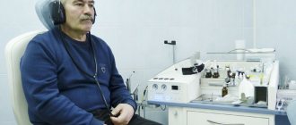
- audiometry;
- electrocochleography – electrical activity of the cochlea;
- X-ray of the cervical spine;
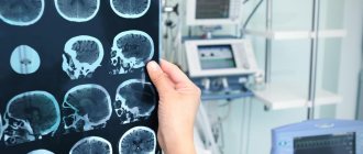
- CT, MRI of the brain.
Examination with a tuning fork is the simplest instrumental test. To determine hearing loss in medicine, 2 types of tuning forks are used: low-frequency C128 and high-frequency C2048. Main types of testing:
- Rinne test - gives a positive or negative answer to the presence of bone and air conduction. After striking the tuning fork, it is applied with a thin stem, first to the bones of the skull behind the ears, then brought to the ear canal.
- Weber test – tests the symmetry of the quality of bilateral hearing. After hitting the tuning fork, it is placed exactly in the center of the head (above the crown). Normally, a person hears equally in both ears. If there are pathologies, on one side hearing may be better/worse/completely absent.
Audiometry is a traditional way to determine the quality of hearing. The device produces a graph that is used for subsequent treatment and/or adjustment of hearing aids. Basic audiometry techniques:
- tonal suprathreshold – detects damage to auditory receptors;
- speech – determines the ability to distinguish spoken speech;
- Ultrasound sensitivity test - detects damage to the nerve trunk.
Lab tests
Used to diagnose infections and related chronic diseases. Basic tests:
- general blood analysis;
- lipid profile;
- determination of hormonal profile;
- blood clotting test.
Causes of sensorineural hearing loss
Sensorineural hearing loss in children can be caused by:
- abnormalities of brain development;
- developmental disorders of the cochlea;
- adverse effects on the mother’s body during pregnancy (intoxication, previous infections, etc.);
- genetic mutations;
- difficult childbirth.
Acquired hearing loss may be associated with the following factors:
- head injuries;
- acoustic injuries (the most common is listening to music on headphones at high volume);
- barotrauma;
- past intoxications;
- viral diseases;
- acute and chronic otitis;
- treatment with ototoxic drugs;
- local and systemic metabolic disorders;
- tumors;
- vascular pathologies;
- work in hazardous conditions;
- autoimmune diseases.
The causes of acute sensorineural hearing loss are often traumatic factors (traumatic brain injury, exposure to toxins, infection, etc.). If treatment begins on time, in the first 3 days, there is a chance of complete restoration of hearing.
Chronic sensorineural hearing loss is most often associated with progressive changes in the structures of the inner ear. Treatment requires the elimination of provoking factors, the use of conservative methods to slow the development of pathology and rehabilitation measures (hearing correction).
Prognosis and prevention
With timely treatment, the prognosis is relatively favorable. The sooner the patient sees a doctor for examination, the greater the chance of restoring or maintaining hearing. Statistics show that in 50% of cases there is a partial or complete restoration of hearing function. In other cases, hearing is compensated through specially selected and tuned devices.
Anyone who has a hereditary predisposition, chronic diseases of a hormonal, vascular or neurological nature, works in unfavorable conditions, or simply takes care of their own health should adhere to the following preventive recommendations:
- Treat any infectious diseases promptly and completely.
- Avoid prolonged stays in rooms with background noise of 70 dB or higher. If this cannot be avoided, use special earplugs with active noise reduction.
- Don't listen to loud music on headphones.
- Avoid drinking alcohol and taking uncontrolled medications.
- Be regularly examined for vascular and hormonal pathologies.
Remember, hearing loss significantly impairs a person’s quality of life. Even with successful prosthetics, you will not be able to restore your natural level of hearing.
How does the disease manifest itself?
Hearing problems can manifest themselves clearly or develop unnoticed by the patient. Acute sensorineural hearing loss is characterized by a sudden loss or progressive decrease in hearing in one or both ears, as well as one or a combination of the following symptoms:
- tinnitus of various types (high-frequency ringing or squeaking, low-frequency noise, etc.);
- vestibular dysfunctions (dizziness, nausea, vomiting, coordination problems, etc.);
- autonomic dysfunctions (sweating, rapid heartbeat, changes in blood pressure, etc.).
At the beginning of the development of chronic pathology, the clinical picture may be erased. The patient should be alert to the following situations:
- during a regular or telephone conversation you constantly have to ask again;
- there are difficulties communicating in an environment with background noise (on the street, in public places, etc.);
- understanding the interlocutor requires observing his lips;
- There are regular problems with identifying the source of noise.
The attention of loved ones should be attracted by oddities in a conversation (for example, a person hears words incorrectly or does not catch the meaning of what was said), watching TV at excessive volume, lack of reaction to events accompanied by noise (outside the patient’s field of view), and others.
Symptoms
When hearing loss is suspected to be due to a condition such as sensorineural hearing loss, symptoms will help determine the correct diagnosis. In this case, typical symptoms are:
- Sudden loss of hearing on one or both sides. The person indicates that he has become worse at understanding speech and perceiving high-frequency sounds;
- The appearance of tinnitus. This is not a permanent symptom. But it may indicate the cause of decreased hearing - acute injury, acute disruption of blood flow in the artery of the labyrinth, intoxication.
Qualified doctors who work in the Nearmedic clinic network necessarily assess the presence of risk factors. To do this, they ask the patient targeted questions:
- Do you have any relatives in your family who suffer from hearing loss?
- Is the work associated with high noise production conditions?
- Have you had any dangerous infections in the near future (measles, mumps, flu, meningitis, etc.)?
- Have you taken drugs from the ototoxic group (see above)?
Only after this comes the stage of objective examination. The otolaryngologist has an otoscope at his disposal during the appointment. This is a device that allows you to precisely examine the outer parts of the ear and the eardrum. It is not possible to look into the internal sections, but based on the condition of the peripheral sections, it is possible to identify some causes of decreased hearing - inflammation, degenerative processes, etc.
At the same time, the doctor evaluates the symmetry of the face. This sign allows us to exclude the involvement of cranial nerves in the pathological process, which is especially often observed with herpes infection.
Where to go if you have symptoms of hearing loss?
The diagnosis of sensorineural hearing loss is carried out by an otolaryngologist or audiologist. The first stage includes a survey, medical history and examination of the ears using a special device.
Laboratory examination involves a general blood test; according to indications, a coagulogram, blood biochemistry, and hormone tests are prescribed. Instrumental diagnostics includes the following options for a comprehensive hearing test:
- tests with whispered and spoken speech;
- tuning fork tests;
- pure tone threshold audiometry;
- impedancemetry;
- recording of auditory evoked potentials;
- otoacoustic emissions;
- extratympanic electrocochleography.
To assess hemodynamics in the structures of the auditory analyzer, duplex scanning of the brachiocephalic vessels is prescribed. To exclude serious causes of hearing loss (complications of injury, vascular pathology, tumor, etc.), CT and/or MRI of the head and neck are performed.
Why is treatment at Nearmedic convenient, reliable and effective?
Doctors at our center:
- They apply an individual approach to each patient, therefore they prescribe the diagnostic and treatment methods indicated only for him, and not everything in a row, as if according to a template;
- They work in a team, so if necessary, they immediately consult patients with related specialists in order to establish a true diagnosis;
- They constantly improve their qualifications at international symposia and congresses, which means they are aware of the latest medical advances in the field of hearing loss.
Nearmedic is a reasonable combination of quality medical services and affordable prices.
How is sensorineural hearing loss treated?
In accordance with clinical recommendations, acute sensorineural hearing loss is treated under the close supervision of an audiologist, who dynamically evaluates the effectiveness of therapy and, if necessary, carries out its correction. Treatment options include:
- hearing-friendly mode;
- hormone therapy;
- complex therapy with drugs to improve microcirculation and rheological properties of blood, antioxidants and antihypoxants.
In chronic sensorineural hearing loss, concomitant diseases are treated first. Twice a year, the patient must undergo courses of drug treatment using drugs to improve intracerebral and labyrinthine circulation and correct metabolism. At the same time, hearing aids are provided using a hearing aid.
The unsatisfactory effectiveness of hearing aids in severe sensorineural hearing loss is the reason for cochlear implantation.
Audiologists at the Ear, Nose and Throat Clinic successfully diagnose and treat sensorineural (sensorineural) forms of hearing loss. The clinic has equipment to conduct a complete diagnosis of hearing pathologies. The treatment program is drawn up taking into account the cause and characteristics of the development of disorders. Hearing aid is provided according to indications. Doctors at our medical center provide care to both adults and young patients.
Modern treatment at Nearmedic
Treatment of sensorineural hearing loss using modern methods is carried out at the Nearmedic center. To do this, doctors use the latest scientific developments in this matter. Specialists have drugs from different groups in their arsenal. Surgical treatment for this pathology is not indicated.
Depending on the characteristics of the course of the disease and identified risk factors, otolaryngologists from the Nearmedic network of clinics develop an individual treatment program for each patient. If acute sensorineural hearing loss is diagnosed, treatment is carried out only in a hospital setting, while maintaining a gentle auditory regimen is mandatory (exposure to loud sounds is excluded).
In the acute period, otolaryngologists must prescribe:
- Infusion therapy to restore normal microcirculation in the inner ear and reduce the degree of intoxication;
- Corticosteroid drugs that reduce the severity of edema and the inflammatory reaction (it is these conditions that are destructive to nerve cells, so they must be eliminated very quickly). In some cases, otolaryngologists may inject corticosteroids through the eardrum and into the auditory tube to increase the effectiveness of treatment;
- Antiplatelet agents to reduce blood viscosity and improve its flow through blood vessels, incl. in the area of the inner ear.
If this is chronic hearing loss, then the treatment measures are of a slightly different nature. These are supporting courses 1-2 times a year, which are aimed at:
- Improving blood flow in brain tissue, incl. in the labyrinth area;
- Normalization of metabolism in nerve cells and tissues.
How is the search for genes leading to RNNS carried out?
More than half of cases of congenital RNHL are caused by homozygous and compound heterozygous recessive mutations in the GJB2 (genetic hearing loss DFNB1: OMIM 220290 ). The frequency of this genetic type of hearing loss is 1: 1000 newborns. Some rare specific mutations in the GJB2 gene exhibit a dominant-negative effect and, in a heterozygous state, lead to autosomal dominant non-syndromic hearing loss (DFNA3A: OMIM 601544 ) or keratitis-ichthyosis-deafness (OMIM 148210 ), Vohwinkel (OMIM 124500 ), Barth-Pumfrey syndromes (OMIM 149200 ), palmoplantar keratoderma with deafness (OMIM 148350) . The Center for Molecular Genetics determines whether the patient has mutations in the GJB2 gene. This study includes a search for the 8 most common mutations in the GJB2 (c.35delG, c.-23+1G>A (IVS1+1G>A), c.101T>C (p.Met34Thr), c.313_326del14, c. 235delC, c.167delT, c.358_360delGAG (p.Glu120del) and del(GJB2-D13S175)), constituting 95% of the total number of chromosomes with a mutation in the GJB2 gene, by allele-specific MLPA; as well as searching for mutations in the GJB2 gene by sequencing the mRNA sequence (exons 1 and 2) and exon-intron junctions of the gene. The approach used allows us to detect more than 99% of mutations in the GJB2 gene. If necessary, a search for an extended 309-kb del deletion (del(GJB6)-D13S1830) in the DFBN1 .
In patients with NNS without mutations in the GJB2 gene, mutations in other genes are observed: more than 100 more genes responsible for NNS have been described. The Center for Molecular Genetics is searching for mutations of the 32 most common genetic forms of NNS and syndromes masquerading as them in patients without mutations in the GJB2 gene by sequencing a panel of 32 genes presented in the Table.
| Gene | Disease | OMIM |
| STRC | DFNB16, c.deafness and male infertility | 603720 |
| MYO7A | Ushera village 1 B, DFNB2, DFNA11 | 276900, 600060, 601317 |
| MYO15A | DFNB3 | 600316 |
| TECTA | DFNB21, DFNA8/12 | 603629, 601543 |
| SLC26A4 | DFNB4, Pendreda village | 600791, 274600 |
| CDH23 | DFNB12, p. Ushera 1 D | 601386, 601067 |
| USH2A | Ushera village 2 A | 276901 |
| TMPRSS3 | DFNB8/10 | 601072 |
| TMC1 | DFNB7/11, DFNA36 | 600974, 606705 |
| COL11A2 | DFNB53, DFNA13, Stickler type III, p. Nancy-Sweeney-Inslee syndrome | 609706, 601868, 184840, 215150 |
| OTOF | DFNB9 | 601071 |
| OTOA | DFNB22 | 607039 |
| PCDH15 | DFNB23, p. Ushera 1 F | 609533, 602083 |
| KCNQ4 | DFNA2A | 600101 |
| LOXHD1 | DFNB77 | 613079 |
| WFS1 | DFNA6/ DFNA14, Tungsten village | 600965, 222300, 614296 |
| ADGRV1 | With. Ushera 2 C | 605472 |
| MYH14 | DFNA4 | 600652 |
| MYO6 | DFNB37, DFNA22 | 607821, 606346 |
| ACTG1 | DFNA20/26, p. Baraitser-Winter type 2 | 604717, 614583 |
| PTPRQ | DFNB84, DFNA73 | 613391, 617663 |
| MYH9 | DFNA17, p. Alporta with leukocyte inclusions and macrothrombocytopenia | 603622, 155100 |
| OTOGL | DFNB84 | 614944 |
| TRIOBP | DFNB28 | 609823 |
| CLDN14 | DFNB29 | 614035 |
| LRTOMT | DFNB63 | 611451 |
| DFNB59/ PJVK | DFNB59 | 610220 |
| TPRN | DFNB79 | 613307 |
| WHRN | DFNB31, p. Usher 2 D | 607084, 611383 |
| ALMS1 | Alstrom s. | 203800 |
| POU3F4 | DFNX3 | 304400 |
| SMPX | DFNX4 | 300066 |
When conducting prenatal (antenatal) DNA diagnostics in relation to a specific disease, it makes sense to diagnose common aneuploidies (Down, Edwards, Shereshevsky-Turner syndromes, etc.) using existing fetal material, paragraph 54.1. The relevance of this study is due to the high total frequency of aneuploidy - about 1 in 300 newborns, and the absence of the need for repeated sampling of fetal material.
Publications on the topic of this section
What type of hereditary hearing loss is most common?
About 75% of all cases of hereditary hearing loss are classified as recessive nonsyndromic hearing loss (RNHL) or recessive nonsyndromic hearing loss .
With a recessive type of inheritance, a child receives from each parent the same variant of the gene that causes this form of hearing loss. A “recessive” gene appears only in conjunction with another gene of the same type and causes RNNS. At the same time, the child’s parents do not suffer from hearing impairment, since they have a normal variant of this gene in a pair of genes received from their parents. However, they are carriers of the gene for recessive non-syndromic deafness . Thus, a child may have hearing impairment, while his parents and all other relatives may have normal hearing at any age.
By non-syndromic form we mean that hearing loss is not accompanied by other signs or diseases of other organs and systems that would be inherited along with hearing loss, which occurs in syndromic forms. For example, Pendred syndrome, the most common syndromic variant of hearing loss, is a recessive disease characterized by a combination of hearing impairment with the formation of a euthyroid goiter. A goiter develops in adolescence and later, so in children the differential diagnosis of Pendred syndrome and non-syndromic recessive hearing loss is extremely difficult.
Autosomal dominant forms of non-syndromic hearing loss are relatively rare and account for no more than 25% of all non-syndromic deafness. With dominant forms, one altered copy of the gene is enough for the disease to manifest itself. In such cases, as a rule, one of the parents of a child with hearing loss is sick, but a new mutation may also arise.
What benefit is expected from identifying genes that lead to RNNS?
This is an opportunity to accurately determine the causes of hearing loss. Genetic analysis makes it possible to make a correct prediction of the recurrence of the disease in the family, as well as to assess the likelihood of the birth of children with hearing impairment in families of relatives. Early detection of a genetic defect helps to timely choose the correct tactics for treatment and rehabilitation of a child with hearing loss, and will protect him from taking unnecessary medications.
Even a healthy person can find out his genotype, since he may be a carrier of the hearing loss gene (with a probability of 1/20). Testing for carrier status of the hearing loss gene is especially important for individuals who have relatives with hearing impairment, as well as spouses in consanguineous marriages.
For a married couple in which both are carriers of a mutation in the hearing loss gene (regardless of whether they already have a child with hearing loss or not), it is possible to conduct prenatal (prenatal) DNA diagnosis of the disease in the fetus in early pregnancy (9 -12 weeks).
