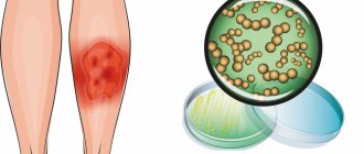
Ringworm (microsporia) is a disease manifested as a fungal infection of the skin, nail plates and hair follicles. The pathogen is a mold fungus of the genus Microsporum. Its colonies form in keratinized substrates. Microsporia remains a relatively common disease - dermatologists identify 60-75 cases for every hundred thousand Moscow residents. The pathology has a pronounced seasonality. The peak incidence occurs at the end of summer and beginning of autumn - the period of breeding of offspring in cats and other animals.
Ringworm
Ringworm (also known as microsporia) is a highly contagious fungal disease caused by the Microsporum fungus. It affects the skin, scalp and vellus hair, and rarely nails. Ringworm on the skin develops in animals and humans. The source of infection is most often a sick animal. At the same time, children are more susceptible to the disease, since they are more likely to come into contact with stray cats and dogs, which may be carriers of the fungus. In addition, the pH of the skin of adults is shifted to the “acidic” side, which prevents the fungus from actively multiplying. According to statistics, symptoms of ringworm are 5 times more common in boys than in girls.
Don't cut your hair bald
In the past, ringworm was treated with older generation antifungal tablets. The active substances accumulated in the skin, hair and throughout the body for a long time, and then only had an effect on the disease. Children of the 21st century are luckier: modern anti-lichen medications are more effective and varied - these are not only tablets, but also ointments. In addition, even if microsporia occurs on the scalp, doctors do not require parents to shave off all their children's hair, as was previously the case. Today, doctors sometimes ask to expose only those places where there are spots. After all, ointments penetrate smooth skin more easily. By the way, they are usually prescribed to children who have a small, newly appeared lesion. If there are several spots and they are actively spreading, you will have to use tablets.
To help your baby overcome shingles faster, try to follow some simple rules. Put aside synthetic and hard clothes, giving preference to cotton and soft ones. It will not irritate or scratch already injured skin. Change and process linen more often using high temperatures - iron or steam, and also wash, boil and iron the baby doll's clothes separately, so as not to infect other family members. And to avoid catching a ringworm yourself, wear disposable latex gloves during all these procedures. And be sure to treat the entire house at the very beginning of treatment: floors with disinfectants, toys with boiling water or chlorine-containing substances, and the sofa and armchairs with an iron. And although from the moment the treatment begins, the likelihood of infection becomes less and less, to be sure, repeat all these manipulations at the end of the course. Until the baby recovers, wash his body and hair with antifungal shampoos, but under no circumstances give him a bath. Otherwise, the infection may spread throughout the body. Carry out bath procedures as follows: wrap the affected areas of the skin in cellophane so that water does not get on them, and gently wipe the baby with a sponge. With one or two outbreaks of ringworm, you will get away with a slight fright and home quarantine, but if the child is covered all over, you will have to go to the hospital under round-the-clock supervision by doctors. Treatment can last from two weeks to several months - it all depends on how early it is started. Only your doctor can decide when to stop taking antifungal medications. If you do not destroy at least one spore of the lichen, the disease will begin again after some time. The criterion for recovery will be three negative fungal tests; they will be taken from the baby during treatment at intervals of two weeks.
Reasons for development
The main causes of ringworm are contact with the pathogen:
- A child aged 4–11 years whose sweat reaction is predominantly alkaline.
- A child or adult with reduced immunity, the presence of scratches, scabs, microtraumas that can become infected.
Most often the carrier is an animal, less often a person. Infection also occurs through objects or personal belongings on which infected hairs, hairs or skin scales remain. Infection is possible through undisinfected hairdressing tools, through soil on which the causative agent of the disease can persist for up to 3 months, during processing of hay. According to statistics, up to 70–80% of cases of the disease occur as a result of contact with stray young cats and kittens.
Incubation period of the disease
The incubation period of the fungal infection that causes the appearance of ringworm ranges from several days to 6–8 weeks and depends on the type of pathogen that has entered the body.
Superficial lichen may appear as the first symptoms within 5-7 days after infection. Infiltrative-suppurative lichen does not give any clinical manifestations for several months. The first signs of the disease appear no earlier than 6-8 weeks after the pathogen enters the body.
Symptoms
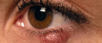
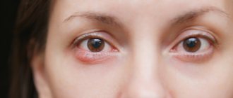
Ringworm on the face and body appears in the form of pink or red circles raised 1-3 mm above the surface of the skin. The lesions are characterized by clear boundaries, while pale areas of skin with red dots are visible inside. These spots quickly increase in size. Their number is growing over time.
The skin inside the lesions becomes covered with a white crust, which peels and itches. Ringworm on the scalp is characterized by split hair and hair loss. As the number of lesions increases, they begin to connect and overlap each other. Ringworm on the body occurs with increased symptoms when the pathogen gets inside existing lesions, and new fungal rings form in them.
Hygiene measures when in contact with a sick person
If you or your child have touched a sick animal or interacted with a sick person, then you need to take immediate action.
- The sooner you wash away particles containing fungus from your skin, the less likely you are to become infected.
- Wash your hands several times with antifungal soap. The simplest remedy, which is available in almost every store, is cinnamon laundry soap, or better yet, soap with birch tar.
- Wash your entire body with this soap. Suddenly, particles of the patient’s skin got under his clothes. Do not use a hard washcloth. It leaves micro-scratches on the skin, into which fungus easily penetrates.
- To wash your hair, you must use an antifungal shampoo. For example, Nizoral. You can also use it as a shower gel.
- A modern remedy with a powerful antifungal effect is Citeal. Dilute it in a small container five times. You will end up with a foaming liquid that can be used to wash your hands and entire body.
- Lavender oil, tea tree oil and turpentine have an antifungal effect. They can be used to treat small areas of skin.
Also, five days after contact, it is advisable to consult a dermatologist. He examines the body with a Wood's lamp. If you do become infected, the disease can be detected in the early stages. This will help you quickly treat her at home and avoid going to the hospital.
Diagnostics
It is not difficult to make a primary diagnosis if you know what ringworm looks like. To clarify the diagnosis use:
- analysis of clinical data;
- results of luminescent analysis;
- microscopic studies;
- sowing material.
In some cases, the source of infection is difficult to determine, since the incubation period ranges from 2 to 45 days. Symptoms may appear on the head, neck, arms, legs, including the palms, soles of the feet, groin areas and folds. Fungal infection causes itching, discomfort in the area of inflammation, and creates psychological discomfort.
Differential diagnosis and treatment of Devergie's disease
Devergie's lichen pilaris (syn.: Devergie's disease; pityriasis rubra pilaris; lichen ruber acuminatus) is a heterogeneous chronic inflammatory skin disease, which is divided into both hereditary forms, transmitted autosomal dominantly, and sporadic, acquired ones. Although the name and description are generally believed to be by Alphonse Devergie, the first case was reported in 1828 by Claudius Tarral. He noted isolated scaly rashes, penetrated in the center by hair, upon palpation of which a very dense roughness was felt on the surface of the skin [1–6].
Devergie's disease (DD) is a rare disease accounting for 0.03% of all skin diseases. The etiology and pathogenesis of dermatosis are still not fully understood; the opinions of modern authors differ. The theory of hereditary predisposition plays a dominant role, although there are other theories [3, 5]. An immunomorphological study of cells in the inflammatory infiltrate in patients with BD was characterized by an increased number of cells with the expression of markers involved in the development of the inflammatory immune response through the mechanism of delayed-type hypersensitivity. This allows us to classify the disease as a chronic dermatoses characterized by hyperproliferation of keratinocytes mediated by T cells [7]. According to K.N. Suvorova et al. (1996), the onset of the disease can be observed in all age periods from 5 to 72 years. The course of the disease is chronic, sometimes for decades, with no seasonality noted [8].
In 1980, Griffiths described 5 clinical forms of the disease, which doctors used for quite a long time:
- Classic adult type of the disease.
- Atypical adult type.
- Classic juvenile type.
- Limited juvenile type.
- Atypical juvenile type.
However, in modern dermatology it is generally accepted that the classic adult type and the classic juvenile types of the disease have identical clinical manifestations and differ only in the age of the patients. For this reason, the current classification of BD includes three clinical forms:
- Classic type.
- Limited juvenile type.
- Type associated with HIV infection [6].
Diagnosis of BD, especially in the initial stage, is difficult, since the clinical picture of the disease develops slowly. The pathological process is represented by a rash consisting of follicular papules with perifollicular erythema surrounding the hair shaft. The papules have a conical shape with characteristic horny spines - “Beignet cones”. This form of nodules forms the pathognomonic symptom of “grater” - a sensation of a rough surface upon palpation (Fig. 1). Localization of follicular hyperkeratosis on the dorsal surface of the I–II phalanges of the fingers is found in the literature under the name “Besnier’s symptom.” Typical for BD is the brick-red or yellowish-red color of the rash. The disease can begin either acutely or gradually. Typically, the rash appears on the upper half of the body in the form of erythematous spots or a single plaque and spreads downwards (Fig. 2) [1–5, 9]. This dermatosis debuts with erythemosquamous lesions on the face and pityriasiform peeling of the scalp, which are accompanied by itching of varying intensity, which can mimic seborrheic dermatitis. Over time, the clinical picture changes; as a result of the fusion of erythemosquamous plaques, the process spreads and gradually takes on the character of diffuse erythroderma, then a typical symptom is the presence of islands of healthy skin against the background of erythroderma (Fig. 3). The presence of islands of healthy skin is of great clinical importance for the diagnosis of BD, sometimes being one of the most important differential signs. This symptom is the presence of small areas of healthy-looking, coin-shaped skin with a diameter of about 1 cm, scattered on an erythrodermic background on any part of the skin. Peeling is heterogeneous: the scales on the upper half of the body are small, on the lower half they are often large-plate. A clinical symptom such as follicular hyperkeratosis on the dorsal surface of the phalanges of the fingers (Besnier's sign) is observed in 50% of cases. Simultaneously with the onset of the disease or later, palmoplantar hyperkeratosis appears, which occurs in 80% of cases with this type of disease [4, 5, 8, 9].
The nail plates are often affected, have a yellowish color, are streaked with longitudinal or transverse grooves, and subungual hyperkeratosis is often pronounced. In some cases, there is deformation of the nail plates of the feet and hands, up to onychogryphosis.
Clinical diagnosis of the disease is based on characteristic signs:
- ostifollicular papules, forming the “grater” symptom;
- perifollicular erythema, with a tendency to merge;
- the presence of “islands of healthy skin” against the background of erythroderma;
- brick-red color of the skin;
- palmoplantar hyperkeratosis;
- nail changes;
- Besnier's sign.
However, quite often rashes with BD resemble psoriasiform elements, and the presence of diffuse erythroderma makes the dermatosis similar to psoriasis and toxicoderma. Psoriasis and BD share not only the similarity of clinical manifestations, but also the mechanisms of development of the inflammatory process. The results of a histological and immunomorphological study conducted by O. R. Katunina (2005) indicate a common pathogenetic pathway for the development of the inflammatory process in the skin in psoriasis and BD. In both cases, activation of antigen-presenting cells occurs, hyperproliferation of keratinocytes, which, acquiring the features of immunocytes, contribute to the maintenance of the pathological process [7].
Diagnostics
We conducted clinical observations of 23 patients with Devergie's lichen pilaris. The clinical diagnosis of each subject was confirmed by histological examination of a skin biopsy. 15 (65.2%) patients were initially diagnosed with psoriasis, 6 (26%) patients were treated for toxicoderma, and only 2 (8.8%) of them were allegedly diagnosed with Devergie pilaris. Due to such a large number of diagnostic errors, we analyzed the main differential diagnostic features of BD.
First of all, our attention was drawn to the good health of all patients with BD, which persists even with diffuse damage to the skin, in contrast to patients with toxicoderma or psoriatic erythroderma, in whom complaints of prodromal phenomena, low-grade fever and weakness prevail. Subjective sensations of patients with BD are mainly focused on changes in the skin, a feeling of tightness and dryness, which contradicts the observations of foreign authors who indicate that approximately 20% of patients experience itching or burning [6]. In the differential diagnosis of BD, one should pay attention to the pathognomonic symptom of “islands of healthy skin.” This sign was recorded in all observed patients. The symptom is easy to distinguish from areas of healthy skin in psoriatic erythroderma, if we keep in mind that the characteristic islands of healthy skin in psoriatic erythroderma do not exceed 1.5 cm in diameter, have clear rounded boundaries and are located against the background of diffuse erythroderma. When establishing a diagnosis of BD, it should be remembered that this is ostiofollicular dermatosis and the primary element is a papule penetrated in the center by hair. However, we must not forget about the follicular form of psoriasis, when in a patient with psoriasis we can also observe pink ostiofollicular papules, riddled with hair in the center. In this case, it can be quite difficult to distinguish these diseases if you do not pay attention to typical psoriatic plaques in characteristic locations: the scalp, extensor surfaces of the extremities. Dynamic observation of the patient will allow you to establish the correct diagnosis. In psoriasis, papules always tend to grow peripherally and coalesce due to infiltration of the dermis, in contrast to BD, in which diffuse erythroderma is formed due to the erythemosquamous process. The absence of infiltration and lichenification, abundant large-plate peeling, characteristic of psoriasis, once again speaks in favor of the diagnosis of BD.
The next very important symptom of BD is the characteristic color of the skin - brick red or carrot, described by numerous authors [3–10]. We noted that 7 patients (30.4%) had a carrot-colored skin tone, and the rest had a brick-red color (n = 16; 69.6%). However, palmoplantar hyperkeratosis, which developed in 19 (82.6%) of the patients we observed, in all cases gave a yellowish-carrot coloration to the skin.
Although damage to the nail plates among the examined patients was diagnosed only in 18 (78.2%) patients, I would like to note that the severity of this symptom most likely depends on the duration of the disease, since the growth rate of the nail plate is much slower than the development of the pathological process. This assumption was confirmed by our observations: in patients with damage to the nail plate, the duration of the disease ranged from 4 to 10 months. Changes in the nail plate were characterized by subungual hyperkeratosis, color changes, and longitudinal and transverse striations. However, onycholysis, characteristic of psoriasis, was not observed in any patient.
As mentioned above, in the majority of patients with BD we observed (n = 21; 91.2%), the correct diagnosis was not initially established. Patients received hyposensitizing and antihistamine drugs, and more than half of them (n = 15; 65.2%) received long-acting steroids, but despite intensive therapy, no positive dynamics in treatment were noted. This fact once again confirms that BD is resistant to conventional therapy. Summarizing the above, we can identify the main differential diagnostic features that allow us to distinguish lichen pilaris from psoriasis:
- the good general condition of patients with BD is maintained even with diffuse damage to the skin, in contrast to psoriasis;
- a characteristic feature is the presence of islands of healthy skin;
- the primary element is the ostiofollicular papule;
- brick-red or carrot-colored skin;
- absence of infiltration, lichenification and abundant large-plate peeling characteristic of psoriasis;
- onycholysis, characteristic of psoriasis, is observed very rarely;
- palmoplantar hyperkeratosis without infiltration, giving a yellowish-carrot tint to the skin;
- torpidity to hyposensitizing, antihistamine and hormonal therapy.
Treatment
Currently, drugs that affect keratinization processes are retinoids. For the first time in 1930, Moore synthesized retinol from carotenoids and began to study its effect on the body. Today it has been proven that vitamin A is involved in the regulation and proliferation of many types of cells from the moment of embryonic development and throughout life. The most effective among all synthetic retinoids in the treatment of BD is Neotigazon at a dose of 0.5–0.7 mg/kg/day [4–6, 10].
Neotigazone (acitretin 10 mg, 25 mg) has pronounced lipophilicity and easily penetrates tissues. It should be borne in mind that due to individual differences in the absorption and rate of metabolism of acitretin, the dose must be selected individually. The initial daily dose is 25 or 30 mg per day for 2–4 weeks. It is better to take capsules once a day in the evening with meals or with milk. As a rule, the therapeutic effect is achieved with a daily dose of 30 mg. In some cases, it may be necessary to increase the dose to a maximum of 75 mg/day. However, one should not count on a rapid clinical effect from the therapy in the treatment of BD, especially in adults. Our opinion coincides with the opinion of foreign authors who believe that symptomatic improvement in BD occurs within 1 month, but significant improvement and, perhaps, resolution of the process is possible within 4–6 months, and according to some data, the average duration of therapy is about 4 years [6]. Our observations of patients with BD established an average duration of therapy of 9 months (Fig. 4).
Given the possibility of developing severe side effects, during long-term treatment the possible risk should be carefully weighed against the expected therapeutic effect. Contraindications to the use of Neotigazon include hypersensitivity to the drug (acitretin or excipients) or to other retinoids; severe liver and kidney failure; severe chronic hyperlipidemia, chronic alcoholism; cholelithiasis, chronic pancreatitis; diseases of the central nervous system, accompanied by increased intracranial pressure. A. A. Kubanova et al. (2005) indicate contraindications to the prescription of retinoids if there is a history of epithelioma (including familial), squamous cell or basal cell carcinoma [10]. Neotigazon has a strong teratogenic effect. The risk of having a child with developmental defects is especially high if Neotigazon is taken before or during pregnancy, regardless of the dose and duration of therapy. The literature indicates various time intervals between the end of taking retinoids and pregnancy necessary to eliminate the risk of a teratogenic effect. According to various authors, this period ranges from 6 months to 2 years [10].
The lipophilic properties of the drug suggest that it passes into breast milk in significant quantities. For this reason, Neotigazon should not be prescribed to nursing mothers. Adverse reactions are observed in most patients taking Neotigazon; they usually disappear after reducing the dose or discontinuing the drug. It should be noted that at the beginning of treatment there is an increase in the symptoms of the disease. Our analysis of Neotigazon therapy for patients with BD showed that the most common side effects are: dry lips (n = 23; 100%), cheilitis and cracks in the corners of the mouth (n = 23; 100%); dryness and flaking of the skin (n = 23; 100%); dry mucous membranes (n = 23; 100%); hair loss (n = 12; 52%); feeling of thirst and dry mouth (n = 5; 21.7%); intolerance to contact lenses is less common (n = 3; 13%); conjunctivitis (n = 2; 8.6%), stomatitis (n = 2; 8.6%), nosebleeds (n = 1; 4.3%), rhinitis (n = 1; 4.3%); disturbance of taste (n = 1; 4.3%). These side effects are usually reversible and disappear after discontinuation of Neotigazon. If severe headaches, nausea, vomiting and visual disturbances occur, the drug should be immediately discontinued and the patient referred to a neurologist. The physician is required to monitor liver function before starting treatment with Neotigazon, every 1-2 weeks during the first month after starting treatment, and then every 3 months while taking a maintenance dose. During treatment with large doses of Neotigazon, a reversible increase in the level of triglycerides and serum cholesterol is possible, especially in patients at high risk (with lipid metabolism disorders, diabetes mellitus, obesity, alcoholism). If liver function does not return to normal, the drug must be discontinued. In this case, it is recommended to continue monitoring liver function for another 3 months.
In patients with diabetes, retinoids may improve or worsen glucose tolerance, so blood glucose levels should be checked more frequently than usual early in treatment.
Due to the risk of developing hypervitaminosis A, concomitant use of vitamin A and other retinoids should be avoided. Since both Neotigazon and tetracyclines can cause increased intracranial pressure, their simultaneous use is contraindicated. With the combined use of methotrexate and Neotigazon, there is a risk of developing hepatitis, so the use of these two drugs simultaneously is also contraindicated.
To reduce the toxic effect of Neotigazon on the body, adjuvant therapy is prescribed: hepatoprotectors, lipotropic agents and gastric enzymes, B vitamins, nicotinic and pantothenic acids. To reduce the risk of hyperlipidemia, foods rich in fat should be avoided during treatment.
According to various authors, alternative treatment methods may include Prednisolone (15–20 mg/day), Diprospan (2.0 IM once every 10 days No. 3) and cytostatics: Prospidin (50–100 mg IM daily per course 2.0–3.0 g) or Methotrexate (15 mg IM once every 7 days) [6, 11].
External treatment while taking retinoids is not of fundamental importance, but it significantly improves the general condition of the patient and increases his quality of life. Both classic ointments and creams and modern dry skin care products from various manufacturers are used. To eliminate massive horny deposits, ointments with 2–5% salicylic acid, 10% urea, and 1–20% malic acid are prescribed.
In addition, physical therapy is used to treat patients with BD. Basically, various baths are used: sulfide baths lasting 6–10 minutes every other day, 12–15 baths per course; positive radon baths for 10–20 minutes every other day; At home, it is possible to use starch baths: 300–500 g of potato starch are diluted in 2–5 liters of cold water and then poured into a bath at a temperature of 36–37 ° C, the duration of the procedure is 15–20 minutes every other day, the course is 15–20 procedures. In children, the duration of the procedure should not exceed 15 minutes; the course is 8–12 baths.
The question of the advisability of using ultraviolet irradiation (UVR) in such patients has not yet been resolved; the opinions of modern authors are contradictory and require further study [6, 12]. According to our observations, in all examined patients, ultraviolet radiation or insolation provoked an exacerbation of the process.
Thanks to the achievements of domestic and foreign science, in recent years there have been significant shifts in the understanding of the clinical and genetic polymorphism of hereditary skin pathology, knowledge in the field of pathomorphology and immunomorphology has significantly advanced, which has expanded the possibilities of accurate diagnosis of BD. The use of synthetic retinoids has made it possible to increase the effectiveness of therapy for this category of patients. However, treatment of BD is long-term and is always accompanied by the development of a number of side effects, which leads to limited possibilities for prescribing the drug to elderly people and patients with a complicated medical history.
Literature
- Berenbein B. A., Studnitsin A. A. et al. Differential diagnosis of skin diseases. M.: Medicine, 1989. 672 p.
- Elkin V.D., Mitryukovsky L.S. Selected dermatology. Rare dermatoses and dermatological syndromes. Handbook of diagnosis and treatment of dermatoses. Perm, 2000. 699 p.
- Mordovtsev V.N. Hereditary diseases and malformations of the skin. M.: Nauka, 2004. 174 p.
- Kalamkaryan A. A., Kubanova A. A., Akimov V. G., Arifov S. S. Devergie pilaris versicolor // Bulletin of Dermatology and Venereology. 1990, no. 6. pp. 20–23.
- Kubanova A. A., Akimov V. G. Differential diagnosis and treatment of skin diseases: Atlas-reference book. M.: Medical Information Agency LLC, 2009. 304 p.
- European guidelines for the treatment of dermatological diseases / Ed. A. D. Katsambasa, T. M. Lotti. M.: MEDpress-inform, 2008. 736 p.
- Katunina O. R. Comparative immunomorphological characteristics of inflammatory infiltrate cells involved in a delayed-type hypersensitivity reaction in patients with psoriasis and Devergie's disease. Author's abstract. diss. Ph.D. honey. Sci. M., 2005. 20 p.
- Suvorova K. N., Kuklin V. T., Rukovishnikova V. M. Pediatric dermatovenerology. Kazan, 1996. 441 p.
- Kubanova A. A., Arifov S. S. Features of the clinical course of pityriasis ruber pilaris Devergi // Bulletin of Dermatology and Venereology. 1990, no. 7. pp. 59–61.
- Rational pharmacotherapy of skin diseases and sexually transmitted infections: Hand. for practicing doctors, ed. Kubanova A.A., Kisina V.I.M.: Litera, 2005. 882 p.
- Holliday AC, Megan NM, Berlingeri-Ramos A. Methotrexate: Role of Treatment in Skin Disease. // Skin Therapy Letter. 2013;18 (3).
- Osorio F., Magina S. Phototherapy and Photopheresis // Expert Rev Dermatol. 2011;6(6):613–623.
A. A. Kubanov*, Doctor of Medical Sciences, Professor Yu. A. Gallyamova**, 1, Doctor of Medical Sciences, Professor
* FSBI SNTsDK MH RF, Moscow ** GBOU DPO RMAPO MH RF, Moscow
1 Contact information
Abstract. Diagnostics of Devergie pityriasis rubra pilaris criteria and approaches to treatment are presented in the article. Application of synthetical retinoids allows increasing effectiveness of therapy of such patients. But treatment of Devergie disease requires long time and is followed by development of several side effects that cause limitations for prescription of medicine to elderly people and to patients with compromised history.
Treatment
If you notice the first symptoms of ringworm, you should immediately go to the hospital. The effectiveness of treatment and the patient’s quality of life during it depend on the timely initiation of treatment.
Prescribed against ringworm:
- antifungal drugs of systemic or local action. For large areas it is better to use ringworm preparations in the form of sprays, for small areas - ointments and creams;
- hormonal drugs. They are prescribed for severe inflammation of the skin in combination with antimycotic agents;
- glucocorticosteroids are necessary in cases where a secondary infection is associated with inflammation.
The main part of treatment is the use of local drugs. The decision on the need to use tablet forms of drugs is made by the doctor based on the clinical picture of the individual patient.
Microsporia: why is it important to see a doctor in time?
There are factors that cause difficulties in treatment:
• Dermatophyte fungi of the genus Microsporum penetrate deeply into the skin and hair follicles.
• Fungi remain viable for a long time under environmental conditions.
• There are no quick ways to get rid of microsporia. It has been proven that the growth of the fungus stops on the 14-20th day of treatment. Both you and the child will have to be patient.
• In the presence of chronic diseases, it is more difficult to defeat the fungus: a suppurative form often develops, recovery is delayed due to a weakened immune system.
• Insufficient dosage of drugs or self-cessation of treatment leads to a rapid return of the disease - relapse. What are the consequences? The emergence of addiction of the fungus to drugs, the long course of the disease and/or the development of complications.
When prescribing treatment, the doctor takes into account all these factors, selecting an individual management plan for each patient.
Prevention
The main measures to prevent ringworm are careful adherence to hygiene procedures:
- Using only your own towels and combs, preventing the child or adult from sharing them and using other people’s hygiene items.
- After visiting swimming pools, bathhouses, and water parks, you should immediately wash with soap, wash and wash the items you used.
- If someone in your home shows signs of ringworm, further infestation should be prevented. His bedding and underwear must be washed separately. All items are thoroughly washed and treated with disinfectants.
- Preventing contact with stray animals, especially cats. According to statistics, about 50% of them are infected with microsporia or are its carriers.
- The patient should be isolated from the team until complete recovery.
- If there is a sick animal in the house, all surfaces must be treated with disinfectant solutions.
With careful prevention, ringworm may not appear in other household members.
How to protect your baby from infection?
Instill in your child the rules of personal hygiene and make sure they follow them: wash your hands after playing outside or with animals, keep your shoes clean, and much more.
Explain to your child that you cannot use other people's things - for example, clothes, a hat or a comb.
Try to exclude your child from contact with stray animals: explain that you cannot pet a street kitten or puppy.
Have you decided to get a pet: a cat, a dog, a hamster or a parrot? Wonderful! However, be sure to visit a veterinarian first so that he can examine the new family member.
If you already have a pet, regularly examine it yourself and take it to the veterinarian.
Do not leave baby strollers on the landing or outside. Homeless, sick animals, having settled comfortably in them for the night, can leave behind a “gift” in the form of fungus.
Abrasions, scratches, puncture or incised wounds are an easy way for pathogens to enter. Therefore, treat any wounds with antiseptics containing iodine, which has a detrimental effect on both bacteria and fungi.
Popular questions about ringworm
How does ringworm manifest in children?
When lichen develops on the skin, pink round spots appear on the infected areas, which rise slightly above the rest of the surface. These spots itch and become covered with a whitish crust that begins to peel off. If the spots are located on the scalp, the hair within their radius breaks off and falls out.
How does lichen manifest itself in a child?
Ringworm is manifested by the development of spots of fungal infection, the number of which is constantly increasing. The spots increase in size and merge into asymmetrical patterns. A flaky crust forms inside the spots, from which scales infected with the fungus fall off. Secondary infection is possible when one spot begins to develop another.
What does ringworm look like in its early stages?
At the initial stage, ringworm looks like a pink spot of regular shape, raised above the surface of the skin by 1-3 mm. The spots can be localized on any part of the body - head, neck, arms, legs, stomach, back and even in the groin.
What is pityriasis rosea and why is it called pink dandruff?
The content of the article
Pink dandruff (Pityriasis rosea - PR) is an acute inflammatory disease of unknown etiology, characterized by the appearance of skin rashes mainly on the torso and limbs. It is an erythematous, scaly disease. The disease occurs seasonally. The etiology is most likely viral. The most likely factor is herpes virus type 7 (HHV7).
Most cases (75%) involve people aged 10 to 35 years, although there have been reports of cases diagnosed in a 3-month-old infant and in an 83-year-old man. The disease occurs throughout the world without any particular racial predilection.
Pityriasis rosea is a disease for which patients visit a dermatologist with a frequency of up to 2% of all visits per year. Occurs mainly in spring and autumn; in summer the level of symptoms decreases significantly.
The disease affects more women than men in a 2:1 ratio. The duration of the disease is 6 to 8 weeks, but is highly variable.
How do you know if pityriasis rosea is going away?
After a course of therapy, the spots disappear without scarring. In place of the plaques, clean skin remains, possibly lighter. Over time, the color evens out. In patients with atypical lichen, ulcers may form.
Complications during self-medication:
- infection of affected areas with bacteria;
- suppuration, ulcers;
- hyperpigmentation.
Hyperpigmentation
To prevent this development of the disease, at the first symptoms of pityriasis rosea, it is recommended to make an appointment with a dermatologist.
Correct treatment of pink dandruff in Gibert's disease
Treatment of pityriasis rosea is primarily based on the diagnostic results and recommendations of the dermatologist based on his knowledge and experience. Independent attempts to cure pathology usually lead to the appearance of accompanying symptoms, sometimes very dangerous.
Considering the symptoms, the dermatologist prescribes:
- ointments with corticosteroids acting in several directions - relieve skin tightness and accelerate the healing of the rash;
- antiallergic – antihistamines – relieve itching;
- immunomodulators – stimulate natural immunity;
- antiseptic solutions – prevent secondary infection;
- dandruff softening ointments.
During treatment, the dermatologist recommends:
- Avoid hot water procedures and swimming. Showering with a soft sponge is allowed;
- Wear loose, natural underwear. Synthetics do not allow the skin to breathe, exacerbating symptoms;
- Eliminate allergens: eggs, chocolate, red fruits.
- Reduce fried, flour, smoked foods;
- Do not use cosmetics.
Many names - one face. Symptoms of pityriasis rosea
The first description of the disease as "ringed roseola" dates back to 1798. Uch. Robert William defined the disease as irregularly shaped spots consisting of small thin scales that never coalesce into scabs and are not accompanied by redness or signs of inflammation.
Other names for this disease: erythema annulatum, tonsurans maculosus, Herba, roseola squamosa (Nicolas, Chapard), pink lichen (Wilson), pityriasis versicolor (Horand), pseudoexanthemata nitoexanthemata, roseola furfuracea herpetiformis (Behrend) - mainly determined the morphological features and variability individual spots.
Pityriasis rosea
Only in 1860, the French physician Camille Melchior Gibert gave the name, which lasted almost 150 years. He wrote: “There is only one thing that can be described under the name of pityriasis rosea, and the characteristic features of this pathology are: small, branched spots, very faintly colored, no larger than a fingernail, numerous and tightly fitting ... the disease usually lasts up to 6 weeks or 2 months... in young people... “. Pityriasis means "bran" or "fine husk" and rosea translates to "pink".
The symptoms of pink dandruff are indeed quite characteristic. First, a heraldic lesion (the so-called mother plate) appears on the skin, most often on the torso. Usually it is several centimeters of red or dark pink erythema, covered with scales inside.
After a few days (and sometimes weeks), much smaller skin lesions of similar morphology are seeded. The rash occurs symmetrically, mainly on the torso and neck. There are no changes in the mucous membrane. Usually the skin lesions do not cause itching.
Possible variations of the rash:
- urticarial form of RL – blisters appear instead of plaques;
- vesicular form of RP - rashes in the form of small bubbles with clear or cloudy liquids;
- papular form of RL – protruding, cavity-free formations.