Author Zybina A.M.
The nervous system integrates the entire organism into a single orchestra, carries out its interaction with the environment, voluntary movements (together with the muscular system), and all manifestations of mental activity. All functions of the nervous system are performed by a network of neurons connected to each other through synapses. Their viability is supported by glial cells.
The nervous system is divided according to its anatomical location into central (CNS) and peripheral (PNS). The central nervous system consists of the brain and spinal cord. PNS - made up of nerves (a bundle of nerve cell processes) and nerve ganglia, or ganglia (a collection of neuron bodies) located outside the nervous system.
Based on their functions, the nervous system is divided into somatic (animal, SomNS) and vegetative (autonomous, ANS) divisions. The SomNS controls voluntary contractions of skeletal muscles. The ANS controls the activity of internal organs. It is divided into two divisions: sympathetic (SNS) and parasympathetic (PNS). Both the SomNS and the ANS have both central and peripheral sections.
Why is the nervous system needed?
The human nervous system performs several important functions at once: - receives information about the external world and the state of the body, - transmits information about the state of the whole body to the brain, - coordinates voluntary (conscious) movements of the body, - coordinates and regulates involuntary functions: breathing, heart rate , blood pressure and body temperature.
More movement
Movement is a natural human need. Find out how much you need to move for a good state of the nervous system.
Vascular pathologies
Circulatory disorders have a particularly severe impact on the brain, which is very sensitive to a lack of oxygen and nutrients. Therefore, the consequences can be very serious:
- Stroke is the death of part of the brain cells due to a sudden disruption of blood flow (destruction or complete blockage of blood vessels). Very often, patients cannot fully undergo rehabilitation, which results in lifelong disability of group 1.
- Atherosclerosis is the loss of elasticity of blood vessels, the formation of cholesterol deposits and blood clots on the inner surface.
- An aneurysm is a thinning of the walls of blood vessels, the formation of a seal that can rupture, leading to serious consequences. Death is often observed.
How is it structured?
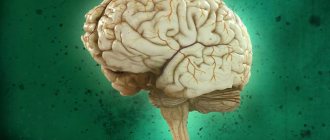
The brain
is
the center of the nervous system
: approximately the same as the processor in a computer.
The wires and ports of this “supercomputer” are the spinal cord and nerve fibers. They permeate all the tissues of the body like a large network. Nerves transmit electrochemical signals from different parts of the nervous system, as well as other tissues and organs. In addition to the nerve network called the peripheral nervous system, there is also the autonomic nervous system
.
It regulates the functioning of internal organs, which is not consciously controlled: digestion, heartbeat, breathing, and the release of hormones.
The brain is the “administrator” of the nervous system and human psyche
Previously, it was believed that a person’s “soul” was contained in his heart. With the development of science, humanity gradually began to study what made us human, the crown of the animal world - the brain.
Our mental system is formed through the interaction of the cerebral cortex and the underlying sections (dietum, midbrain, brainstem). Each area is responsible for one or another function. But the most interesting thing is that when one section of neurons fails, its work can be partially replaced by another, which is called neuroplasticity.
The frontal lobe is involved in the formation of emotions, memory, speech and behavior. Evolutionarily, this part is the most developed in Homo sapiens, since it began to develop with the transition of primates to upright walking and the activation of fine motor skills of the upper limbs. Therefore, the frontal lobe is responsible for many functions. To understand what effect the frontal lobe has on a person’s mental state, we should mention the so-called frontal syndrome, which is observed in people with organic brain damage due to external influences, vascular, oncological and other pathologies. They experience disinhibition in behavior, self-control disappears, and a tendency toward brutal, antisocial behavior, as well as outbursts of aggressiveness, appears. In addition to behavior and emotions, memory is impaired, a person cannot concentrate on any task, and the function of cognition of the outside world suffers. In severe cases, the core of the personality is lost - we stop seeing the person as he was before.
There is a part of the brain called the hypothalamus, which is responsible for regulating autonomic functions. It communicates with the endocrine system of the body and is directly connected with the hormone regulator gland – the pituitary gland. The latter secretes special substances that give a signal to the pituitary gland to release hormones: gonadotropic (affecting the gonads), thyroid-tropic (thyroid), adrenocorticotropic (adrenal glands), somatotropic (tissue growth) and prolactin (breast).
What can harm the nervous system?
Toxic substances
disrupt the electrochemical processes in the cells of the nervous system and lead to the death of neurons.
Heavy metals (for example, mercury and lead), various poisons (including tobacco and alcohol
), as well as some medications are especially dangerous for the nervous system.
Injuries occur when the limbs or spine are damaged. In the case of bone fractures, the nerves located close to them are crushed, pinched or even severed. This results in pain, numbness, loss of sensation or impaired motor function. A similar process can occur when posture is impaired
.
Due to the constant incorrect position of the vertebrae, the nerve roots of the spinal cord that exit into the foramina of the vertebrae are pinched or constantly irritated. These pinched nerves
can also occur in joint or muscle areas and cause numbness or pain. Another example of a pinched nerve is the so-called tunnel syndrome. In this disease, constant small movements of the hand lead to a pinched nerve in the tunnel formed by the bones of the wrist, through which the median and ulnar nerves pass. Some diseases, such as multiple sclerosis, also affect nerve function. During this disease, the sheath of nerve fibers is destroyed, causing conduction in them to be disrupted.
Instead of a preface
“All illnesses come from nerves!” Is this true or not? Exaggeration or sad reality? And what kind of diseases are these that are caused by nerves? For clarity, I will allow myself to cite one case from my medical practice.
I work in a clinic, where, as you know, in addition to local doctors, doctors of other specialties - “narrow specialists” - also see patients. This, by the way, is Western technology, when it is not a person who is treated, but his illness, and accordingly, each disease has its own specialist. But we are already accustomed to this and cannot even imagine how it could be otherwise.
So, one day a middle-aged man came to an appointment; after a conflict at work, a stomach ulcer opened up, a rash appeared on his hands, his blood pressure rose, and he couldn’t sleep at all! And instead of sleep, sadness appeared, and anxiety. His stomach was treated by a gastroenterologist, his skin rash by a dermatologist, and his blood pressure by a therapist. And everything would have been fine, as they say, but when the man remembered the conflict, everything was repeated again, and the doctors, sensing something was wrong, referred him to a psychotherapist.
It should be said that the human brain is designed like the most perfect computer; or rather, the computer is designed in a similar way to the nervous system. In the human body, the channels for inputting information are the eyes, ears, skin, there are receptors that react to the biochemical composition of the blood, etc. So, the brain reacts very sensitively to a word, and a rude word can cause a whole storm in the body, just like this happened in our case: the central nervous system responded to psychological stress with a powerful release of biologically active substances as its defense, and one of these substances was histamine, which caused stomach ulcers and neurodermatitis.
Moreover, with each memory of what happened, the brain reacted with a new release of the same active substances, as if it were not a simple memory, but a repeated event.
Yes, this is how our nervous system works: memories are as real to the brain as the events that gave rise to them. To help that man, it was necessary to influence the central nervous system, which triggered a defensive reaction in the form of the release of certain substances that acted on the stomach, and on the skin, and on blood pressure, and on sleep, and on mood, and acting like you understand, not in the most successful way. Which is what was done. Along with sadness, all other illnesses soon disappeared.
It turns out that the nervous system is really the head of everything? Of course, that's why she's a head! And you, of course, have experienced how much it hurts at least once in your life. The central nervous system is, of course, just a part of the nervous system, but without a doubt it is its main part. That is why it is so carefully packed by the skeletal system. And we will talk about this later.
So what needs to be done to maintain the health of our most advanced computer - the nervous system? Well, first of all, it should be said that what a person does not know, he does not own. Secondly, today any of us should have an idea of how the human body works. You need to rely on doctors, but do not forget that they treat diseases, and the science of health is different. And here everyone needs to work for themselves: no one can take responsibility for your own health for you.
Now is the time when doctors share with you, dear reader, all their knowledge and accumulated experience so that you take on the task of maintaining your own health. This is exactly what your humble servant, a psychotherapist, is going to do for you. I hope that this will be not only useful for you, but also interesting. Are you interesting to yourself?
You need to know your own body in order to help it, treat it with care, so that the problems of the body do not overshadow the abilities and needs of the soul for creativity and love. Therefore, do not consider it difficult to find out how the holy of holies of our body - the nervous system - works, how to avoid its breakdowns, and if they do happen, eliminate them. Together with the doctor. We're together right?
How to keep your nervous system healthy?
1. Eat a healthy diet
.
All nerve cells are covered with a fatty sheath called myelin. To prevent this insulator from breaking down, your diet must contain sufficient amounts of healthy fats, as well as vitamin D and B12. In addition, foods rich in potassium, magnesium, folic acid and other B vitamins are useful for the normal functioning of the nervous system. 2. Give up bad habits
: smoking and drinking alcohol.
3. Don't forget about vaccinations
.
A disease such as polio affects the nervous system and leads to impaired motor functions. You can protect yourself from polio through vaccination. 4. Move more
. Muscle work not only stimulates brain activity, but also improves conductivity in the nerve fibers themselves. In addition, improved blood supply to the entire body allows the nervous system to be better nourished.
Note to parents
If the baby does not get enough sleep, the functioning of his nervous system is disrupted. Healthy sleep is very important for a child - learn the basic rules.
5.
Train your nervous system daily
.
Read, do crossword puzzles, or go for a walk in nature. Even composing an ordinary letter requires the use of all the main components of the nervous system: not only peripheral nerves, but also the visual analyzer, various parts of the brain and spinal cord. 15 minutes
to handwriting texts , trying to write the letters as neatly as possible.
Structure of the central nervous system
The CNS consists of the brain and spinal cord, each of which has white and gray matter. White matter consists of pathways, myelinated and unmyelinated axons. Myelin is white, which gives the corresponding shade to the tissue. Gray matter consists of neuron cell bodies. It can be located in the nervous system in the form of a tube (spinal cord); nuclei, or ganglia (clusters of neuron bodies in the thickness of the white matter), as well as the cortex (gray matter on the surface of the white matter).
The spinal cord is located in the spinal canal and its mass is 40 g. On its lateral surface, the dorsal roots, carrying afferent (sensitive, to the brain) information, enter from the back, and the anterior roots, carrying efferent (motor, from the brain) information, exit from the front. The section of the spinal cord corresponding to each pair of roots is called a segment. The segments are named according to where the roots exit the spine. The spinal cord has 8 cervical, 12 thoracic, 5 lumbar, 5 sacral and 1 coccygeal segments. In general, the number of spinal cord segments corresponds to the number of vertebrae. Exceptions are the cervical region, where there are 8 segments per 7 vertebrae; and coccygeal, where there is 1 segment for 3-4 vertebrae (Fig. 1).
Rice. 1. Structure and location of spinal cord segments.
In a cross section of the spinal cord, there is gray matter in the center surrounded by white matter. The gray matter has the shape of a butterfly, in the center of which is the spinal foramen, filled with cerebrospinal fluid (CSF). The butterfly consists of approximately 13 million neurons and has anterior and posterior horns (Fig. 2b, 3). The middle horns are also well defined in the middle sections of the spinal cord. Sensitive (sensory) information to interneurons (interneurons) enters the dorsal horns along the dorsal root. The anterior horns contain motoneurons (motor neurons) that send motor information to the muscles; it is their axons that form the anterior root. The middle horns contain neurons of the central sections of the ANS.
The spinal cord works on a reflex principle. A reflex is a stereotypical response of the body to any (external or internal) influence. The simplest reflex is monosynaptic. To implement it, two neurons are enough. An example of such a reflex is the knee reflex. When the receptor is irritated, the impulse is transmitted along the dendrite to the body of the neuron located in the nerve ganglion near the spinal cord. The axon of this neuron enters the spinal cord through the dorsal roots and forms a synapse with the motor neuron in the anterior horn. The axon of the motor neuron exits through the anterior roots and goes to the effector organ, where it changes the activity of the organ itself (Fig. 50a). The polysynaptic reflex includes an additional link in the form of one or more interneurons between the ganglion and motor neurons. Interneurons can additionally process information, compare it with other stimuli and the internal state of the body, making a decision about how to respond to the stimulus.
Rice. 2. Reflex arc (a) and histological section (b) of the spinal cord.
Rice. 3. Diagram of the structure of a section of the spinal cord.
The white matter of the spinal cord includes pathways. It is divided by the butterfly into anterior, posterior and lateral funiculi (Fig. 3).
In the posterior cords there are ascending tracts through which information is transmitted from the PNS to the spinal cord and further to the brain. In the anterior horns of the spinal cord there are descending tracts through which information goes from the brain to the spinal cord, and from the latter to the PNS. In the lateral horns, the ascending tracts are located posteriorly, and the descending tracts are located anteriorly.
The brain is located in the skull and consists of 5 sections. Its average mass is 1.5 kg and it contains up to 100 billion neurons. There are 12 pairs of cranial nerves (cranial nerves) that arise from the brain.
The medulla oblongata is the junction of the spinal cord and the brain. Its length is approximately 25 mm. In the lower part of the medulla oblongata one can still distinguish a butterfly; in the upper parts the bodies of neurons are collected into nuclei. The IX-XII pairs of the cranial nerves (Fig. 5) depart from the medulla oblongata and the corresponding nuclei lie in it. These nerves control movement and sensation in the throat, tongue, and neck. The medulla oblongata contains the largest center of the parasympathetic nervous system, which, through the X nerve (vagus, vagus nerve), controls the activity of all internal organs. The medulla oblongata contains centers for regulating breathing and vital reflexes, such as sneezing and coughing. Here is the olive kernel, which is responsible for balance. All tracts from the spinal cord to the brain pass through the medulla oblongata.
The hindbrain consists of the pons and cerebellum. The pons serves as a continuation of the medulla oblongata. It contains a lot of white matter that connects the cerebellum to the rest of the brain. This white substance forms a ridge on the underside of the bridge, making it easy to distinguish. The pons, together with the medulla oblongata, form the bottom of the 4th ventricle of the brain (a continuation and expansion of the spinal canal). V-VIII ChMN depart from the bridge. Here lie the auditory and vestibular nuclei, nuclei that innervate sensitivity and facial muscles (including facial muscles). The pons contains the locus coeruleus, which is responsible for regulating sleep.
Rice. 4. Main parts of the brain.
Rice. 5. Cranial nerves. I-olfactory, II-visual, III-oculomotor, IV-trochlear, V-trigeminal, VI-abducens, VII-facial, VIII-vestibular-cochlear, IX-glossopharyngeal, X-vagus, XI-accessory, XII-hyoid.
The cerebellum is well developed in humans due to upright posture and fine motor skills of the hands. This part of the brain is responsible for maintaining posture, balance, motor learning, and some motor reflexes. The cerebellum has a cortical structure. The cerebellar cortex consists of three layers and is divided into two hemispheres by the vermis. Under the cortex there is white matter, among which there are 3 pairs of cerebellar nuclei. To carry out its functions, it receives information from the vestibular apparatus, olive and other parts of the human motor system.
Rice. 6. External structure (a) and histological section (b) of the cerebellar cortex.
The midbrain consists of the cerebral peduncles and the roof (Fig. 7). The aqueduct of Sylvius passes through the center of the spinal cord and connects the third and fourth ventricles. The third and fourth pairs of cranial nerves depart from the midbrain. These nerves control the movements of the eyeballs. The third nerve contains parasympathetic fibers that control pupil width. The midbrain contains elements of the motor system: the red nucleus and the substantia nigra. On the roof of the brain is the quadrigeminal region. The colliculus receives visual information, and the inferior colliculus receives auditory information. This is necessary for the implementation of the orientation reflex.
Rice. 7. Appearance (a) and section (b) of the midbrain.
The medulla oblongata, pons, and midbrain together form the brainstem. The reticular formation runs through the entire trunk, regulating the overall level of brain activity.
The diencephalon consists of the thalamus, hypothalamus, pituitary gland and pineal gland. The third ventricle of the brain is located here. It serves as the origin of the second cranial nerve. The pituitary gland is the gland through which the nervous system controls the humoral one. The pineal gland is also a gland that regulates circadian rhythms. The thalamus filters information entering the cortex and removes unimportant repetitive sensory stimuli (heartbeat, gastrointestinal function, nose in the field of view, touch of clothing, etc.) In addition, the thalamus contains nuclei of the limbic system (forms mood), motor and associative kernels. The hypothalamus controls the activity of the pituitary gland and also regulates the internal state of the body. It contains the centers of hunger, thirst, sexual behavior, pleasure, displeasure, etc. Thus, the main function of the hypothalamus is to maintain homeostasis of the entire organism.
The telencephalon (forebrain) consists of the cerebral cortex and basal ganglia (nuclei). The first and second ventricles of the brain are located symmetrically under the cortex. Its area is about 220 cm2, it forms grooves and convolutions (Fig. 8). It consists of 6 layers. The hemispheres are connected to each other by the corpus callosum - a ridge of white matter. The cerebral cortex processes sensory information, the formation of voluntary movements, memory and higher nervous activity. The first cranial nerve approaches the olfactory bulbs. The basal ganglia are nuclei of gray matter located in the thickness of the white matter. They play an important role in voluntary movements, motor learning and the formation of emotions.
Rice. 8. Structure (a) and histological sections (b, c) of the cerebral cortex.
Congenital diseases
A number of diseases of the nervous system arise due to gene disorders (mutations). Another common cause is embryonic developmental disorders or injuries received during birth. The most famous pathologies:
- Epilepsy is a disease accompanied by convulsions and seizures.
- Canavan syndrome is a brain disorder that causes delays in intellectual development. Treatment is impossible.
- Tourette's syndrome is a disease in which a person begins to move involuntarily and even shout words. Usually goes away as you get older.
- Spinal muscular atrophy is a lesion of the neuronal cells of the spinal cord, which impairs the development of muscle tissue, which can be fatal.
- Huntington's chorea - tics, dementia. The pathology is genetically predetermined, but begins to manifest itself only in old age.


