What is vertebrogenic cervicalgia?
The term "cervicalgia" means pain in the neck , and the word "vertebrogenic" indicates a connection with the spine. In terms of duration, this syndrome can be acute or chronic.
Information for specialists: according to the International Classification of Diseases, vertebrogenic cervicalgia is recorded with code M 54.2. The diagnosis must include the severity of symptoms, stage of the disease and treatment regimen. It is also advisable to indicate changes in the spine (spondylopathy or osteochondrosis).
The nature of pain during cervicalgia depends on the causes of its occurrence.
Pain of vertebrogenic nature in the cervical spine is divided:
| For spondylogenic or vertebral cervicalgia: | For discogenic or true cervicalgia: |
| In this case, the affected bone tissue irritates the nerves, which causes pain and tonic spasms of the neck muscles. Usually the cause is an infectious process, neoplasms or osteoporosis. | This is a consequence of the destruction of the intervertebral disc. The disease is fraught with pinching of nerve endings and loss of elasticity of the cartilage layer. |
According to the manifestations of the course, they distinguish:
- Vertebrogenic cervicalgia in acute form . It is characterized by severe pain and severe accompanying symptoms. Usually a complication of dorsopathies and injuries. In general, symptoms last about 10 days
- Chronic . With this type of vertebrogenic cervicalgia, there is constant mild pain that lasts for over 3 months. The triggering mechanism is any tumors, indolent infections and pathologies affecting the carotid artery.
Depending on the location, the disease occurs in the following forms:
- Cervicobrachialgia. The vessels of the hands are affected, which causes loss of sensation in these limbs.
- Cervicocranialgia. The pathology contains negative changes in blood vessels, which is dangerous due to the deterioration of local blood flow.
In the international classification ICD-10, the disease is coded M54.2.
Neck pain: cervicalgia or cervical dorsopathy?
Back pain affects 80–100% of the world's inhabitants at one time or another in their lives. Moreover, 70% of them, at least once, lose their ability to work for this reason. It is obvious that back pain occurs in accordance with the localization of dorsopathy (occipito-atlanto-axial, cervical, cervicothoracic, thoracic, thoracolumbar, lumbar, lumbosacral, sacral and sacrococcygeal), often spreading to several regions or without a specific distribution zone. According to international terminology, local pain in the neck, which is one of the variants of back pain, is called cervicalgia, when the musculo-ligamentous apparatus is involved and the pain radiates to the head - cervicocranialgia, when radiating to the arm - cervicobrachialgia. When the roots of the cervical spinal cord are compressed or irritated, radiculopathy develops; changes in the structure of the spinal cord at the cervical level lead to the development of cervical myelopathy [4,14]. Among back pain syndromes, neck pain (including cervicalgia, cervicocranialgia and cervicobrachialgia) ranks second (30.2%) after low back pain (42.0%), and in the vast majority of cases leads to temporary disability in people under 45 years of age, which is a serious socio-economic problem throughout the world. On average, the duration of pain is about 10 days, in 70% of patients the pain syndrome regresses within 1 month, and in 10% of cases the pain is chronic and lasts more than 3 months. [1,18]. In some cases, neck pain may be the only manifestation of cervical dorsopathy, but more often it is combined with other symptoms, as well as with signs of dorsopathy of other localization. By analogy with the term “pain in the lower back”, which is consistently used in the literature, which is reflected in the ICD-10, a number of publications, mainly popular science, mention “pain in the upper back”, which is quite justified. Neck pain is considered one of the manifestations of “office syndrome” - a complex symptom complex that includes disorders manifested in various organs and systems, and develops in office workers due to exposure to various environmental factors in the work environment, which affects up to 60–70 % of people of working age [8]. The causes of neck pain are very diverse. There are vertebrogenic and non-vertebrogenic etiological factors. Vertebrogenic pain syndrome in most cases is caused by degenerative-dystrophic changes in the elements of the vertebral motion segment (VMS). It should be noted the anatomical features of the cervical vertebrae, the bodies of which have uncinate processes and uncovertebral joints (Luschka joint). A distinctive feature of the cervical vertebrae is the presence of transverse processes that form a canal where the vertebral artery passes. The bodies of the upper cervical vertebrae are connected by an additional joint (Cruveilhier joint), which is formed by the tooth of the second vertebra and the anterior arch of the atlas [14]. With radiculopathy, neck pain occurs due to mechanical compression of the roots and aseptic inflammation in the radicular zone and perineural structures. In this case, edema, ischemia, and microcirculatory disorders develop, caused by the release of proinflammatory cytokines from the nucleus pulposus of the disc into the epidural space, such as phospholipase A2, leukotriene B4, thromboxane B2, tumor necrosis factor-α [12,23]. Under these conditions, the excitability of nociceptors increases, foci of pathological ectopic impulses appear, which leads to sensitization of spinal and supraspinal nociceptive neurons. The pain is of a mixed nature; in addition to the nociceptive one, there is also a neuropathic component. The occurrence of neuropathic pain is associated with the phenomenon of central sensitization. A decrease in the excitation threshold of neurons in the dorsal horns of the spinal cord causes the perception of various non-painful peripheral impulses as painful stimuli. When the pain syndrome is chronic, a long-term imbalance of the nociceptive and antinociceptive systems leads to the generation of pain even in the absence of painful stimuli [11]. Due to the anatomical and biomechanical characteristics of the cervical spine, pathological changes are most often observed in the lower cervical vertebrae and surrounding structures, which bear the greatest load. As a rule, radiculopathy affects the C5–C7 roots. Radicular pain syndrome is characterized by the presence of acute shooting pains, paresthesias and other sensory disturbances that radiate along the dermatome and are combined with motor disturbances in the area of the affected root [3,32]. The development of reflex muscular-tonic syndrome is associated with arthrosis of the facet joints, hypertrophy of the ligamentous apparatus, and involvement of the cranial muscles and muscles of the upper shoulder girdle in the pathological process. Provoking factors are inadequate prolonged static or dynamic load, hypothermia, which leads to reflex muscle tension, which themselves become a source of pain. Reflex pain syndromes in the neck area are usually characterized by an acute course and unilateral localization. The pain intensifies when the neck muscles are stretched and is aching and pulling. There is a restriction of movement in the cervical spine; muscle tension and areas of local compaction are objectively detected. There are no symptoms of “loss”. The occurrence of muscle-tonic syndromes is often associated with tension in certain muscles. Syndrome of the anterior scalene muscle, described by N.S. Naffziger, is characterized by compression of the neurovascular bundle in the space of the scalene muscles or between the anterior scalene muscle and the first or accessory cervical rib. The clinical picture is represented by pain in the neck, slight limitation of movements in the cervical spine. Upon examination, attention is drawn to the forced position of the head, which is tilted forward and slightly towards the tense muscle, swelling of the arm, motor and sensory disorders, mainly in the area of innervation of the ulnar nerve. In pectoralis minor syndrome, described by IS Wright, compression of the neurovascular formations between the pectoralis minor muscle and the coracoid process of the scapula is observed. The occurrence of this syndrome can be caused by strong abduction of the arm. Patients experience pain in the neck, arm, and chest area. Sensory disorders in the area of innervation of the ulnar nerve are objectively detected; motor disorders predominate in the muscles innervated by the median nerve. The Wright test and the occurrence of a systolic murmur in the area of the axillary artery when abducting and raising the arm help in diagnosing the syndrome. The syndrome of glenohumeral periarthrosis is caused by various reasons, including reflex and neurodystrophic processes in spinal osteochondrosis, a variant of myofascial pain syndrome, and prolonged immobilization of the shoulder joint. Some authors consider glenohumeral periarthrosis as part of a complex regional pain syndrome. In the clinical picture, there is intense pain in the shoulder joint, limited movement in the joint is determined, as a result of which the term “frozen shoulder” is sometimes used, and pain is noted on palpation of the muscles surrounding the shoulder. Often, against the background of muscular-tonic syndrome, myofascial pain syndrome is formed. Significant factors leading to the development of myofascial pain syndrome at the cervical level are antiphysiological postures, head position, pathology of the shoulder joints, hypothermia, and diseases of the circulatory system [1]. With this option, during palpation it is possible to identify trigger points in spasming muscles, pressure on which causes local and referred pain. It should be remembered that non-vertebrogenic causes of neck pain can be diseases of internal organs, spondylitis, less often metastatic tumors, tumors of the thoracic and abdominal cavities, and psychogenic disorders [1,12,14]. The narrowing of the intervertebral foramina due to degenerative-dystrophic changes in the SDS causes compression of not only the roots, but also the vertebral arteries, which leads to the development of vertebral artery syndrome. It often occurs in young and middle-aged people. The term “vertebral artery syndrome” is collective and combines a complex of cerebral, vascular, and autonomic symptoms. The main pathogenetic mechanisms of vertebral artery syndrome are compression of the artery trunk, autonomic plexus and narrowing of the lumen of the vessel due to reflex spasm, which contribute to a decrease in blood flow to the posterior parts of the brain with subsequent cerebral circulatory failure [9]. Its clinical picture is very variable, which made it possible to identify several different clinical variants. The most common is vestibulo-atactic syndrome. Transient disturbances of cerebral circulation in the vertebrobasilar vascular system in the form of drop attacks, Unterharnscheidt syndrome, visual, motor and sensory disorders, and bulbar disorders are possible. When the sympathetic plexus of the vertebral artery is irritated, posterior cervical sympathetic syndrome is observed, in which there are cochleovestibular, visual and autonomic disturbances. Collecting an anamnesis, determining the characteristics of the pain syndrome and provoking factors, the psychological state of the patient, identifying reflex syndromes and focal neurological symptoms is the key to the correct choice of further tactics for managing the patient. An important instrumental method is radiography of the cervical spine, which can identify degenerative changes and determine the degree of their severity. It should be remembered that the main goal of this study is to exclude space-occupying formations of the vertebrae, osteoporosis, and spondylitis. If there are necessary indications, the patient is sent to X-ray computed tomography (XCT) and magnetic resonance imaging (MRI), with the help of which it is possible to establish in detail the severity of pathological changes in the intervertebral disc, vertebrae, musculo-ligamentous apparatus, and spinal cord substance. Identification of clinical signs of vertebral artery syndrome in a patient requires examination of the condition of the extra- and intracranial sections of the vertebral artery, assessment of the function of the blood flow of the vertebral-basilar vascular system. For this purpose, Doppler ultrasound and duplex scanning of blood vessels are used. If a tumor, metastatic or inflammatory lesion of the vertebrae is suspected, radioisotope scintigraphy is used to detect local accumulation of radiopharmaceuticals in areas of bone tissue damage. The presence and severity of osteoporosis helps to establish densitometry. To determine the level of damage to the structures of the spinal cord and peripheral nervous system, including to clarify the nature of radiculopathy, electroneuromyography (ENMG) is performed. Treatment of patients with neck pain requires a particularly careful and individual approach. Taking into account the multicomponent nature of the pain syndrome (nociceptive, neuropathic, psychogenic pain components) and the course of the disease, therapy should be comprehensive and include both medicinal and non-medicinal methods. Determining the correct algorithm for managing a patient with pain is the key to successful treatment. Formation of the correct motor stereotype, limiting the load in the acute period, using temporary immobilization, maintaining the patient’s emotional background, and adequate therapeutic exercises prevent the chronicization of the pain syndrome, the formation of pain behavior and promote a speedy recovery. First of all, it is necessary to take measures to reduce or relieve pain. When choosing drug treatment, one should take into account the patient’s age, concomitant pathology and its current drug correction, the effectiveness and safety of the drug, the presence of side effects, and the cost of the drug. Non-steroidal anti-inflammatory drugs (NSAIDs) are at the forefront among drugs for pain relief. Non-selective NSAIDs, moderately selective NSAIDs and specific COX-2 inhibitors are used. One of the well-known NSAIDs used to quickly relieve pain is lornoxicam (Xefocam). Our choice of the drug Xefocam was based on the fact that its analgesic properties are due to the balanced inhibition of both COX-1 and COX-2 with complex suppression of the production of proinflammatory prostaglandins. Significant in the mechanism of action of the drug is the inhibition of the release of free oxygen radicals from activated leukocytes, which enhances its analgesic and anti-inflammatory activity. A valuable property of lornoxicam in the treatment of pain with a multicomponent mechanism, including a neuropathic component, is its effect on the central nervous system (CNS), expressed in the activation of the neuropeptide opioid system, increasing the level of endogenous morphines (dynorphin and B-endorphin), which is clinically manifested by an improvement in the condition patients. In terms of the strength of the analgesic effect, the drug Xefocam in a therapeutic dose is equivalent to 20 mg of morphine, 40 mg of ketorolac or 100 mg of tramadol, while it does not have an opiate-like effect on the central nervous system, does not cause drowsiness and breathing problems, or drug dependence. Important features of lornoxicam are the stimulation of proteoglycan synthesis and the prevention of articular cartilage degeneration. Xefocam is 99% bound to plasma proteins, but the drug actively penetrates into the joint cavities, where it remains in sufficient therapeutic concentration for a long time (10–12 hours), which allows taking the drug only 2 times a day. The short plasma half-life (approximately 4 hours) of lornoxicam ensures the restoration of protective physiological levels of prostaglandins between doses of the drug necessary to protect the gastric mucosa and maintain normal blood flow in the kidneys, which significantly reduces the severity of side effects without causing a cumulative effect after repeated doses. Xefocam is completely metabolized in the liver, with one third of its inactive metabolites being excreted by the kidneys, and two thirds by the liver and intestines, which reduces the load on these organs and improves the tolerability of the drug. The pharmacokinetics of lornoxicam is almost the same in elderly people and young or mature people, which does not require any dose adjustment in the elderly. For the treatment of pain, a dose of Xefocam 8–16 mg/day is used, prescribed in 2 doses. The duration of use depends on the dynamics of the clinical manifestations of the disease. There are tablet and injection forms of the drug Xefocam, containing 4 and 8 mg of the active substance. Of particular interest is the tablet drug Xefocam Rapid, which is intended for quick and effective relief of acute pain. The pharmacokinetics of Xefocam Rapid corresponds to the intramuscular route of administration of lornoxicam. The analgesic effect of the drug appears after 10–15 minutes, which is achieved thanks to the unique composition and design of the tablet. The active substance is placed in microgranules coated with a buffer substance. The latter, reacting with gastric juice, creates a slightly alkaline environment in which lornoxicam quickly dissolves and is absorbed into the blood. This form of the drug is convenient to use because it avoids the parenteral route of administration, which distinguishes Xefocam Rapid from other NSAIDs. Numerous domestic and foreign clinical studies conducted using various pain rating scales indicate that the drug Xefocam is superior in its analgesic and anti-inflammatory effects to a number of other NSAIDs, and the onset of action of the drug Xefocam Rapid when administered orally is indeed comparable to that when administered parenterally NSAIDs. There is known experience with the use of Xefocam in surgery [24,25]. The use of lornoxicam and morphine during the first postoperative day using the “patient-controlled analgesia” (PCA) method revealed almost the same consumption (20 and 22 mg, respectively). When comparing lornoxicam at a dose of 8 mg and pethidine at a dose of 50 mg, it was found that while the analgesics were equally effective, the tolerability of the former was significantly better. According to W. Frenzel and FW Kursten, the frequency of development of undesirable phenomena in the whole group is 24.9%, while their dose -dependent nature is traced. The most common undesirable phenomena include gastrointestinal complications in the form of dyspepsia, abdominal pain, nausea, diarrhea, vomiting. Studies performed on volunteers using loroxicam for 2 weeks. In a dose of 4 and 8 mg/day, did not reveal reliable changes in hemostasis indicators [22]. Good tolerance of the drug is noted in studies conducted in Germany, Norway, Russia and other countries [10,17,31]. The drug has proven itself well in the treatment of back pain. The study by F. Rainer et al. It was shown that the Loroxics at a dose of 16 mg, administered orally with acute pain in the lower back, had an anesthetic effect in 94% of patients 1 hour after taking the drug, and the average duration of this effect was 8– 9 hours [16]. In the study by W. Kullich and G. Klein, after intravenous administration of xfocami at a dose of 4 or 8 mg/day. Patients with acute lower back pain marked a clinical improvement. At the same time, it was possible to register an increase in the level of endogenous opioids, dinorfin and β -endorphin, which indicates that Xephokami really activates the central mechanisms of analgesia [28]. The effectiveness with prolonged use of Xephocam was demonstrated in two parallel randomized double blind studies. Xephoka at a dose of 8 mg/day. and diclofenac at a dose of 100 mg/day, prescribed in 2 doses for 14 days, were more effective than a placebo, and had a comparable analgesic effect [15]. A unique combination of the action of Loroxycam, manifested in the form of a pronounced inhibiting of the COO with prostaglandin -depressive effects with simultaneous active stimulation of the production of physiological endorphin, makes the drug one of the most effective and safe modern analgesics, which is confirmed by numerous clinical, including placebo -controlled, research [21,26,30]. If the patient has persistent intense pain syndrome, focal neurological symptoms at the beginning of therapy, it is advisable to prescribe corticosteroids for 3-5 days, followed by rapid cancellation. Conducting blockades of muscle groups or trigger points by introducing drugs is an effective method of stopping the pain syndrome, but requires a sufficiently high qualification of a specialist. Preparations are widely used to reduce the severity of the local muscle -tonic syndrome - muscle relaxants (Baclofen, thizanidine, tolperisone). In addition to NSAIDs and muscle relaxants who come to the forefront in the treatment of pain in the neck, in the future, to relieve pain, manual therapy, physiotherapy exercises, massage and traction are used. In chronic pain, neuropathic pain, drugs are the forefront in the treatment of patients that suppress the peripheral and central sensitization and activate the antinociceptive system. These include antidepressants, preference is given to selective inhibitors of the reverse capture of serotonin and norepinephrine, and anti -vocalsants. Treatment with these drugs is usually long, the effect is observed after several weeks [1.5,7,20]. In the complex treatment of acute vertebrogenic pain, B vitamins are traditionally used, their effectiveness in combination with NSAIDs is especially high [7]. Studies confirm the effectiveness of the use of group B vitamins with nociceptive and neuropathic pain. Not only the metabolic, but also the neurotrophic effect of vitamin B1 (thiamine) is described - the most important component of the physiological system of nervous impulses. It was established that vitamins B6 and B12 (pyridoxine and cyanocobalamin) play an important role in the processes of myelinization of nerve fibers. Pyridoxine is involved in the synthesis of mediators not only of the peripheral nervous system, but also the central nervous system [2,6,19,27,29]. One of the drugs containing a complex of B vitamins is neurobion, the treatment of which should begin with intramuscular administration, and after 10 days to continue orally at a dose of 2 tablets/day. for 1-2 months. Depending on the effectiveness of therapy. The inclusion of neurobion in the complex therapy of pain allows you to achieve a more pronounced effect while using the NSAID, reduce the duration of the pain episode, the duration of therapy, and the frequency of relapse. It is known that the bulk of people suffer from vertebrogenic pain, which cannot be attributed to any of the well -known nosological units. In most cases, the pain exists for no more than a few weeks, and for 1 month. Most patients of working age begin work, even if they experience some pain [13]. That is why the timely and effective relief of acute pain is relevant, since otherwise it can transform into chronic, and the restoration of the previous quality of patients of patients will require significantly greater efforts. Literature 1. Pain syndromes in neurological practice / Ed. V.L. Golubeva. M.: Medpress - Inform, 2010. 330 p. 2. Burchinsky S.G. The possibilities of complex neurotropic pharmacotherapy for neuropathic and neuralgic syndromes // Health of Ukraine. 2009. No. 4. S. 14–15. 3. Wein A.M. Pain syndromes in neurological practice. M.: Medpress, 1999. 365 p. 4. Veselovsky V.P. Clinical classification of vertebro -neurological syndromes. Kazan, 1995. 144 p. 5. Danilov A.B. An algorithm for diagnosis and treatment of pain in the lower back in terms of evidence -based medicine // Atmosphere. Nervous diseases. 2010. No. 4. S. 11–18. 6. Danilov A.B. Treatment of acute back pain: B vitamins or NSAID? // RMJ. 2010. Special issue "Bolievo Syndrome". S. 35–39. 7. Danilov A.B. The use of group B vitamins for back pain: new analgesics? // RMJ. 2008. Special issue "Bolievo Syndrome". S. 35–39. 8. Danilov A.B., Kurganova Yu.M. Office syndrome // RMG. 2011. No. 30. S. 1902–1908. 9. Kalashnikov V.I. Vertebrate artery syndrome: clinical options, classification, principles of diagnosis and treatment // International neurological journal. 2010. No. 1 (31). S. 93–99. 10. Kovalchuk V.V., Efimov M.A. Comparative characteristics of the effectiveness and tolerance of short therapy courses with various non -steroidal anti -inflammatory drugs in the treatment of patients with dorsalgia // Zhurb. neurol. and a psychiatrist. them. S.S. Korsakov. 2010. No. 1. P. 55–58. 11. Kukushkin M.A. The mechanisms of development and the principles of etiopathogenetic therapy of chronic pain // Zhurn. neurol. and a psychiatrist. them. S.S. Korsakov. 2012. No. 2. S. 89–94. 12. Levin O.S., Makotrova T.A. Vertro -carbon cervical radiculopathy // breast cancer. 2012. No. 12. S. 621–627. 13. Podchufarova E.V. Pain in the lumbosacral region: diagnosis, treatment // breast cancer. 2004. No. 10. S. 581–584. 14. Popelansky Ya.Yu. Diseases of the peripheral nervous system: Guide for doctors. M.: Medicine, 1989.464 p. 15. Mayrhofer F., Siegmeth W., Kolarz G. et al. A multICentre, RANDOMISED, Double - Blind Study Comparing Lornoxicam with Convention Diclofenac in Patients Low Back Pain // Annals of Experimental and Clinical Medicine. 1994. Vol. 1. No. 5-6. P. 283–290. 16. Rainer F., Klein G., Mayrhofer F. et al. A promective, MultICentre, Open - Label, UNCONTROLLLED PHASE II Study of the Local Toleability, Safety and Efficacey Chlortenoxicam in Pati Ain // EUR. J. Clin. Res. 1996. Vol. 8. P. 1–13. 17. Aabakken L., Osnes M., Frenzel W. GastroinTestinal Toleability of Lornoxicam Compared to that of Naproxen in Healthy Male Volunteers // Aliment. Pharmacol. Ther. 1996. Vol. 10. No. 2. P. 151–156. 18. Adams R., Victor M., Ropper A. Pain in the Back, Neck and ExtremeMits // Principies of Neurology. 1997. P. 194–225. 19. Hosseinzadeh H., Moallem Sa, Moshiri M., Sarnavazi MS et al. Anti - Nociceptive and Anti - Inflammatory Effects of Cyanocobalamin (Vitamin B12) AGAINST ACUTE and Chronic Pain and Inflammation in Mice // Arzneimittelforschung. 2012. Vol. 62, No. 7. P. 324–329. 20. Carey T., Evans A., Hadler N. et al. Acute severe low back pain. A Population - Based Study of Prevalence and Care-seeking // Spine. 1996. Vol.21. P. 339–344. 21. Bolukbasi N., Ersanli S., Basegmez C. et al. Efficacy of Quick - Release Lornoxicam Versus Placebo For Acute Pain Management After Dental Implant Surgery: A Randomid Placebo - Controlled - BLIND TRIAL // EUR. J. Oral Implantol. 2012. Vol. 5, No. 2. P. 165-173. 22. Frensel W., Kursten FW Lornoxicam. A novel Highly Potent Anti - Inflammatory and Anaalgesic Agent. Clinical Investigator's Brochure. HAFSLUND NYCOMED PHARMA, 1995. 23. Gatchel RJ, Gardea Ma Lower Back Pain: Psychosocial Issues. Their Importance in Predicting Disability, Response to Treatment and Search for Compensation // Neurol. Clin. 1999. Vol. 17. P. 149–166. 24. ILIAS W., Jansen M. Pain Control AFTER HYSTERECTOMY: An Observer - Blind, Randomid Trial of Lornoxicam Versus Tramadol // Br. J. Clin. Pract. 1996. Vol. 50, No. 4. P. 197–202. 25. Rosenow de, van krieken I., Stolke D. et al. Intravenous Administration of Lornoxicam, A New NSAID, and PetHidine for Postoperation Pain: A Placebo Controlled Comparison // Clin. Drug. Invest. 1996. Vol. 11. P. 11–19. 26. Kidd B., Frenzel W. A Multicenter, Randomized, Double Blind Study Comparing Lornoxicam with Diclofenac in Osteoarthritis // J. Rheumatol. 1996. Vol. 23, No. 9. P. 1605–1611. 3 2012. AUG 24. [EPUB. ahead of print]. 28. Kullich W. and Klein G. Influence of the Nonsteroidal Antiinflammatory Drug Lornoxicam IV on the Endogenous Opitides dynorphin and Beta --endorphin. Aktuelle rheumatol. 1992. Vol. 17, No. 4. P. 128–132. 29. Reyes - Garcia G., Castillo --henkel C., Medina - Santillan R. et al. Mechanisms of Anaalgesic Action of B Vitamins in Formalin --induced Inflammatory Pain // Proc. West. Pharmacol. Soc. 2002. Vol. 45. P. 144-146. 30. Ravic M., Johnston A., Turner P. Clinical Pharmacological Studies of Soome Possible Interactions of Lornoxicam with Other Drugs // Postgrad. Med. J. 1990. Vol. 66. P. 30–34. 31. Rose P., Steinhauser C. Comparison of Lornoxicam and Rofecoxib in Patients with Activated Osteoarthritis (Color Study) // Clin. Drug. Investig. 2004. Vol. 24. No. 4. P. 227–236. 32. Rowe LJ Imaging of Mechanical and Degenerative Syndromes of the Lumbar Spine // Clinical Anatomy and Management of Low Back Pain. Oxford: Butterworth - Heinemann. 1997. P. 275–313.
Causes of the disease
Vertebrogenic cervicalgia is a fairly common disease. There are a large number of factors that provoke this problem.
The main reasons are:
- Excessive physical activity.
- Prolonged uncomfortable position.
- Inactive lifestyle.
- Sedentary work at the computer.
- Depression and stress.
- Poor food.
- Diseases of the cervical spine.
- Metabolic disorder.
- Mental disorders.
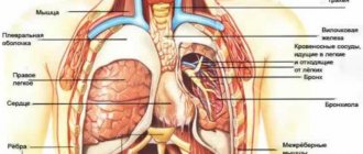
It must be taken into account that a lot of nerve endings and blood vessels pass through the neck . Therefore, all problems in the cervical spine result in acute pain, and sometimes a malfunction of the internal organs.
Complications of previous diseases may also be the cause.:
- Osteoporosis changes bone tissue.
- Rheumatoid arthritis affects the joints.
- Intervertebral hernia destroys the structure of the vertebrae.
- Spondylosis changes all structures of the spine.
As a rule, the causes of neck pain are degenerative changes in the cartilage of the spine
Pain in vertebrogenic cervicalgia is often caused by a functional disorder of the cervical vertebrae and concomitant pathologies.
Video: “Neck pain due to osteochondrosis: what to do?”
Possible consequences
Left untreated, it can lead to serious problems with brain function. This is explained by the fact that damaged vertebrae in the cervical region impair blood supply to the head.
Naturally, arterial stenosis causes the following problems:
- Excruciating headaches.
- Frequent fainting.
- Coordination of movements is impaired.
- Can paralyze limbs.
To avoid complications, treatment should be started in a timely manner.
Diagnosis of vertebrogenic headache
To establish the correct diagnosis, the doctor must carefully interview the patient and conduct a thorough examination. If cervicocranialgia is suspected, additional research methods are prescribed:
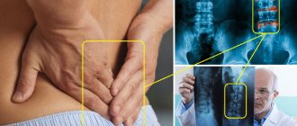
- X-ray of the cervical spine. The photographs clearly show pathological processes that explain the existing complaints and manifestations of the disease;
- CT scan. It is carried out in less clear cases to more accurately determine pathological areas;
- MRI. An informative method indicated for patients who have any contraindications to x-ray examination;
- Angiography of the vessels supplying the brain (x-ray examination using contrast agents);
- Dopplerography, which allows you to evaluate the properties of blood flow through the vessels and find areas with impaired hemodynamic processes.
Symptoms
The main manifestation of this disease is pain . Cervicalgia is accompanied by a lot of symptoms, but muscular-tonic syndrome is more often observed. In this case, the neck muscles become denser, and if you press on them, the pain intensifies.
Other symptoms include:
- The movement of the head is accompanied by a specific crunching sound.
- Throbbing headache, as well as dizziness.
- Restricted neck mobility.
- When you tilt your head back, fainting occurs.
- Partial loss of vision and hearing.
- The pain spreads to one of the arms and shoulder.
- The gait becomes unsteady.
- The upper limbs become weak.
- Tingling, as well as numbness of the face, hands and back of the head.
- Nausea, but no vomiting.
- When coughing or sneezing, the pain worsens.
Such symptoms are typical for any person, regardless of gender and age . In chronic cervicalgia, the symptoms are less pronounced, in contrast to the acute disease. In case of hypothermia or spinal injury, the pain is intense and severely hinders movement. When the disease occurs against the background of a concomitant disease, the pain is usually mild.
Video: “All causes of neck pain”
Treatment
Did you know that...
Next fact
Therapy is prescribed after a thorough examination. It should be noted that some drugs and physical procedures have strict contraindications. The prognosis for recovery is very favorable.
Drugs
To eliminate neck pain, a complex of several groups of drugs is prescribed. Painkillers with an anti-inflammatory effect : Celebrex, Xefocam, Ibuprofen. Medicines are used in the form of tablets, injections and ointments.
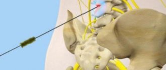
In the hospital, epidural blocks and botulinum toxin is injected into the muscles. If there is severe pain, they resort to novocaine blockades. Lidocaine is sometimes used. In case of acute pain syndrome, hormonal drugs (glucocorticosteroids) are sometimes prescribed, which have a powerful anti-inflammatory effect.
To tone blood vessels and improve microcirculation, Sermion and Trental are prescribed. Treatment is not complete without muscle relaxants , which relieve muscle spasms and reduce pain.
In the presence of osteoporosis , agents are used that activate bone tissue regeneration - fluorides and anabolic steroids. Drugs that prevent bone destruction are also needed - Myocalcic, estrogens.
Sometimes there is a need to take antidepressants: Sertraline, Diazepam.
Physiotherapy
- Electrophoresis.
- Ultraviolet irradiation.
- Electroneurostimulation.
- Diodynamic currents.
- Balneotherapy.
- Electroacupuncture.
Surgical
Any surgery in the neck area is quite risky due to the increased concentration of blood vessels and nerves in this area.
Indications for surgical intervention are considered:
- Acute damage, when damage to internal organs is noted.
- Increasing paresis with the threat of necrosis of nerve endings. In this case, the pain syndrome decreases, but weakness increases
The decision to carry out an operation must be motivated and soberly balanced.
Exercise therapy and massage
For vertebrogenic pathology, therapeutic exercises are necessary, which contain a complex of movements with muscle tension. The purpose of exercise is to correct muscle dysregulation .
The special complex has a common core of physical exercises that restore the motor pattern. They also promote post-isometric relaxation, stretch muscles and activate self-mobilization of the spine.
The exercises of the complex are treated with certain movements that are aimed at correcting changes in certain muscle groups of the spine.
It should be noted that exercise therapy is usually used for prevention or during remission to prevent exacerbation.
Basic exercises:
- Position sitting on a chair. The back is straight, arms are lowered. About 15 head tilts are done in different directions.
- The starting position is the same. Tilt your head back and hold for a few seconds. The movement is repeated up to 10 times.
- In a sitting position, raise your shoulders up as much as possible with a slight delay.
- Slowly rotate your head with a gradual increase in amplitude.
Such exercises can be done at any time and in any conditions. They are especially recommended for people leading a sedentary lifestyle.
Self-massage is very useful for this disease. It helps reduce pain and relieve vascular spasm.
Method of performing movements:
- Lightly stroking the head from the crown to the back of the head.
- Stroking the lateral cervical regions with fingertips. Be sure to do it from top to bottom and on both sides at the same time.
- Lightly massage the shoulders with your palms.
- Rubbing and kneading the trapezius muscle of the neck to the shoulder.
- Dashed movements in the direction of the spinous process of the 7th cervical vertebra.
- Stroking the back of the head, neck area and shoulder girdles.
At the moment of exposure to painful areas, pain, warmth and a feeling of stinging may occur. The main thing is to avoid severe pain and monitor the sensations. It is better to master self-massage with the help of a vertebrologist.
Therapy at home
At home, you can only reduce the symptoms of cervicalgia. You can use some folk remedies on your own, which are quite effective and time-tested.
The most popular of them : baths with certain herbal infusions, rubbing with a decoction of barberry root.
These 2 methods will not harm, but will not eliminate the cause of the disease. The most important thing is that they will relieve neck discomfort. Herbal baths warm up and reduce pain. Rubbing relaxes the cervical region, as the components of barberry have a gentle effect.
Naturally, if symptoms of a pinched nerve ending in the neck appear, you should immediately apply an analgesic ointment to the painful area. You can take a Diclofenac or Ibuprofen tablet.
It is imperative to exclude drafts and limit neck movements . Wrapping your neck in a woolen cloth will help reduce discomfort.
As for nutrition, there are no special diets for this syndrome.