In this article we will tell you:
- Causes of iridocyclitis
- Main types of iridocyclitis
- Symptoms and complications of iridocyclitis
- Diagnosis of iridocyclitis. Treatment
- Prevention of iridocyclitis
It happens that eye diseases concern one particular department, tissue or structure of an organ. So, with cataracts we talk about clouding of the lens, with keratitis - with inflammation of the cornea, with macular degeneration - with damage to the retina. Something else happens when several departments are involved in the pathological process at once. Such diseases are called combined, and it is to this group that the disease discussed in this article, iridocyclitis, belongs.
Iridocyclitis
is an inflammation of both the ciliary (ciliary) body and the iris of the eyeball.
The name of the disease comes from the Greek words iris - rainbow, and kykios circle, eye. Iridocyclitis is also called anterior uveitis. The disease is distinguished from uveitis proper by the localization of the inflammatory process: with ordinary uveitis, the choroid of the eye (uveal tract) is affected. The combination of lesions characteristic of ocular iridocyclitis occurs due to the anatomical similarity: the blood supply and innervation of the departments are closely related, and when one of them is affected, the inflammation quickly spreads to the other. Sometimes there are inflammatory processes in only one of the mentioned sections - the iris or the ciliary body. In these cases, we will talk about individual diseases: cyclitis and iritis.
You can get iridocyclitis at any age. Somewhat more often it affects young people 20-40 years old.
Causes of iridocyclitis
There are many reasons why iridocyclitis can develop. Among them are both exo- and endogenous. With the exogenous nature of the disease, the reason for its development lies in external factors. Endogenous iridocyclitis is a consequence of internal problems in the body and is more common.
The blood-ophthalmic barrier is normally responsible for the health of the eyes: it traps harmful bacteria, microbes, poisons - all those foreign objects that can cause diseases of the eye structures. When the permeability of this natural filter increases, harmful substances enter directly into the iris and then into the ciliary body, turning both sections into foci of inflammation.
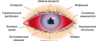
The following are the main groups of causes of iridocyclitis:
- Infections: bacterial, viral, fungal.
- Helminthic infestations.
- Allergy.
Herpes, influenza, ARVI measles, tuberculosis, some sexually transmitted diseases (syphilis, gonorrhea, chlamydia), staphylococcal and streptococcal infections, malaria, brucellosis: all these ailments can affect the permeability of the blood-ophthalmic barrier and cause inflammation.
Seasonal hay fever or year-round allergies (to dust, food, household chemicals, medications, etc.) can also cause the disease.
- Chronic foci of inflammation in the oral or nasal cavities, otitis.
- Rheumatic diseases.
In persons suffering from rheumatism and syndromes associated with it, the incidence rate can reach up to 40%.
- Metabolic diseases (diabetes, sarcoidosis, gout, etc.).
- Injuries (contusions, wounds) and postoperative complications.
Risk factors for the occurrence of this disease may include:
- traumatic, stressful situations;
- hypothermia of the body;
- too intense physical activity;
- bad habits, unbalanced diet;
- immune failures.
It happens that even specialists fail to find out the cause of the disease: then they talk about iridocyclitis of unknown etiology.
Risk factors
The risk of developing iritis increases if: 6
- There is a specific genetic change. This change is referred to as HLA-B27 and causes the immune system to not function fully.
- There is a sexually transmitted infection. Syphilis and HIV are associated with a significant risk of iridotskilitis.
- I have an autoimmune disease. Iridocyclitis often develops in people with ankylosing spondylitis and rheumatoid arthritis. About 40% of cases of iridocyclitis occur against the background of a systemic disease.
- There are pockets of chronic infection of the ear, throat, nose or mouth.
- There are bad habits. Tobacco smoke has a negative impact on eye health.
Main types of iridocyclitis
According to the nature of the appearance and course of iridocyclitis, they are divided into:
- acute (begin abruptly. Symptoms of acute iridocyclitis of the eye are pronounced, easily diagnosed, duration – 3-6 weeks);
- chronic (they are more sluggish in nature compared to acute forms, symptoms are more difficult to relieve, duration is up to several months);
- recurrent - periods of attenuation of the disease are replaced by exacerbations (more often in winter).
According to the nature of inflammation, iridocyclitis is:
- serous. Iridocyclitis of this form is characterized by the accumulation of serous secretion in the chambers of the eye.
- hemorrhagic. Such iridocyclitis is characterized by severe damage to the vessels supplying blood to the ciliary body and iris.
- exudative. The main difference between this form is the accumulation of pus in the form of a crescent on the lower part of the iris.
- fibrinous-plastic. Iridocyclitis of this type occurs with the formation of fibrin threads. With fibrinous iridocyclitis, synechiae form in the anterior chamber of the eye, and fibrinous exudate accumulates.
What it is?
Iridocyclitis is an intraocular inflammation that affects the iris and the anterior part of the ciliary body.1 It is a fairly rare disease - about 0.3 cases per 1000 population. In the structure of eye pathology they occupy 5-12%.2
If iridocyclitis is not chronic, it can be cured in about 1-3 months. If we talk about all forms of intraocular inflammation, this pathology is one of the most common forms. Quite often, acute iridocyclitis develops into chronic recurrent forms.
Complications may also occur, including glaucoma and cataracts. To avoid such a scenario, it is important to diagnose iridocyclitis early and treat it.
Symptoms and complications of iridocyclitis
The main symptoms of iridocyclitis are nonspecific: they are inherent in many ophthalmological (and not only) diseases. Patients with inflammation of the ciliary body and iris complain of:
- headache;
- blurred vision;
- photophobia;
- lacrimation;
- blurred vision;
- hyperemia.
Signs characteristic of acute iridocyclitis are:
- sharp pain in the eyeball, radiating to the head;
- increased pain when touching the eye in the projection area of the ciliary body;
- thickening of the iris, narrowing and immobility of the pupil;
- increased pain at night;
- swelling of the eyelids;
- the vessels visible along the outer edge of the iris are first bright red, later violet.
In chronic iridocyclitis, smoothing of the iris pattern, pronounced exudation, and the formation of protein tubercles, adhesions, and synechiae are observed. The pain is less pronounced than in the acute form.
Untimely or ineffective treatment of the disease often leads to complications. The inflammatory process can spread to other parts - then the patient develops chorioretinitis, scleritis, keratitis, vasculitis, endo- or panophthalmitis. In addition, complications of iridocyclitis include:
- the appearance of cataracts, glaucoma;
- retinal disinsertion;
- adhesions, pupil fusion;
- corneal dystrophy.
In difficult cases, we can talk about irreparable loss of vision and even removal of the eyeball.
Iridocyclitis: treatment methods
Drops that dilate the pupil are prescribed as first aid. The specialist prescribes medications that act on the causative agent of infection, increase immunity, and eliminate inflammation. Usually these are drugs that contain antibiotics. Corticosteroids, cytostatics, immunomodulators, enzyme agents, and absorbable exudates can be used.
With timely initiation of therapy, the prognosis is favorable. If treatment is prescribed late or the disease is severe, then various consequences are possible: sympathetic inflammation of the second eye, glaucoma, atrophy of the eyeball, blindness.
Diagnosis of iridocyclitis. Treatment
Patients with any form of the disease should undergo treatment under the supervision of a specialist. Self-diagnosis and self-medication in this case are unacceptable: after all, the consequences can be too severe.
To clarify the diagnosis, the ophthalmologist will prescribe general tests: blood tests for various parameters, X-ray and ultrasound examinations, and, if necessary, tests for allergens and rheumatism. Local examination will consist of examination with a slit lamp, palpation, measurement of intraocular pressure, and biomicroscopy of the eye.
The doctor will develop a treatment plan. Most likely, it will be carried out in a hospital. The set of drugs will depend on the cause of the disease. In addition to medications, a specialist may prescribe a course of physiotherapeutic procedures, for example, electrophoresis.
Diagnosis
The diagnosis is established on the basis of a comprehensive clinical and laboratory examination of patients and consultations with specialists in various fields.
Differential diagnosis
with I. it is carried out with conjunctivitis and an acute attack of glaucoma (tsvetn. Fig. 3). In the initial stage of I., pericorneal vascular injection can be mistaken for conjunctival, which can lead to improper treatment, the development of synechiae and other complications.
The main differences between I. and an acute attack of glaucoma are presented in the table.
Table. Differential diagnosis of acute iridocyclitis and acute attack of glaucoma
| Signs | Acute iridocyclitis | Acute attack of glaucoma |
| Nature of vascular injection | Inflammatory, pericorneal | Stagnant |
| Cornea condition | Transparent, smooth surface | Diffusely cloudy, rough, as if worn out |
| Anterior chamber depth status | Normal or uneven | Small |
| Iris condition | Hyperemic, pattern blurred | Not changed |
| Pupil condition | Narrow | Wide |
| Nature of pain | Severe pain in the eye without radiating | Pain in the eye radiating to the jaw, teeth and back of the head |
| Intraocular pressure | Normal or decreased, sometimes increased | Sharply increased |
Serol, reactions (Wassermann reaction, RSK) to syphilis, Toxoplasmosis, histoplasmosis, brucellosis are only of relative importance, indicating the general infection of the body. The method of identifying a focal reaction in the eye in response to subcutaneous or intradermal injection of specific antigens - tuberculin, toxoplasmin, staphylococcal and streptococcal allergens - has the greatest diagnostic significance.
In the diagnosis of viral infections, immunofluorescent examination of scrapings of the conjunctival epithelium is used—the method of fluorescent antibodies. The most reliable diagnostic value is the determination of specific antiviral antibodies in the aqueous humor of the anterior chamber. To diagnose herpetic I., a method is used to detect a focal reaction in the eye in response to repeated intradermal administration of herpetic polyvaccine, as well as fluorescent gray l. a method based on the detection of viral antigen in eye tissues after intradermal injection of herpetic antigen.
When identifying bacterial and tissue sensitization, serol, reactions and intradermal tests with microbial antigens and uveopigment antigen are used. This microprecipitation method is most widely used. In order to identify cellular sensitization to the antigens of tuberculosis, toxoplasmosis, staphylococcus, streptococcus and herpes virus, in vitro methods of blastotransformation of lymphocytes (see), inhibition of migration of blood leukocytes and leukocytolysis are used (see Leukolysis).
To assess immunol, the reactivity of the body, the determination of immunoglobulins G, M and A in tear fluid and serum is of particular importance.
Certain forms of iridocyclitis
Traumatic iridocyclitis
develops after penetrating wounds, contusion of the eyeball, burns, corneal ulcers, as well as intraocular operations. It complicates approx. 60% of all penetrating wounds. Develops as a result of the introduction of exogenous infection, chemical. reactions when foreign bodies enter, pronounced processes of proliferation and mooring, and the occurrence of autoimmune reactions. A severe complication of traumatic I. is the development of sympathetic inflammation and bilateral phacogenic I.
The clinic of traumatic I. is characterized by the same signs of inflammation as endogenous I. The severity of inflammatory phenomena in traumatic I. can be different. In mild forms of I., inflammatory phenomena gradually subside and disappear after 10–15 days. In severe forms that occur with the phenomena of exudation and proliferation, the course of I. can be long and there is a danger of developing sympathetic inflammation.
Phacogenic iridocyclitis
develops mainly after penetrating injuries or operations associated with damage to the lens. In rare cases, it may be caused by endogenous factors.
In the differential diagnosis of traumatic and phacogenic I., disruption of the integrity of the lens capsule and the presence of large, loose white precipitates (so-called lens precipitates) on the posterior surface of the cornea are of particular importance. In some cases, bilateral phacogenic inflammation occurs, which differs in wedge, pattern, course, and outcome from sympathetic inflammation. In the pathogenesis of phacogenic I. the development of sensitization to lens protein is of great importance. In this regard, extraction of traumatic cataracts or removal of remnants of the lens substance is a necessary condition for the successful treatment of phacogenic I.
Sympathetic iridocyclitis
has an autoimmune nature, therefore, timely and correct treatment with corticosteroids of traumatic I., occurring with autoimmune reactions, is of particular importance in its prevention (see Sympathetic ophthalmia).
Bibliography
Angel V.I. The state of nonspecific reactivity of the body and some biochemical indicators in endogenous iridocyclitis, Ophthalm, journal, No. 3, p. 193, 1973; Garkavi R. A. On the issue of phacogenic uveitis, in the same place, No. 2, p. 76, 1960; Zaitseva N. S. et al. Pathogenesis of endogenous uveitis in the light of experimental analysis, Vestn, ophthalm., No. 5, p. 45, 1974; Zaitseva N.S. et al. The significance of some immunological indicators in the diagnosis and clinic of endogenous uveitis, ibid., No. 3, p. 52, 1978; Zolotareva M. M. Selected sections of clinical ophthalmology, p. 5, Minsk, 1973; Kovalevsky E.I. Pediatric ophthalmology, p. 189, M., 1970; Lebekhov P.I. Perforated eye wounds, L., 1974; Multi-volume guide to eye diseases, ed. V. N. Arkhangelsky, vol. 1-2, M., 1960-1962; Podgornaya N. N. and Sokolovsky G. A. Fluorescein angiography of the iris, Vestn, ophthalm., No. 3, p. 9, 1973; Samoilov A. Ya., Yuzefova F. I. and Azarova N. S. Tuberculous eye diseases, p. 109, M., 1963; Stukalov S. E. Immunological studies in ophthalmology, Voronezh, 1975; Campinchi R.e. A. L'uveite, phenom^nes immunologiques et allergiques, P., 1970; Dinning WJ a. Perkins ES Immunosuppressives in uveitis, Brit. J. Ophthal., v. 59, p. 397, 1975; Perkins ES Recent advances in the study of uveitis, ibid., v. 58, p. 432, 1974; Schlaegel TF Progress in uveitis, 1959—1969, Surv. Ophthal., v. 15, p. 25, 1970; S mi th RE, Godfrey WA a. K imura SJ Complications of chronic cyclitis, Amer. J. Ophthal., v. 82, p. 277, 1976; System of ophthalmology, ed. by S. Duke-Elder, v. 9, L., 1966; W i tm er R. Diagnostik der Uveitis, Ther. Umsch., Bd 26, S. 342, 1969; Woods A.C. Endogenous uveitis, Baltimore, 1956.
O. B. Chentsova.