Right-sided lower lobe pneumonia develops more often than left-sided pneumonia. This is due to the peculiarities of the anatomical structure of the respiratory tract on the right. The right lower lobe bronchus has an oblique direction, so microorganisms penetrate it faster into the lung tissue. At the Yusupov Hospital, pulmonologists use modern examination methods to diagnose pneumonia and determine the source of inflammation. For treatment, effective drugs are used, registered in the Russian Federation, which have a minimal range of side effects.
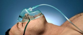
Patients with mild right-sided lower lobe pneumonia are hospitalized in a therapy clinic. In life-threatening conditions, they are transferred to the intensive care unit. If indicated, they undergo artificial ventilation of the lungs using modern portable and stationary ventilators. All severe cases of right-sided lower lobe pneumonia are discussed at a meeting of the Expert Council, in which professors and doctors of the highest category take part.
Causes
The vast majority of right-sided pneumonia caused by microorganisms is an independent disease. Less common is pneumonia, which is a manifestation of an acute infectious disease. Right lower lobe pneumonia is predominantly caused by the following pathogens:
- pneumococci;
- staphylococci;
- hemophilus influenzae;
- mycoplasma.
Less commonly, right-sided lower lobe pneumonia develops under the influence of chlamydia and Klebsiella. In patients who are in the hospital, the cause of right-sided lobar pneumonia can be infection with gram-negative microorganisms (enterobacteria, Pseudomonas aeruginosa, acinetobacter), Staphylococcus aureus and anaerobes. In case of right lower lobe pneumonia in patients with immunodeficiency, in addition to gram-negative bacilli and pneumococci, pneumonia is often caused by cytomegaloviruses, fungi, and mycobacteria. The main cause of aspiration right-sided lower lobe pneumonia is the entry into the respiratory tract of microflora of the stomach or oropharynx (anaerobic bacteria, gram-negative microflora and Staphylococcus aureus).
The main causative agents of atypical inflammation of the lower lobe of the right lung are mycoplasma, chlamydia, and legionella. In viral-bacterial right-sided lower lobe pneumonia, respiratory viruses cause pneumonia only in the initial period of the disease. The main pathogens that determine the clinical picture, severity and outcome of the disease remain the bacterial flora.
Features of the disease
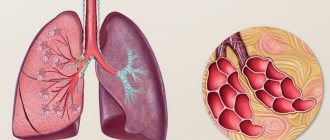
The human lungs are a paired organ; they are divided into the left and right lungs. Each lung consists of lobes: the right lung has three lobes, the left lung has two. Each lobe is divided into segments, which are ventilated by a separate segmental bronchus and have a corresponding branch of the pulmonary artery. Pneumonia most often develops in the right lung. The main bronchus of the right lung has a vertical direction and is a continuation of the trachea. It branches into each lobe of the lung. The bronchus of the middle lobe is the longest and narrowest, so the inflammatory process most often occurs in this area.
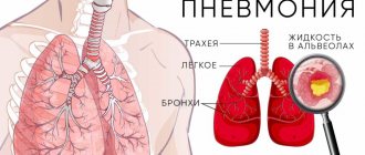
Right-sided middle lobe pneumonia involves inflammation in the middle lobe of the right lung with the involvement of lung tissue and alveoli in the infectious process. If only one segment of the lung is affected, they speak of segmental or focal pneumonia. When two or more segments are affected, lobar pneumonia develops. If the infectious process spreads to the pleural membrane, pneumonia is called lobar: the disease has a severe course and can lead to serious complications (respiratory failure, toxic shock, cardiopulmonary failure, coma).
Right-sided middle lobe pneumonia is often bacterial in nature. Its most common pathogens are:
- staphylococcus,
- hemophilus influenzae,
- Pneumococcus,
- streptococcus,
- mycoplasma,
- chlamydia,
- legionella.
Signs
The following syndromes are characteristic of pneumonia of the lower lobe of the right lung:
- intoxication (pallor of the skin, loss of appetite, weakness, general weakness, muscle and headaches, shortness of breath, palpitations);
- syndrome of general inflammatory changes (fever, feeling hot, chills);
- syndrome of inflammatory changes in the lung tissue (cough with sputum production, increased vocal tremors and bronchophony, shortening of percussion sounds, changes in the frequency and nature of breathing, moist rales and crepitus);
- syndrome involving other organs and systems in the pathological process.
An X-ray sign of right-sided lower lobe pneumonia is the presence of large areas of clearing, involving the lower lobe of the right lung. At the Yusupov Hospital, patients undergo radiography in two projections or large-frame fluorography. Bacteriological examination of sputum or bronchial washings is carried out before prescribing antibiotics. It helps to detect the pathogen and determine its sensitivity to antibiotics.
To clarify the nature of pneumonia, immunological studies are used. Viral and viral-bacterial pneumonia are detected using virological and serological studies. Patients can undergo complex procedures at partner clinics.
A general blood test allows you to determine the severity of the inflammatory process in the right lobe of the lower lung. In patients with pneumonia, the white blood cell count and erythrocyte sedimentation rate increase. With severe intoxication syndrome, the number of red blood cells decreases and toxic granularity of neutrophils appears. C-reactive protein appears in the blood, fibrinogen content and the level of sialic acids increase.
Bacterial pneumonia is characterized by leukocytosis; with viral pneumonia, the number of leukocytes in the blood decreases. In mycoplasma and ornithosis inflammations of the lungs, leukopenia is combined with a very high erythrocyte sedimentation rate. For patients with a prolonged course of right-sided lower lobe pneumonia and complications of pneumonia, the immunological reactivity of the body is determined. In order to find out the degree of involvement of other organs in the pathological process, an electrocardiogram is recorded, echocardiography is performed, and indicators of external respiratory function are determined using spirometry.
Diagnostics
To identify pneumonia and its type, an integrated approach is used:
- anamnesis collection. The doctor finds out when the first symptoms of the disease appeared. The patient must tell in detail about his feelings, report the presence of chronic diseases, if any;
- physical examination. On auscultation, fine bubbling rales are heard. Bronchophony and weakened vesicular breathing are recorded. During percussion, dullness of the pulmonary sound is noted;
- radiography. In case of right-sided middle lobe pneumonia, the X-ray image shows a darkened area in the middle lobe of the right lung, the presence of single or multiple foci;
- general blood analysis. In patients with pneumonia, there is an increase in the number of leukocytes, an increase in neutrophils, an acceleration of ESR, a decrease in the level of hemoglobin and platelets;
- bacteriological analysis of sputum. Microscopy of the smear is performed and cultured on a special nutrient medium to determine the pathogen.
To diagnose the disease, specialists at the Yusupov Hospital use high-precision equipment, which allows them to reliably determine the type and nature of the disease. High-quality and reliable diagnosis is a key factor in prescribing the correct therapy.
Types of right-sided pneumonia
Depending on the clinical picture, right-sided pneumonia can be of several types:
- lobar;
- focal;
- interstitial;
- purulent.
Purulent pneumonia of the right lung is the most dangerous pathology, which can be fatal.
According to the localization of the inflammatory process, right-sided pneumonia can be:
- focal;
- segmental;
- share;
- drain;
- total or polysegmental;
- basal.
In addition, right-sided pneumonia occurs:
- upper lobe;
- middle lobe;
- lower lobe.
Lower lobe pneumonia on the right is accompanied by a significant increase in temperature, chills, severe cough with pain in the right chest and viscous sputum. It usually occurs with mild symptoms.
Causes of development of bilateral pneumonia
The most common cause of bilateral pneumonia is an infection in the lungs. Most often these are the following types of infectious pathogens:
- Bacteria. As a rule, these are pneumonia caused by streptococci, pneumococci, staphylococci and other types of bacterial pathogens. In medical practice, bacterial pneumonia occurs most often. A serious problem is nosocomial infections that affect the patient's respiratory system during aspiration, ventilation, surgery, or taking cytotoxic drugs.
- Viruses. Viral pneumonias are less common than bacterial ones. Most often, pneumonia is caused by influenza viruses, respiratory syncytial virus, and coronaviruses (SARS-CoV, SARS-CoV-2, MERS-CoV). With a viral infection, the interstitial tissues of the lungs often become inflamed with the formation of infiltrates in the alveolar sacs. Damage occurs as a result of the immune response to infection. During a viral infection, white blood cells (in particular, lymphocytes) activate cytokines (the so-called cytokine storm), which leads to the accumulation of fluid in the alveoli and further respiratory failure.
- Fungi. Fungal pneumonias are much less common than bacterial or viral ones. Often, fungal infection of the lungs is observed against the background of intensive antibacterial therapy. Pneumonia caused by fungi also occurs in people with weakened immune systems (eg, HIV-infected patients), as well as patients with chronic obstructive pulmonary disease (COPD).
Source: PheelingsMedia / Depositphotos
It is noteworthy that the likelihood of developing bilateral pneumonia in some categories of people is higher than in others. Let's consider the main risk factors for the development of bilateral pneumonia:
- Age – people over 65 and children under 5 years old.
- Lack of vitamins and nutrients (people with morbid thinness).
- Pulmonary diseases, such as asthma, COPD, cystic fibrosis, tuberculosis and others.
- Smoking.
- Presence of chronic diseases. In particular, these are heart and vascular diseases, lung pathologies, and diabetes.
- Immune system dysfunction. It is noteworthy that the danger lies in both a weakened immune system and a hyperimmune response. People suffering from autoimmune diseases are also at risk.
- Patients taking immunosuppressants—drugs that suppress the immune system.
- People who have problems swallowing.
- People who have recently had a viral or bacterial respiratory tract infection.
Treatment of pneumonia
To treat pneumonia, the most accurate diagnosis indicating the causative agent of the disease is necessary. If the cause of the disease is a virus, the doctor will prescribe antiviral therapy, and if fungi, antifungal therapy. If the bacteriological nature of pneumonia is confirmed, the patient will be prescribed antibacterial therapy, taking into account a large selection of modern medications that can destroy the pathogen and stop inflammation.
In the case of prescribing antibacterial drugs, i.e. antibiotics, there are two possible ways to take the medicine: in the form of tablets for mild pneumonia (often the patient is allowed treatment at home with unconditional regular monitoring by the attending physician) and intramuscularly/intravenously in a hospital in case of severe disease. Medicines will also be prescribed to combat symptoms and generally strengthen the body: expectorants, vitamins, drink plenty of fluids; Additionally, it is advisable for the patient to follow a certain diet to restore the body’s strength and support the immune system.
Pneumonia is considered a well-studied disease and, despite its sometimes severe course (if it is a hospital-acquired form, or if the patient has not sought help for a long time in a community-acquired condition), in the hands of competent specialists it can be easily cured.
The pulmonology department of our center is rightfully considered one of the leading in the country, thanks to modern diagnostic and treatment equipment and the composition of the team of specialists: our doctors develop unique treatment and rehabilitation programs, train colleagues from other centers, and publish the results of their research in leading international publications.
The department has all possible means of diagnosing and treating lung diseases: both modern and time-tested. Moreover, in our center, the only one in Russia, clinical trials are conducted, and it is our specialists who confirm the effectiveness of new diagnostic and therapeutic equipment, which is being registered for future use in all pulmonology departments of the country. This is a great trust in the center’s qualifications, which we justify year after year, first of all, to our patients.
Symptoms of right-sided pneumonia
Right-sided pneumonia of a bacterial nature is manifested by the following symptoms:
- cough;
- secretion of sputum;
- increased temperature;
- leukocytosis;
- blue skin in the area of the nasolabial triangle;
- rapid breathing and heart rate.
Viral right-sided pneumonia manifests itself:
- muscle weakness;
- rapid fatigue;
- fever;
- dry cough with little sputum production.
Acute pneumonia
The dominant role in the etiology of acute pneumonia belongs to infection, primarily bacterial. Typically, the causative agents of the disease are pneumococci (30-40%), mycoplasma (6-20%), Staphylococcus aureus (0.4-5%), Friedlander's bacillus, less often - hemolytic and non-hemolytic streptococcus, Pseudomonas aeruginosa and Haemophilus influenzae, fungi and their associations ; Among the viruses are influenza virus, RS virus, adenoviruses. Purely viral acute pneumonia is rare; usually ARVI facilitates the colonization of lung tissue by endogenous or, less commonly, exogenous bacterial microflora. In ornithosis, chicken pox, whooping cough, measles, brucellosis, anthrax, salmonellosis, the development of acute pneumonia is determined by the specific causative agent of this infection. Microorganisms enter the lower parts of the respiratory tract through the bronchogenic route, as well as hematogenous (in infectious diseases, sepsis) and lymphogenous (in case of chest injury) routes.
Acute pneumonia can occur after exposure to the respiratory tract of light chemical and physical agents (concentrated acids and alkalis, temperature, ionizing radiation), usually in combination with secondary bacterial infection with autogenous microflora from the pharynx and upper respiratory tract. Due to the long-term use of antibiotics in the development of acute pneumonia, the role of opportunistic microflora has become more significant. There are cases of allergic (eosinophilic) acute pneumonia caused by helminthiases and medications. Acute pneumonia can occur uncomplicated and with complications; mild, moderate or severe; with the absence or development of functional disorders.
Various factors that reduce the resistance of the macroorganism predispose to the occurrence of acute pneumonia: prolonged intoxication (including alcohol and nicotine), hypothermia and high humidity, concomitant chronic infections, respiratory allergies, nervous shock, infancy and old age, prolonged bed rest. The penetration of infection into the lungs is facilitated by impaired patency and drainage function of the bronchi, inhibition of the cough reflex, insufficiency of mucociliary clearance, defects in pulmonary surfactant, decreased local immunity, including phagocytic activity, levels of lysozyme and interferon.
In acute pneumonia, inflammation affects the alveoli, interalveolar septa and vascular bed of the lungs. Moreover, in different parts of the affected lung, different phases can be simultaneously observed - flushing, red and gray “hepatization”, resolution. Morphological changes in acute pneumonia vary depending on the type of pathogen. Some microorganisms (staphylococcus, Pseudomonas aeruginosa, streptococcus) secrete exotoxins that cause deep damage to the lung tissue with the appearance of multiple small, sometimes merging foci of abscess pneumonia. In acute Friedlander pneumonia, extensive infarct-like necrosis occurs in the lungs. Interstitial inflammation dominates in pneumonia of pneumocystis and cytomegalovirus origin.
Symptoms of pneumonia
- pain in the side, especially when breathing deeply,
- dyspnea,
- dry cough or cough with sputum,
- increased fatigue, lethargy,
- headache,
- elevated temperature (up to 39-40°) or temperature below normal (especially in the elderly),
- nausea, vomiting.
If you do not contact a specialist when the first signs of the disease appear, the symptoms will begin to worsen and pneumonia will progress, affecting an increasing volume of the lungs. Therefore, you should not delay making an appointment with a doctor, especially if you or your loved one are at risk:
- children under 5 years old, adults over 65 years old,
- people with reduced immunity due to HIV infection or other serious diseases,
- diagnosed with asthma, COPD, diabetes mellitus or cardiovascular failure,
- heavy smokers,