General information
Pneumothorax is an accumulation of air in the pleural cavity, which leads to collapse of the lung with subsequent displacement of the mediastinum to the healthy side.
The dome of the diaphragm lowers and the vessels of the mediastinum are compressed, which negatively affects the functioning of the cardiovascular and respiratory systems. As a result of pneumothorax, gas can enter between the layers of the parietal and visceral pleura through any defect in the chest or on the surface of the lung. After air enters the pleural cavity, the pressure inside the pleura increases (it should be below atmospheric pressure), which leads to partial or complete collapse of the lung. ICD-10 code: J93 Pneumothorax.
Intestinal pneumothorax is mistakenly confused with pneumatosis intestinalis , which is characterized by the appearance of cysts containing gases or air.
Pathogenesis
Primary spontaneous pneumothorax occurs as a result of rupture of subpleurally located emphysematous bullae, which are formed against the background of congenital defects of elastic pulmonary structures or against the background of cysts that have developed abnormally in their terminal bronchioles.
Until now, the pathogenesis of bullae formation remains unknown. It is believed that they are formed as a result of the degradation of elastic fibers in the lungs, which is caused by the activation of macrophages and neutrophils due to smoking. There is a shift in the balance between antiproteases, proteases and the antioxidant oxidation system. Formed bullae provoke inflammatory blockage of the small airways, which leads to an increase in intra-alveolar pressure and air enters the pulmonary interstitium.
The air moves towards the root of the lung, causing emphysema . Due to increased pressure in the mediastinum, the parietal mediastinal pleura ruptures with the development of pneumothorax. Increased intrapleural pressure prevents fluid from leaking into the pleural cavity.
As a result of a large primary spontaneous pneumothorax, a sharp decrease in the vital capacity of the lungs occurs, an increase in the alveolar-arterial oxygen gradient with the subsequent development of hypoxemia of varying severity. Hypoxemia develops against the background of ventilation-perfusion imbalance and the formation of a shunt from right to left. The clinical picture will directly depend on the severity of these disorders. Maintaining normal gas exchange does not allow the development of hypercapnia .
Rupture of lung tissue can occur in the area of pleural fusion during forced breathing or coughing. Secondary spontaneous pneumothorax develops when a pathological focus breaks into the pleural cavity in people suffering from destructive diseases of the pulmonary system ( lung gangrene , lung abscess , tuberculosis cavity , pulmonary infarction ). A similar process can occur in patients with histiocytosis X , neoplastic diseases of the mediastinum and lungs, chronic obstructive diseases ( bronchial asthma ).
Most often, right-sided pneumothorax is recorded, much less often - bilateral.
Classification, types of pneumothorax
By distribution there are:
- unilateral pneumothorax (right or left side);
- bilateral pneumothorax.
According to the development of complications:
- uncomplicated;
- complicated (bleeding, emphysema, pleurisy).
By volume of air in the pleural cavity:
- Small (less than 20% of the lung tissue is in a state of collapse). When X-rayed, a small pneumothorax appears as a small rim of air along the peripheral edge of the lung and, as a rule, does not require specific treatment in the absence of symptoms of respiratory failure.
- C middle (the lung collapses to 1/2 the distance between the border of the heart and the lateral edge of the pleural cavity). Average pneumothorax requires treatment: with the help of repeated aspirations, a needle and a syringe with a volume of 50-100 ml, puncture of the pleural cavity is performed with air aspiration.
- Big . For large pneumothorax, a special drainage tube must be inserted, focusing on the same anatomical landmarks as during aspiration. Help is to connect the drain to a siphon system, which functions as a one-way valve. You can immediately use a drainage tube equipped with a special valve.
Pneumothorax in connection with the environment is divided into:
- Open pneumothorax . A pressure equal to atmospheric pressure is created in the pleural cavity due to communication between the pleural cavity and the external environment. The most important condition for the expansion of the lung is negative pressure in the pleural cavity, which is why with an open pneumothorax the lung collapses. In a collapsed lung, gas exchange cannot occur. Open pneumothorax completely shuts off the lung from the respiratory process, the blood ceases to be fully enriched with oxygen.
- Closed pneumothorax . Communication with the external environment is completely absent. A small volume of gas enters the pleural cavity, which does not increase. Closed pneumothorax is considered the easiest, because air trapped in the pleural cavity can gradually resolve on its own. In this case, the lung does not expand.
- Valvular pneumothorax . It develops during the formation of a valve structure that can allow air to pass in only one direction: from the external environment or the lung into the pleural cavity. The valve structure prevents the reverse flow of air and with each act of breathing there is an increase in pressure in the pleural cavity. Valvular pneumothorax is considered the most dangerous, because. in addition to shutting off the lung from the respiratory process, irritation of the nerve fibers in the pleura itself occurs, which inevitably leads to pleuropulmonary shock and displacement of organs in the mediastinum, which negatively affects their work and compression of large arteries and veins.
Pneumothorax can also be:
- complete (the lung collapses completely);
- parietal (the lung does not expand completely, a small volume of air is in the pleural cavity, is considered a “closed type”);
- encysted (formed against the background of adhesive lesions of the visceral and parietal pleura, can be completely asymptomatic, and is considered less dangerous);
- bilateral complete pneumothorax (due to critical impairment of respiratory function, if assistance is not provided in a timely manner, it can lead to a very rapid death).
Tension pneumothorax is a life-threatening condition caused by the accumulation of air entering under high pressure in the pleural cavity. The intense form most often develops as a result of traumatic injury to the chest, but can be a consequence of certain diseases of the respiratory system. In the absence of timely treatment, tension pneumothorax leads to death due to excessive compression of the mediastinal organs, heart and large vessels.
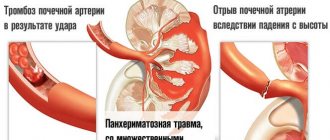
Traumatic pneumothorax develops after exposure to trauma to the chest area with subsequent rupture of the pleural layers (with knife or gunshot wounds). Traumatic pneumothorax can also develop with a closed chest injury: the pleural layer is damaged by a broken rib.
Spontaneous pneumothorax is not associated with iatrogenic treatment, diagnostic procedures, or trauma. Spontaneous collapse of the lung is read as idiopathic, spontaneous. The reasons can be very different ( cystic fibrosis , ankylosing spondylitis , Beck's sarcoidosis , etc.).
Causes of pneumothorax
It is customary to distinguish two groups of reasons.
Mechanical damage to the lungs and chest. These include:
- penetrating wounds and other open traumatic injuries of the chest;
- closed chest injuries with damage to the lung tissue by rib fragments;
- artificially induced pneumothorax, which is applied for diagnostic purposes during thoracoscopy and for therapeutic purposes in pulmonary tuberculosis;
- Iatrogenesis - complications after diagnostic and therapeutic procedures (intercostal nerve block, installation of a subclavian catheter, puncture of the pleural cavity).
Diseases of the chest and lungs:
- Of a specific nature. Lung collapse occurs as a result of a breakthrough of the cavernous lesion during tuberculosis , rupture of the cavern.
- Non-specific. Collapse develops as a result of rupture of air cysts in patients with emphysema (bullous disease) of the lungs, as well as when a lung abscess breaks into the pleural cavity (pyopneumothorax), or with spontaneous rupture of the esophagus.
Causes of spontaneous pneumothorax (secondary):
- endometriosis ;
- bullous disease;
- malignant neoplasms;
- infectious diseases ( SARS );
- interstitial lung lesions;
- inflammatory diseases of connective tissue ( Marfan disease , polymyositis , ankylosing spondylitis ).
First aid for spontaneous pneumothorax of the lung and treatment
Pneumothorax is a life-threatening condition characterized by the presence of air in the pleural cavity.
Pneumothorax can be spontaneous (spontaneous), iatrogenic and traumatic. The consequences of traumatic pneumothorax include: hemothorax, recurrent pneumothorax and the formation of pneumomediastinum. Depending on the connection with the environment, open, “tension” (valvular) and closed pneumothorax are distinguished. Open is characterized by “depressurization” of the respiratory system due to a chest injury. The normal pressure in the lung changes, which leads to its “sticking together” and the cessation of gas exchange. Symptoms of closed pneumothorax include sharp chest pain, inability to breathe normally, rapid heartbeat, and cold sticky sweat. With valve pneumothorax, air passes in one direction - into the pleural cavity and cannot escape back - the valve “triggers”. Valvular pneumothorax is characterized by compression of the lung and large vessels, irritation of the nerve endings of the pleura, which can cause respiratory failure. Possible causes of pleural pneumothorax:
- infectious diseases (tuberculosis, Pneumocystis pneumonia);
- pathologies of the respiratory system (cystic fibrosis, asthma, obstructive pulmonary disease);
- connective tissue diseases (scleroderma, rheumatoid arthritis, polymyositis);
- thoracic endometriosis;
- oncological diseases;
- chest injuries;
- closed chest injuries;
- complex diagnostic and therapeutic measures.
Treatment of open pneumothorax is carried out exclusively in a hospital setting. Emergency care for pneumothorax is provided in an ambulance.
Symptoms of pneumothorax of the lungs
With all forms of valvular pneumothorax, a typical clinical picture is observed, characterized by the development of severe life-threatening disorders of respiratory and circulatory functions.
The patient's condition deteriorates very quickly, and characteristic symptoms of pneumothorax appear:
- motor excitement;
- increasing expiratory shortness of breath with difficult and prolonged exhalation;
- cyanotic mucous membranes and skin;
- uneven, rapid breathing with intermittent inhalation and pain in the chest.
In this case, breathing sounds are sharply weakened, in some cases they are not heard at all.
Breathing and shortness of breath acquire a specific, peculiar character. The patient tries to hold his breath while inhaling, because... when exhaling, the chest decreases in volume and compression of the already compressed lungs occurs, both on the affected and healthy sides. There is an increase in blood pressure followed by a rapid decrease.
The pulse is tense at the initial stages, then becomes rapid with weak filling. There is marked smoothness of the intercostal spaces on the affected side and a slight excursion of the chest.
When hemothorax is attached, a box sound will be detected by percussion. Obstruction of venous outflow is evidenced by strongly pronounced and dilated veins in the neck. A sharply noticeable air emphysema , which can affect the head, neck, limbs and the entire torso. To assess the dynamic course of the process, a chest X-ray is performed.
Prognosis and prevention
With timely assistance, the outcome of the disease is favorable. The prognosis for traumatic pneumothorax depends on the nature of the damage to the chest organs. In any case, the patient will need a long recovery period under the supervision of a doctor. Relapses of the pathology are possible only in patients with severe diseases of the respiratory system. In this case, the patient is recommended to undergo preventive examinations by a pulmonologist with the necessary studies.
Preventive measures include timely treatment of diseases of the respiratory system and clinical observation in the presence of chronic lung pathology.
You can get answers to all your questions at an appointment with our specialist. Sign up for a consultation at a time convenient for you. And remember, prevention is always better and cheaper than treatment.
Related Services: Asthma Symptoms Chronic Bronchitis
Tests and diagnostics
In critically ill patients, pneumothorax can be diagnosed based on information from the medical history and physical examination. The patient registers:
- severe chest pain;
- rapid increase in shortness of breath ;
- hypotension;
- rapid heartbeat, tachycardia ;
- contralateral displacement of the trachea;
- pulsus paradoxus;
- reduction in the severity of breathing sounds.
All of the above signs are not highly specific and characteristic exclusively of pneumothorax. In secondary spontaneous pneumothorax, the severity of shortness of breath does not correspond to the scale of pneumothorax, and latent emphysema may be the cause of weakened breathing.
Acute changes in ventilation parameters, such as an increase in airway pressure or a decrease in tidal volume, can either be associated with pneumothorax or be a manifestation of another pathology. This is why chest X-ray remains the gold standard for diagnosing pneumothorax.
Radiography
On a radiograph, the main sign of pathology is an area of clearing, which is located along the peripheral edge of the pulmonary field and separated by a clear boundary from the collapsed lung. The area is devoid of pulmonary pattern. X-ray examination reveals the communication between the environment and the pleural cavity.
The main radiological sign of pneumothorax is an area of clearing, devoid of a pulmonary pattern, located along the periphery of the pulmonary field and separated from the collapsed lung by a clear boundary corresponding to the image of the visceral pleura. X-ray examination can reveal the connection of the pleural cavity with the external environment.
With open pneumothorax, during inspiration there is an increase in the gas bubble, collapse of the lung and subsequent displacement of the mediastinal organs to the unaffected side. The diaphragm dome moves downwards.
With a closed form of pneumothorax, the picture on the radiograph depends on the amount of gases collected in the pleural cavity and the level of intrapleural pressure. If the volume of air in the pleural cavity is small and the pressure is below atmospheric, and the lung is only slightly collapsed, then on exhalation it collapses, and on inhalation it increases in volume.
If the pressure is higher than atmospheric pressure, then the lung will sharply collapse, its respiratory excursion will be barely noticeable, and the mediastinal organs will also shift to the healthy side, the diaphragm will descend downwards. If atmospheric and intrapleural pressure are equal, then the lung collapses only partially, while the respiratory excursion is maintained and the mediastinal organs shift only slightly to the healthy side.
With valvular pneumothorax, the configuration of the collapsed lung in the act of breathing and its size do not change. The lung completely collapses, allowing the organs of the mediastinum to sharply shift to the healthy side, and on exhalation, partially return to the affected area. With prolonged injection of gases into the pleural cavity during valve pneumothorax, tension pneumothorax is formed. A sharp shift of the mediastinum to the healthy side is recorded, the diaphragm thickens and descends, air may appear in the soft tissues of the chest.
With total pneumothorax, air fills the entire pleural cavity, the shadow of the mediastinum fills the entire unaffected side, and the dome of the diaphragm moves downward.
When examined in a lateral position, even a small accumulation of gas can be detected. On the affected side, deepening of the costophrenic sinus, flattening and smoothing of the contours of the lateral, lateral surface of the diaphragm are recorded. If blood enters the pleural cavity along with air, a picture of hemopneumothorax with the formation of a horizontal boundary between the two environments.
CT scan
Thanks to this method, it is possible not only to confirm the diagnosis, but also to assess the size of the pneumothorax and the condition of the lung parenchyma. The study helps to differentiate the pathology from bullous diseases , which helps prevent improper drainage and the formation of a parenchymal-pleural fistula. Thanks to CT, latent pneumothorax has become more frequently diagnosed, which does not manifest itself clinically and is not detected by radiography. The incidence of hidden pneumothorax in patients with polytrauma reaches 64%, and in the general population ranges from 2-15%.
Ultrasound of the chest
Ultrasound examination is the most accessible diagnostic method. The method has several advantages compared to computed tomography and radiography:
- the ability to examine the patient at the bedside;
- no radiation;
- visualization of images in real time;
- the ability to assess the dynamic development of the process.
Ultrasound is considered the most sensitive and specific method in diagnosing lung collapse compared to radiography, since it can be used to assess the reexpansion of the lung after thoracostomy drainage. A significant disadvantage is the need for training in correct visualization and interpretation of results by the attending doctor, who does not work as a radiologist in his main specialty.
With subcutaneous emphysema, ultrasound waves penetrate very poorly into the chest. The method is considered extremely useful when it is necessary to exclude pulmonary collapse after catheterization of the central vessels and pleural procedures, eliminating the need to delay portable radiography.
Publications in the media
Atelectasis (collapse) of the lung is a loss of airiness in an area of the lung, occurring acutely or over a long period of time. In the affected collapsed area, a complex combination of airlessness, infectious processes, bronchiectasis, destruction and fibrosis is observed.
Etiology and pathogenesis • Obstruction of the bronchial lumen by plugs of viscous bronchial secretion, tumor, mediastinal cysts, endobronchial granuloma or foreign body • Increased surface tension in the alveoli due to cardiogenic or non-cardiogenic pulmonary edema, surfactant deficiency, infection • Pathology of the bronchial walls: edema, tumor, bronchomalacia, deformation • Compression of the respiratory tract and/or the lung itself, caused by external factors (myocardial hypertrophy, vascular abnormalities, aneurysm, tumor, lymphadenopathy) • Increased pressure in the pleural cavity (pneumothorax, effusion, empyema, hemothorax, chylothorax) • Restriction of chest mobility ( scoliosis, neuromuscular diseases, phrenic nerve palsy, anesthesia) • Acute massive pulmonary collapse as a postoperative complication (unrecognized and unsanitized obstruction of the main bronchus).
Genetic aspects are determined by the underlying disease (cystic fibrosis, bronchial asthma, congenital heart disease, etc.). Risk factors. Surgeries on the chest organs, COPD, tuberculosis, smokers, obese people and people with short and wide chests.
Pathomorphology • Capillary and tissue hypoxia causes fluid transudation. The alveoli are filled with bronchial secretions and cells, which prevents complete collapse of the atelectasis area. • The addition of infection causes fibrosis and bronchiectasis.
The clinical picture varies depending on the rate of development of bronchial occlusion, the extent of atelectasis and the presence of infection.
• Diffuse microatelectasis, small atelectasis, slowly developing atelectasis and middle lobe syndrome (chronic atelectasis of the middle lobe of the right lung due to compression by lymph nodes) may be asymptomatic.
• Extensive atelectasis due to acute occlusion is characterized by the following symptoms •• Pain on the affected side, sudden shortness of breath and cyanosis •• Cough •• Hypoxia with a significant decrease in paO2 with a tendency to its recovery during the first 24–48 hours due to weakening of blood flow in the atelectasis area • • Percussion: dullness of percussion sound over the area of atelectasis •• Auscultation ••• absence of breath sounds - with occlusion of the airways ••• bronchial breathing, if the airways are patent ••• moist rales with focal obstruction •• Decreased chest excursion •• Displacement apical impulse.
• Chronic atelectasis •• Shortness of breath •• Cough •• Percussion: dullness of percussion sound •• Auscultation: moist rales •• If infected: increased amount of sputum, rise in body temperature •• Recurrent bleeding from the affected area is possible.
Age characteristics • Early childhood: aspiration mechanism, pneumonia • Children: among the most common causes are mediastinal cysts, vascular anomalies • Elderly: among the most common causes are lung tumors, cicatricial stenosis, bronchiectasis.
Special studies • Chest X-ray in two projections •• triangular-shaped intense homogeneous shadow with clear boundaries, with the apex directed to the root of the lung, with a decrease in the volume of the affected area of the lung •• With atelectasis of the lobe or lung - persistent displacement of the mediastinum to the affected side, dome the diaphragm on the affected side is raised, the intercostal spaces are narrowed •• diffuse microatelectasis - an earlier manifestation of oxygen intoxication and acute respiratory distress syndrome: a “ground glass” picture •• rounded atelectasis - rounded shading with a base on the pleura, directed towards the root of the lung (“comet-shaped” tail of blood vessels and airways). More often occurs in patients exposed to asbestos and resembles a tumor •• right-sided midlobe and lingular atelectasis merge with the borders of the heart on the same side (Arman-Delisle sign) • Bronchoscopy is indicated to assess the patency of the airways • EchoCG to assess the condition of the heart in cardiomegaly • CT or MRI of the chest.
Treatment
• The regimen depends on the patient's condition. Physical activity should be encouraged.
• Acute atelectasis (including acute postoperative massive collapse) •• The main cause of atelectasis should be eliminated, sanitation bronchoscopy should be performed, especially in cases of obstruction of the bronchial lumen with viscous sputum or vomit •• In case of foreign body aspiration - endoscopic removal.•• Adequate oxygenation, respiratory hydration mixtures •• In severe cases, mechanical ventilation with positive expiratory pressure or the creation of continuous positive pressure in the respiratory tract in persons with neuromuscular weakness •• Postural drainage (the head of the bed is lowered so that the trachea is below the affected area), breathing exercises, early postoperative mobilization of the patient •• Physiotherapeutic procedures, massage •• Broad-spectrum antibiotics are prescribed from the first day.
• Chronic atelectasis •• Postural drainage, breathing exercises (spirosimulator) •• Ventilation of the lungs with positive expiratory pressure or creation of constant positive pressure in the airways in persons with neuromuscular weakness •• Broad-spectrum antibiotics for purulent sputum •• Surgical resection of an atelectatic segment or lobe with recurrent infection and/or bleeding from the affected area •• If the obstruction is caused by a tumor, then the choice of treatment method is determined by the nature and extent of the tumor, and the general condition of the patient.
• Bronchodilators (salbutamol, fenoterol) - auxiliary value.
Complication : lung abscess (rare).
Prevention • Smoking cessation • Prevention of aspiration of foreign bodies and liquids, incl. vomit • In the postoperative period, the use of long-acting painkillers should be limited • Early postoperative mobilization of the patient • Breathing exercises.
ICD-10 • J98.1 Pulmonary collapse
Treatment of pneumothorax of the lungs
The main step in open pneumothorax is the application of a special occlusive dressing that will tightly and completely cover the wound. Additionally, measures are taken to maintain the normal functioning of the respiratory and cardiovascular systems.
Blood loss is replaced and pain relief is provided if necessary. In stationary conditions, surgical treatment and subsequent suturing of the wound defect in the chest are carried out with the formation through drainage of constant aspiration of air and outflow of exudate from the pleural cavity.
The extent of surgical intervention is determined by the nature of the damage with the maximum possible preservation of healthy lung tissue. After surgery is completed, a permanent drain is installed.
In the open form of pneumothorax, therapy is aimed at eliminating the underlying cause followed by drainage. If the damage is too extensive and there is no way to straighten the lung, temporary obstruction of the bronchial tube is performed using a special plug made of foam rubber or a similar material. Thanks to the plug, the flow of air into the pleural cavity is stopped and all the necessary conditions are created for the complete expansion of the previously collapsed lung. During bronchial obstruction, the visceral pleura fuses with the parietal pleura, which makes it possible to completely eliminate pneumothorax.
How long does it take for a lung to expand?
In case of spontaneous primary pneumothorax, oxygen inhalation is performed to accelerate the resorption of gases in the pleural cavity, which speeds up the process by 4 times. With standard breathing of normal air, air is absorbed at a very low rate - only 2% per day.
In 70% of patients with primary spontaneous pneumothorax of moderate volume, simple aspiration of air from the pleural cavity is considered effective. If more than 2.5 liters of gases are aspirated in a patient over 50 years of age, then doctors will most likely fail.
If everything went well, then 6 hours after aspiration there is no gas in the pleural cavity and the patient can be sent home the next day if his condition is stable. If after aspiration through the catheter the lung does not expand, then the catheter is connected to the Helmich valve or underwater draft, which are used as a drainage tube.
Treatment of open pneumothorax
First aid before the arrival of the ambulance consists of applying a bandage to cover the opening in the chest. For this purpose, a thick bandage made of gauze and cotton wool or a cellophane bandage is used.
Qualified assistance in a hospital setting: puncture, air evacuation and restoration of negative pressure in the pleura. Important: both at the stage of emergency care and in a hospital setting, it is necessary to use painkillers to reduce the patient’s suffering.
Procedures and operations
Pleural puncture with manometry allows us to clarify the type of spontaneous pneumothorax. Intrapleural pressure indicators with closed spontaneous pneumothorax are positive or slightly negative. In the open form of pneumothorax they tend to zero, in the valvular form they tend to increase and are positive.
The aspirated fluid obtained as a result of puncture from the pleural cavity is sent to the laboratory to study the cellular composition and analyze the microflora . Thoracoscopy is performed to determine the size and location of the pleural fistula.
First aid for pneumothorax
When pneumothorax occurs (closed, open or valvular), emergency care is required. If possible, assistance should be qualified and provided in a specialized hospital. In some cases, competent and timely provision of first aid can save a human life. In case of open pneumothorax or suspected lung collapse, a certain sequence must be followed.
Emergency care algorithm for spontaneous pneumothorax:
- Place the victim on an elevated surface to ensure the most favorable position for the respiratory system.
- Apply an occlusive dressing to the wound surface, which during emergency care can be any means that ensures the tightness of the affected part in the chest cavity. Available means include plastic film, adhesive tape, and rubberized fabric. The occlusive dressing is fixed with bandages or any fabric pre-treated with a disinfectant or iodine. Proper application of a bandage helps prevent the entry of an infectious agent onto the wound surface and prevent the development of a bacterial infection. The area around the wound must be treated with baby cream or Vaseline. In a hospital setting, the doctor will treat the peri-wound surface with a special ointment and apply a special hydroactive napkin.
- The patient must be anesthetized; the use of narcotic analgesics is allowed for severe pain.
- To remove air and drain the pleural cavity with drainage, the patient undergoes a pleural puncture in a hospital setting.
- Hormonal medications ( Dexamethasone ) are used to maintain normal blood pressure levels.
- If necessary, resuscitation measures are carried out.
Clinical manifestations of pathology
Symptoms depend on the type of pneumothorax and the degree of compression of the lung. The disease begins suddenly.
General symptoms:
- sticky cold sweat;
- shallow breathing;
- cyanosis of the skin;
- sharp chest pain, rapid heartbeat;
- weakness, decreased blood pressure;
- subcutaneous emphysema;
- an attack of dry cough is a sign of a tension pneumothorax;
- fear;
- the patient takes a forced position to alleviate the condition.
The further condition and life of the patient largely depends on the correct actions at the stage of providing first aid for spontaneous pneumothorax of the lung.
On our website Dobrobut.com you will find more information on this issue. You can make an appointment by calling the above numbers.
Pneumothorax in newborns
The most common cause of pneumothorax is overstretching of the alveoli with their subsequent rupture. It can occur spontaneously or against the background of a developmental defect (congenital lung cyst, congenital lobar pneumonia ). With aspiration pneumonia, air leak syndrome develops in the first 24-26 hours of a child’s life.
In the first day of a child’s life, one of the factors predisposing to the development of pneumothorax is pulmonary hypoplasia , as a result of which the surface of the alveoli decreases and the compliance of the lung tissue decreases.
Pulmonary hypoplasia often occurs with:
- oligohydramnios of various origins ( renal dysplasia , Potter's syndrome , prolonged leakage of amniotic fluid, renal agenesis );
- malformations of the chest ( asphyxial dystrophy of the chest );
- compression of the lungs (with chylthorax , pleural effusion , diaphragmatic hernia );
- weakness of respiratory intrauterine movements (with neuromuscular diseases, oligohydramnios).
Symptoms of pneumothorax in newborns
The pathology can be completely asymptomatic. In patients receiving mechanical ventilation, spontaneous deterioration of the condition may occur:
- severe cyanosis;
- asymmetry of the chest with unilateral lung damage;
- bradycardia.
At the slightest suspicion of pneumothorax, an x-ray of the newborn's chest is performed. Gas tests reveal mixed or respiratory acidosis and hypoxemia.
It is necessary to carry out differential diagnosis with diseases such as:
- lobar emphysema ;
- congenital diaphragmatic hernia ;
- cystic, adenomatous malformation;
- esophagopleural fistula;
- bronchogenic cysts.
Frequent complications of pneumothorax in a newborn are:
- pulmonary or heart failure ;
- infectious lesion;
- hemopneumothorax;
- bronchopleural fistula;
- intraventricular hemorrhages.
Treatment of pneumothorax in newborns
- Puncture of the pleural cavity. The manipulation is used for rapid symptomatic treatment of pneumothorax until permanent drainage is installed. A puncture is sufficient for infants who are not on mechanical ventilation.
- Respiratory therapy.
- Traditional mechanical ventilation.
For persistent bronchopleural fistula, selective intubation of the contralateral lung, pleurodesis with povidone-iodine or fibrin glue is used as a reserve.
Prevention of pneumothorax in newborns
- administration of surfactant to premature infants;
- early administration of surfactant with short-term mechanical ventilation (rapid extubation on nCPAP);
- traditional high-frequency mechanical ventilation using TBP less than 0.5 s;
- use of volumetric ventilation.
Carrying out primary resuscitation of a newborn with asphyxia can provoke damage to the lung tissue with subsequent release of gases into the pleural cavity. Rupture of the visceral pleura occurs spontaneously in some cases.