- What does the biopsy show?
- Is it possible to do without a biopsy?
- Types and methods of biopsy
- Does a biopsy hurt?
- Do I need special preparation for a biopsy?
- Is a biopsy safe? What are the consequences and complications?
A biopsy is a diagnostic procedure performed to obtain a tissue sample (biopsy) from a “suspicious” location, such as a tumor or polyp. A biopsy is necessary to confirm the diagnosis of cancer.
What does the biopsy show?
All cells of the body have a characteristic structure, depending on what tissue they belong to. As a malignant tumor develops, the structure of the cells is disrupted, and these changes can be seen under a microscope.
A doctor examining samples of tissue or cells obtained through a biopsy can tell definitively whether a patient has cancer. While other tests suggest cancer with varying degrees of probability, a biopsy helps to establish an accurate diagnosis.
Indications and contraindications for the study
Typically, a biopsy is prescribed to patients when there is a suspicion of the development of cancer. Morphological examination of biopsy specimens is necessary for the following indications:
- pathology of the digestive tract, soft tissues, retroperitoneum, mediastinum, chest walls, pleural cavity of the lungs, hematoma;
- “cold” thyroid nodule or cyst;
- malignant neoplasm;
- tumors of unknown etiology in the adrenal glands;
- abscess and focal lesions of the spleen;
- liver pathologies.
Morphological examination of biopsy specimens is contraindicated in the following cases:
- there is a threat of miscarriage;
- suspected melanoma;
- the patient’s written refusal to carry out the procedure;
- availability of non-invasive diagnostic testing;
- severe blood clotting disorder.
It is worth noting that during the period of tissue collection for MIB, complications may occur: air embolism, tumor dissemination and, in rare cases, death.
Types and methods of biopsy
A doctor can obtain a biopsy, a tissue sample for examination, in a variety of ways. Depending on this, there are several types of biopsy:
- razor;
- puncture;
- trephine biopsy;
- incisional;
- excision.
Imprint smears, scrapings, shave biopsy
Sometimes it is enough to obtain only a few cells for a biopsy. For example, for early detection of cervical cancer, a smear-imprint is performed from the mucous membrane of the cervix. The material obtained in this way is quite enough to perform laboratory research.
You can also make smears of nipple discharge if breast cancer is suspected.
During a shave biopsy, the doctor cuts off a layer of a certain thickness from the surface of an area of skin using a sharp instrument. A bleeding surface is left on which a pressure bandage is applied.
Needle biopsy
The name of the method comes from the Latin word punctio - “injection”. In turn, puncture biopsy is divided into types: fine-needle, thick-needle (trephine biopsy), aspiration.
Fine needle biopsy
This type of needle biopsy is used when it is necessary to obtain a small number of cells. The doctor inserts a thin needle into the suspicious area and removes a small amount of tissue.
Core needle biopsy
This type of biopsy is optimal in many cases, since it does not require an incision, and at the same time allows you to obtain a fairly large amount of tissue. Core needle biopsy is often used for suspected breast, liver, prostate and a number of other tumors.
Trephine biopsy is used to take samples of skin and bone marrow. The doctor uses a special instrument that resembles a needle, only thicker, in the form of a hollow cylinder with sharp edges. It is immersed in the desired place, and as a result it is filled with a column of fabric.
Aspiration biopsy
In an aspiration biopsy, tissue is removed using a vacuum aspirator—a special cylinder that creates negative pressure. It is connected to a needle. During the procedure, the doctor may obtain several fragments of suspicious tissue at once.
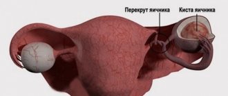
Aspiration biopsy is often used in gynecological practice.
Scan-guided biopsy
Sometimes a suspicious formation is almost impossible to palpate through the skin due to its small size, but can be detected during radiography, ultrasound, or MRI. In this case, the biopsy is performed under the guidance of an x-ray or other image, which helps the doctor guide the needle and control the position of its tip.
During a stereotactic biopsy, images in at least two planes are used, which helps to accurately determine the position of the suspicious lesion and the needle in three-dimensional space. Biopsy under scanning control can be fine-needle, thick-needle, or aspiration.
Biopsy during surgery
During surgery, your doctor may remove part of the tumor (incisional biopsy) or all of it (excisional biopsy). This allows you to obtain the maximum amount of tissue for research. But this type of biopsy has a drawback: the diagnosis is established after the patient has been operated on.
If the surgeon removes the entire studied formation or organ during a biopsy, the procedure is also a therapeutic measure. If the formation (for example, a polyp) turns out to be benign, after its removal a complete cure occurs.
Biopsy during endoscopy
When examining some organs, such as the gastrointestinal tract, an endoscope is used - a thin tube with a video camera and a light source at the end. Through it, you can insert special endoscopic forceps or a needle to take a biopsy from the esophagus, stomach or intestines. This type of biopsy is also called targeted biopsy.
If a tissue sample from the colon is needed, an endoscope is inserted through the anus, a procedure called fibrocolonoscopy or sigmoidoscopy (depending on which part of the colon needs to be examined). If material needs to be obtained from the stomach, esophagus, duodenum, the endoscope is inserted through the mouth, and the study is called fibrogastroduodenoscopy (FGDS).
A biopsy can also be performed during bronchoscopy, cystoscopy (endoscopic examination of the bladder) and other types of endoscopy.
Algorithm for conducting morphological examination of biopsy specimens
Content:
- Algorithm for conducting morphological examination of biopsy specimens
- Indications and contraindications for the study
- Methods for taking biopsy samples
- Preparation for taking a biopsy sample
Histological examination is carried out using a high-resolution light microscope. The laboratory technician performs the necessary preparation of the sample, including dehydration and embedding of the sample in paraffin blocks. Next, using a microtome (an instrument for preparing sections of biological tissue), the laboratory assistant prepares a specimen for research (several micrometers thick).
The separated tissues are placed on a special glass and a special staining procedure is carried out, which allows for maximum visualization of individual cells. After the histologist receives the results of the study, he writes his conclusion, on the basis of which the attending doctor makes or confirms the diagnosis and prescribes appropriate treatment.
In clinical practice, urgent morphological examination is often carried out - when tissue is excised for histology during the operation. Urgent diagnosis is done when it is necessary to quickly determine the course of a surgical intervention. The urgent diagnostic method involves flash freezing of tissues (dehydration is not performed). This is followed by standard cell microscopy. It is worth noting that the duration of an instant study is at least half an hour.
Another option for conducting MIB is cytology. Cytology analysis does not imply excision of tissue, but sampling of cells for examination, when a piece of the mucous membrane is not available to make a preliminary diagnosis. This research method is most often used in gynecology - a smear of the cervical mucosa. It is prescribed to detect tumors in the early stages. It is worth noting that in practice, biopsy is much more effective and efficient than cytology.
Do I need special preparation for a biopsy?
Usually no special preparation is required. The clinic requires you to sign a written consent to perform medical procedures (biopsy). The doctor will tell you what the procedure is, how it will be performed, what the risks are, and answer your questions.
If necessary, local anesthesia is administered before the biopsy using an injection or spray. Sometimes medicated sleep or general anesthesia is used. In this case, you will be asked not to drink or eat for a certain time before the procedure.
How is a biopsy performed?
The patient is in a lying position during the manipulation. First, the doctor performs anesthesia. In most cases, local anesthesia is sufficient. The specialist performs a control ultrasound and determines the exact location of the tumor. After the anesthesia takes effect, the doctor will collect tissue from the pathological area. Most often, the patient does not experience pain, but may feel slight pressure, burning, and tingling. When the material is collected, the doctor removes the needle and treats the puncture site with an anesthetic. In some cases, it is recommended to apply cold for 30-40 minutes.
Is a biopsy safe? What are the consequences and complications?
This depends on the type of biopsy. If it is performed during surgery, then the risks are due to the surgery itself. During a puncture biopsy, the needle may enter a vessel or neighboring organs (for example, the gallbladder during a liver biopsy), bleeding, infection, and pain for some time after the procedure. If the biopsy is performed by an experienced specialist in a well-equipped clinic, there are virtually no risks.
At Euroonko you can perform various types of biopsies. We employ highly qualified doctors and use modern equipment.
Book a consultation 24 hours a day
+7+7+78
How the procedure is performed
Biopsy differs in the methods of collecting material (biopsy):
- Puncture. The material is taken using a hollow surgical needle directly from the lesion. Suitable for biological fluids or cell collection. A less traumatic modification is possible - fine-needle biopsy.
- Aspiration. The collection takes place using a special vacuum device.
- Imprint smears or scrapings of material from the mucous membrane.
- Endoscopic. The sample is taken using special endoscopic equipment. Used to study the gastrointestinal tract, uterus and other organs.
- Sighting. Pathological tissue is examined after collection with special forceps.
- Excision. A surgical method of completely removing the affected organ or just the tumor.
- Incisional. During surgery, part of the tumor is removed.
- Stereotactic. Suitable for collecting material from hard-to-reach areas, the process is controlled using visualization.
- Laparoscopic. The procedure is performed during diagnostic laparoscopy (examination of the abdominal cavity) or thoracoscopy (chest cavity).
The biopsy usually takes place on an outpatient basis under local anesthesia. Sometimes performed during surgery (intraoperative). Its results may influence further patient management tactics.
How the material is examined
Once the tissue is collected, it is sent to a laboratory for analysis. There, a histological (if possible) and cytological examination is carried out.
- Histology. After special processing with a microtome (special knife), very thin sections of tissue are made, which, after staining, are examined under a microscope. The structure of the tumor is studied, the presence of pathologically altered cells that absorb dyes more intensely than normal cells is determined.
- Cytology. Smears containing cellular material are examined under a microscope. The size, degree of differentiation and other characteristics of the cells are determined. This is a less informative study than a histological one, since the cells do not always fall into the field of view.
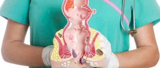
Purpose of cervical biopsy
The main goal of the procedure is the collection and detailed histological examination of the biopath for the presence of atypical cells. Histology is the study, using diagnostic optics, of a previously prepared and stained section of a biopath extracted from the cervix. The study of the material helps to determine the causes of pathological changes in organ cells, identify exophytic condylomas, and oncological processes. The results of the biopsy will help make a final diagnosis and select the most effective treatment regimen.
Advantages of punch or needle biopsy
This is a reliable method of obtaining tissue samples that helps determine whether a tumor is developing and what its nature is.
A puncture biopsy is less traumatic than surgery, which involves incisions in the skin and local or general anesthesia.
The procedure is generally painless and the results are as precise as surgically removing a tissue sample.
Recovery time is short, patients do not require hospitalization.