Diagnosisforeign body of the eye"is well known to almost everyone. It is unlikely that there is at least one person who has never in his life experienced unpleasant sensations caused by a small insect, a speck of dust, an eyelash getting into the eye, that is, what is called a “foreign body”.
Foreign bodies can be superficial, i.e. located on the surface of the eye or intraocular - penetrating into the eye cavity and damaging its membranes.
Most superficial foreign bodies are removed from the eyes as a result of intense blinking and increased tear production. If this does not happen, then it is necessary to seek specialized medical help as soon as possible.
Metal particles getting into the eye are especially dangerous. Often they are so small that they do not cause significant discomfort and the victim does not seek medical help.
However, after a few days, he begins to notice that his vision has noticeably deteriorated. This is due to the oxidation of the metal. The most dangerous in this regard are copper particles, the oxides of which have a toxic effect on the cornea, lens and retina. Therefore, it is so important to promptly contact an ophthalmologist in case of a foreign body in the eyes.
Symptoms of a foreign body in the eyes
Signs of a foreign body in the eye can vary in intensity from slight, almost imperceptible discomfort to unbearable intense pain. It depends on the type of foreign body and its location. The main symptoms of a foreign body in the eye are:
- Burning;
- Scratching in the eye;
- Eye irritation;
- Painful sensations;
- lacrimation;
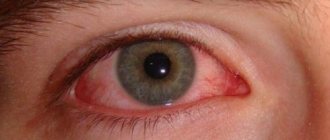
- Redness of the eye;
- Photophobia;
- Difficulty opening the affected eye;
- Deterioration of vision.
- Diagnosis of foreign bodies in the eye
If there is a suspicion of a foreign body in the eye, an ophthalmological examination is performed using a special slit lamp. If necessary, the doctor turns the upper eyelid inside out and checks for foreign particles underneath it.
To detect intraocular foreign bodies, an examination is carried out using an ophthalmoscope, an ultrasound of the eyeball is performed, as well as radiography in two projections.
When can only a doctor help?
If you are unable to remove debris from your eye, then you should consult a doctor. Specialized medical care is also needed when:
- the eye is inflamed
- discomfort does not go away for a long time
- the eye is red and the redness does not disappear (it’s common for the eye to turn red when you rub it or try to get a speck out; you should sound the alarm if the redness lasts for a long time)
- you feel constantly watery or feel like you have a scratch on your eye
- you experience blurred vision
- the eye hurts and the pain intensifies.
In all of the above cases, you should run to the doctor. An ophthalmologist will give advice and help get rid of discomfort and, if necessary, prescribe treatment.
Treatment of a foreign body in the eye
Removal of foreign bodies of the eyes, even those located superficially (in the conjunctiva, sclera or cornea), should only be performed by an ophthalmologist. Attempts to cope with this task on your own can lead to infection of the eyes or further injury to its structures. Superficially located foreign bodies are removed on an outpatient basis under local anesthesia. Most often, the procedure is performed using a special microscope - a slit lamp.
After it, anti-inflammatory and antibacterial eye ointments and drops are necessarily prescribed to prevent the development of a pronounced inflammatory reaction.
Removal of intraocular foreign bodies is carried out in the operating room using quite complex equipment, a microscope and various surgical instruments. Since penetrating wounds of the eyeball threaten the loss of visual function, and even the eye itself, the operation is performed immediately, i.e. for emergency reasons.
What not to do if a foreign object gets into your eye
The most valuable thing you have is your health and the health of your eyes in particular. The ability to see is a gift to be treasured. With eye diseases or partial blindness, life loses its colors and you may face a lot of difficulties. That is why it is extremely important to protect your eyes from damage and foreign objects. You should take all precautions even when you remove the smallest speck.
Sometimes, if you fail to follow hygiene rules and behave incorrectly, you can injure your eye or cause an infection. And then simply removing a speck will turn into a whole epic for you.
Here are a few forbidden tricks:
- rinse eyes with plain tap water
- try to remove the speck with your finger
- touch the eye with dirty hands
- rubbing your eyes (simply rubbing your eyes with your hand can scratch your cornea)
- panic and make sudden movements
If you are very worried and your hands are shaking, it is better not to touch your eyes so as not to cause irreparable harm. Also, avoid driving when you feel something is wrong with your eyes.
Always touch your eyes only with cleanly washed hands, otherwise the consequences can be extremely unpleasant - you can infect your eye and get conjunctivitis, stye and keratitis as a reward.
What to do if there is a foreign body in the eye
If a foreign body gets into the eye, you should adhere to the following recommendations:
- Do not rub and generally touch the affected eye as little as possible. If you wear contact lenses, do not remove them. Any touch to the eye can change the original position of the foreign body and push it deep into the tissues of the eye.
- Try to keep the affected eye closed. The harder and more often you blink, the more irritated your eye will be.
- You should not attempt to remove a foreign body yourself or trust this procedure to random persons nearby. This is quite dangerous and fraught with serious consequences.
- Contact a medical facility as soon as possible for specialized ophthalmological care.
- Be sure to tell your doctor what substances or materials you were working with at the time of your injury.
Team of doctors Ochkov.Net
Removing visible debris
It's easier to remove something from the eye that you see. First you need to find a well-lit place and take a mirror with you.
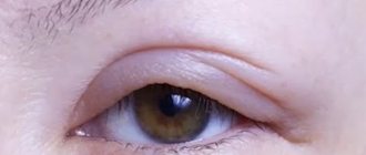
- Gently open the affected eye using your thumb and index finger.
- Place your index finger on the eyebrow growth line, and your thumb just below the lower eyelid. This way you can see what's going on in the eye and find out what kind of debris is bothering you on the cornea (located in front of the iris and pupil) or on the sclera (the white part of the eye).
- When you find a speck, take a damp swab or napkin and carefully remove the dirt. The eyes are a sensitive organ and you may instinctively close your eyes. If you are unable to perform the manipulation yourself, ask a family member to help you.
- Another option is to put special drops or saline solution into your eyes. The drops moisturize the surface of the eye and the water “washes away” the dirt.
If the method of simply dripping drops into your eyes does not help you, then use a damp cloth. Both methods can also be used when you do not see the speck, but only feel it.
First aid for a person with an eye injury: how to provide?
Any eye injury is dangerous; you should not hesitate to see a doctor. Treating a damaged eye at home is strictly prohibited - this can provoke negative consequences, including loss of vision. But first aid is necessary.
What does this help consist of? The location of the injury must be:
- rinse to remove contaminants (if the eye injury is not penetrating);
- stop bleeding with a bandage/bandage;
- numb the injury site (apply ice).
What measures should I take before deleting?
The very first thing you need to do before you start cleaning your eye from a foreign object that has gotten into it is to take care of hand hygiene. If you perform the procedure with dirty hands, you can get a lot of problems, including infection of the eye organs. Also, cleaning the eye without prior hand hygiene can lead to the introduction of new foreign bodies into the cornea and conjunctiva. Hand hygiene before removing debris from the eye will not take up too much of your time. It is enough to wash your hands with soap and clean water, then dry them thoroughly with a dry towel. It is not recommended to wipe your hands with alcohol and alcohol-containing substances, since the contact of alcohol and the surface of the eye in 100% of cases causes severe irritation of the visual organs. This will only interfere with your procedure.
Lens displacement
Displacement of the lens occurs with blunt trauma to the eye. Here, damage to the ligament of Zinn occurs, unevenness of the anterior chamber appears, which leads to trembling of the iris (iridodonesis). The edge of the dislocated lens is visually determined. With ophthalmoscopy, we see two optic discs.
Displacement of the lens is combined with cataracts of traumatic origin, secondary glaucoma, and iridocyclitis. In the case of complete separation of the ligament of Zinn, the lens is dislocated and displaced into the anterior chamber of the eye, and sometimes into the vitreous body. At the same time, the anterior chamber of the eye deepens, the pupil narrows. In such cases, iridocyclitis and an acute attack of glaucoma suddenly appear.
The main symptom of lens displacement is iris trembling, or iridodonesis. Iris tremors can sometimes be seen with the naked eye. But it is better to observe this in the light of a slit lamp. But iris trembling may not always occur. In such cases, symptoms such as different depths of the anterior and posterior chambers of the eyes due to pronounced pressure, as well as movement of the vitreous anteriorly, to where the support of the lens is weakened, help to identify the displacement of the lens.
Lens subluxations most often occur in the upper inner quadrant. When the lens is dislocated, degenerative processes develop, followed by clouding, and in rare cases, its complete resorption.
Treatment of the eye after injury
Since with uncomplicated displacement of the lens, vision decreases insignificantly, no treatment is required. But in case of complications as a result of eye injury (lens opacification, secondary glaucoma), it is required to remove it and replace it with an artificial lens.
Hemorrhage into the vitreous body of the eye: symptoms and treatment
A blow to the vitreous area results in hemorrhage. Blood in the retrolental space of the vitreous expands it, and blood in the orbicular space leads to the formation of a specific rim (stripe) that surrounds the periphery of the lens behind.
Retrolental hemorrhage takes longer to resolve than orbicular hemorrhage. Sometimes minor hemorrhages may be invisible and are discovered later, when they “descend” to the lower part of the anterior chamber.
Hemophthalmos is understood as massive hemorrhage into the vitreous body, occupying a significant part of the latter.
Approximately on the third day after injury, the blood in the vitreous body undergoes a process of hemolysis with the loss of hemoglobin by red blood cells, as a result of which they become colorless and later disappear. And the hemoglobin of erythrocytes takes on the form of grains, which are subsequently absorbed by phagocytes. Hemosiderin is formed, which has a toxic effect on the retina. Sometimes the blood does not completely resolve and a blood clot is formed and replaced by connective tissue moorings. The clinical picture of hemophthalmos is dominated by loss of visual acuity from the state of light perception to complete blindness. Focal illumination and biomicroscopy make it possible to see behind the lens a dark brown granular, sometimes with a reddish tint, mass of blood that permeates the vitreous body. Ophthalmoscopy shows the absence of a fundus reflex. Later, when the blood clot dissolves, one can observe deformation of the vitreous with its liquefaction. Hemophthalmos must be distinguished from partial hemorrhage into the vitreous body, which quickly and completely resolves.
Hemophthalmos leads to the development of degenerative processes in the vitreous body.
Treatment
In case of vitreous hemorrhage, bed rest and a cold bandage are prescribed on the affected eye. Calcium preparations (tablets, eye drops and intramuscular injections), hemostatic agents (Vicasol) are used. To speed up the resorption of hemorrhage, heparin is used (subconjunctivally on days 1–2) and enzyme preparations and potassium iodide. Treatment of hemophthalmia: bed rest with the head end elevated. Use a binocular bandage for 2–3 days. Calcium chloride, pilocarpine 1% 2 times a day, glucose with ascorbic acid are used, and a solution of dicinone (12.5%) is injected subconjunctivally. Then, after 2-3 days, absorbable drugs are used: dionin, potassium iodide, lidase. Corticosteroids (under the conjunctiva) and fibrinolysin are also indicated. In the late period of treatment, ultrasound and physiotherapy help a lot. If there is no positive effect from therapy, then it is necessary to suction the vitreous, and sometimes excise part of it. The removed vitreous body is replaced with luronite (a hyaluronic acid preparation).
We must not forget that with somatic diseases (cardiovascular diseases, atherosclerosis, hypertension, blood diseases, endocrine pathology), the development of hemophthalmos is possible. But in these diseases, hemophthalmos occupies a small part of the vitreous body.
The prognosis depends on the area of hemorrhage. If the hemorrhage covers 1/8 of the vitreous body, it often resolves. With an area of 1/8-1/4 of the vitreous, moorings are formed, which leads to retinal detachment. In terms of restoring visual functions and preserving the organ in total hemophthalmia, when a blood clot occupies more than ¾ of the vitreous, the prognosis is unfavorable. Irreversible destructive changes occur in the vitreous body (organization of a blood clot, formation of adhesions). Tractional retinal detachment and atrophy of the eyeball may develop.
Actions after removal
Even a small speck can cause serious harm. Therefore, after removing it, it is necessary to examine the eye in good daylight.
In case of redness or swelling, you should:
- Instill drops of antibacterial action: “Tobrex”, “Floxal”, “Tsiprolet”. They relieve inflammation and prevent the development of infectious eye diseases.
- Use eye drops such as Taufon or Albucid. They eliminate eye redness and promote rapid healing of the cornea.
In addition to drug therapy, traditional medicine methods can be used to relieve discomfort:
- The eyes are washed with strong black tea. A cotton pad is generously moistened with warm tea leaves and wiped over the eyes. A separate disk is used for each.
- Three times a day, one drop of infusion of sage, chamomile or calendula is instilled.
- Place a freshly cut cucumber or potato slice on closed eyelids and leave for twenty minutes.
- Dissolve a little honey in a tablespoon of boiled water. Moisten a cotton pad with the resulting solution and wipe your eyes.
Traditional medicine methods are applicable only if the foreign body is removed without complications.
Correct instructions for removing debris
In fact, there is nothing difficult about correctly and without negative consequences removing a speck that has gotten into the eye. You can start the procedure immediately after you make sure that your hands are clean and dry. Follow the algorithm below to ensure that the foreign body is removed without pain:
1. Turn on the warm water supply, place your palms together to form a handful, and scoop a small amount of water into it. Open your eyes wide and rinse your face generously in this position. If a foreign object that gets onto the surface of the eye is an ordinary speck of dust, already at this stage it will be washed far beyond the organ. You will feel relief immediately after this.
2. If washing with your eyes open does not bring the expected result, then there is not a speck of dust in the eye, but something more serious. Special measures will be required. Stand in front of the mirror so that you can clearly see the affected eye. Gently pull back the lower eyelid and see if there is a foreign object there. Usually, specks that get into the eye accumulate there. If this is the case in your case, take a cotton swab dipped in water and carefully remove the foreign object from under the eyelid. Once this is done, wash your face with warm water with your eyes open. This completes the procedure.
3. Is there anything under the lower eyelid? Most likely, a foreign object fell not under the lower eyelid, but under the upper eyelid. This is a more serious problem, which is difficult to cope with on your own, but still possible. Take your upper eyelid and gently move it in different directions until the speck falls out on its own. Often the cause of discomfort is the eyelashes themselves, which tuck under the upper eyelid. If you straighten the eyelashes of the upper eyelid, then the discomfort instantly disappears.
4. If there is nothing under the upper and lower eyelids, you can use a simple trick. Place some warm water in a bowl large enough to fit your head. Open your eyes and plunge into a basin filled with water so that your eyes are completely inside. Now blink hard several times, looking alternately in different directions. If a speck lies on the surface of the eye, blinking in warm water will definitely help to cope with the situation and get rid of discomfort.
5. A metal object lying on the surface of the eye in direct visibility can be removed with a magnet. To do this, you need to bring the magnet to your eye so that it grabs the foreign object. However, this is a very controversial method, which should be used only if the speck lies on the surface of the eye and is not stuck into it. Otherwise, great harm will be caused to the cornea and other elements of the eye.
If none of the above methods brings the desired effect, then everything is more serious than you might think. In this case, it is recommended to immediately consult an experienced ophthalmologist. He will conduct an examination, detect a foreign body and take the necessary measures to remove it. Under no circumstances should the problem be left to chance, as this can lead to inflammation of the eye tissue and other troubles.
Diagnostics
Trying to find the source of discomfort, the ophthalmologist examines the fundus, membranes of the eye, and the cavity of the upper and lower eyelids.
A slit lamp examination will help find a foreign body in the eye. For internal damage to the eye, an ophthalmoscope, x-ray or ultrasound of the eye is used.
If the grain of sand cannot be seen using a regular examination, then the ophthalmologist uses special eye drops with a dye. This helps to see the location of the speck and remove it in a timely manner.
1 Help with foreign body entry and removal from the eye
2 Help with foreign body entry and removal from the eye
3 Help with foreign body entry and removal from the eye
Mechanical eye injuries: why can’t you delay getting help?
Any damage to these complex and sensitive organs leads to negative consequences, among which the most dangerous are:
- wrinkling of the eyeball;
- traumatic cataract;
- decreased visual acuity;
- complete/partial blindness;
- phacogenic glaucoma;
- retinal disinsertion;
- eyesore.
Prompt medical care will help avoid these problems. But it is important to follow the rules of first aid and under no circumstances self-medicate. It is also important to remember the safety rules when handling traumatic objects and chemically active substances in everyday life and at work.
Types of foreign bodies
Foreign bodies entering the eye can be divided into:
- superficial - remain on the outer surface of the eyeball and can be detected during examination with the naked eye;
- intraocular - through the outer membranes they penetrate into the internal structures of the eye.
Superficially located foreign bodies in most cases are removed from the eyes as a result of frequent blinking and reflex lacrimation. If this does not happen, you should contact an eye clinic as soon as possible for specialized help.
According to their properties, foreign bodies can be divided into:
- magnetic (iron-containing) - particles of steel and other alloys containing iron;
- non-magnetic - fragments of glass, wood, stone, metals that do not have magnetic properties.
Metal foreign bodies are especially dangerous because they can be small in size and can oxidize, causing a toxic effect on eye tissue. Whatever the object that gets into the eye, it must be promptly identified by an ophthalmologist and removed.