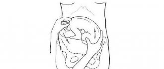
Types of breech presentation of the fetus
Breech presentation of the fetus is divided into types:
- gluteal;
- foot
In a breech presentation, the baby's buttocks are the first to appear during labor. In this case, its legs are located along the body and straightened at the knees. In such a situation, we can say that the woman is lucky, since in this case she practically does not need additional help, and the risk of complications is minimized. With a mixed breech presentation, the legs and pelvis may appear at the same time.
Leg presentation is considered more unfavorable. According to statistics, up to 15% of children with breech presentation are born this way. In the foot position, the legs, not the pelvis, are the first to appear in the birth canal. Usually, in case of leg presentation, a caesarean section is performed.
How to avoid danger?
Don't panic when you hear from your doctor that you have a large fetus. Just try to determine the cause with your doctor. Continuous observation in the hospital may be required until delivery.
If the reason is heredity or high consumption of sweets, the doctor will prescribe a special diet. Food should be wholesome, healthy, but not contribute to weight gain.
The main thing is not to worry or be afraid of childbirth. Your doctor will discuss the progress of labor with you in advance. They will prescribe a planned caesarean section, depending on the indications, or take a wait-and-see approach.
If within four hours from the onset of labor there are signs of a discrepancy between the baby's head and pelvis, surgery will be performed urgently.
Causes of breech presentation
There can be many reasons that cause breech presentation of the fetus, but the most common are:
- polyhydramnios - usually in this case the baby is highly mobile and simply does not have time to take the desired position by the time of birth;
- oligohydramnios - in this case, the child’s ability to change his position is limited, as a result of which the baby simply cannot take a physiological position;
- pregnancy with twins - in this case it is very difficult for babies to move due to lack of space; cephalic presentation of the fetus in multiple pregnancies is rare, regardless of the week of pregnancy;
- entanglement with the umbilical cord - sometimes a very active baby can wrap himself so tightly in the umbilical cord that it becomes completely impossible for him to take the correct position for childbirth;
- uterine pathology - some diseases of the uterus (for example, fibroids) can directly affect the position of the fetus.
In some cases, in the presence of the problems described above, breech presentation of the fetus may pose the following dangers:
- premature birth;
- hypoxia - if the child in the breech position has pinched the umbilical cord;
- difficult childbirth with the risk of injury to both mother and child.
Is there a danger with a large fetus?
The most crucial moment is childbirth. During the delivery process, carrying a large baby can cause some difficulties that can affect the health of both the baby and his mother.
Sometimes the size of the baby's head and the mother's pelvis may not match. It is very difficult for the head of a large baby to pass through the birth canal, no matter how strong the labor is. A large fetus can cause disruption or cessation of labor.
After the birth of the head, problems may arise with the removal of the shoulder girdle. The neonatologist must then check the condition of the baby’s collarbones and arms. A baby may have a cerebral hemorrhage due to a discrepancy between the sizes of the mother’s head and pelvis. Natural delivery becomes difficult.
Diagnosis of breech presentation
It is almost impossible to independently suspect a breech presentation of the fetus without understanding obstetrics and gynecology. In addition, such a situation before a certain period should not bother the expectant mother at all. Typically, the first symptoms of breech presentation in the uterus are determined by an obstetrician during an examination at 32-34 weeks. In this case, the doctor pays attention to the area where the child’s heartbeat can be heard and palpates the abdomen. If the symptoms indicate a breech presentation of the fetus, the specialist will prescribe an additional examination, which will either confirm or refute his conclusion.
There are several methods that experienced obstetricians use to determine the presence of breech presentation in the fetus.
- Firstly, this is a visual inspection and palpation of the abdomen. Usually, it is not difficult for a specialist to determine the position of where the child’s head is and where his feet are. In addition, the obstetrician may resort to listening to the heart. If its sounds are heard above the navel, then the baby is in a breech position. This is the simplest and most accessible method of research and diagnosis.
- Secondly, the obstetrician can determine the presentation of the fetus and the location of the head at the moment of the onset of labor. Typically, this method is resorted to if a woman is admitted for childbirth urgently and she does not have any information about previous examinations and the position of the fetus - pelvic or cephalic. In this case, the specialist feels the position of the child through the vagina.
- Thirdly, ultrasound is considered the most reliable and reliable method. For breech presentation, it is best to perform an ultrasound at 32-34 weeks, when the baby takes its final position.
Waiting for hour "X"
During a routine examination, the doctor, even without the use of technology, is able to approximately determine the position of the baby in the tummy: head down or butt.
The diagnosis is clarified using ultrasound, simple and three-dimensional echography. Early diagnosis of the type of malpresentation will allow you to develop a corrective program or prepare for natural childbirth with an incorrect position or cesarean section according to indications, which will protect you from many injuries and complications. Until the 32nd-34th week, the baby can be in any position. He has plenty of room for a life-changing acrobatic flip and preparation for birth. Sometimes, causing mommy and doctors to worry, the baby turns over just before the contractions begin, and sometimes even with their onset.
Treatment
In fact, in the absence of pathology, breech presentation of the fetus is not some kind of terrible diagnosis that needs to be feared. In addition, in some cases, correcting this situation can be quite simple.
Remember that any exercises and manipulations aimed at giving the child the desired position must be fully agreed with a specialist. Otherwise, you risk causing irreparable harm to your unborn baby.
- Exercise "Turns". To turn, lie on your back and slightly bend your knees. After this, you need to smoothly turn on your side and lie in this position for about 2 minutes. Then the manipulation must be repeated, but on the other side. The number of repetitions in such an exercise should not exceed 8-10.
- Exercise “Lifting the buttocks.” This exercise is contraindicated in women with placenta previa or a uterine scar. Its essence is to lift the pelvis and place a soft cushion under it. You need to spend 10-15 minutes in this position.
All exercises during breech presentation must be performed carefully. Otherwise, you can provoke premature birth. During pregnancy, you should carefully listen to your doctor’s recommendations and do not neglect visits to the gynecologist. Remember that with a breech presentation of the fetus, the woman needs more careful monitoring.
If the methods described above did not give a positive result, then at approximately 35-38 the doctor can perform an “obstetric revolution”. In this case, the specialist, using special manipulations, pressing on the woman’s stomach, gives the fetus the desired position. However, most babies rotated in this way soon return to the pelvic position.
With a breech presentation, the likelihood of birth by cesarean section increases, but many women successfully give birth naturally. A contraindication for vaginal birth in this case may be a fetus that is too large or entangled in the umbilical cord.
Publications in the media
Breech presentation is classified based on the position of the legs and buttocks of the fetus • Pure breech presentation. The buttocks are presented, the legs of the fetus are bent at the hip joints, extended at the knee joints and extended along the body. Observed in 60–65% of cases of breech presentation • Mixed breech presentation. The legs are completely bent at the knee joints and pressed against the fetal abdomen. Observed in 25–35% of cases of breech presentation • Foot presentation. One or both feet or (extremely rarely) the fetal knees are presented •• A distinction is made between complete and incomplete breech presentations. With a complete breech presentation, both legs are presented; with an incomplete breech presentation, one leg or knees of the fetus are presented •• Observed in 5% of cases of breech presentation.
Statistical data . The incidence is 2.7–5.4% of all pregnancies. In 15–30% of cases it ends in the birth of a child with low body weight (less than 2500 g). Etiology • Pelvic narrowing, abnormal pelvic shape • Uterine malformations • Myomatous nodes in the lower segment of the uterus • Fetal malformations , eg anencephaly, hydrocephalus • Placenta previa • Uterine atony, a large number of births in history, uterine tumors, uterine scar, narrow pelvis • Low fetal weight or prematurity, multiple births • Excessive fetal mobility with polyhydramnios.
Clinical picture • High position of the uterine fundus, due to the location of the pelvic end of the fetus above the entrance to the pelvis • Using Leopold's techniques (see Normal pregnancy), it is determined that the fetal head (a round dense voting formation) is located in the fundus of the uterus, and the buttocks (large, irregularly shaped) non-ballot presenting part) - above the pelvic inlet • The fetal heartbeat is heard above the navel or at its level. Special examinations • Vaginal examination during labor •• With breech presentation, the presenting part is softer than with cephalic presentation. You can palpate the groove between the buttocks, the sacrum, the genitals of the fetus •• With a purely breech presentation, you can find the inguinal fold •• With a mixed breech presentation, the foot is palpated next to the buttocks. With the help of palpation of the sacrum, the position and appearance are clarified •• In case of leg presentation, in order not to mistakenly mistake the leg for a dropped handle (for example, in transverse positions), it is necessary to remember the distinctive features of the fetal limbs ••• The leg has a calcaneus, the fingers are straight, short, the thumb is not set back ••• The big toe of the foot cannot be pressed against the sole, unlike the thumb of the handle, which is easily pressed against the palm; you can “say hello” to the handle ••• The knee differs from the elbow by the movable patella, the foot meets the shin at a right angle •• By the location of the popliteal fossa, you can determine the position of the fetus. In the first position, the popliteal fossa faces to the left, in the second - to the right • Ultrasound easily diagnoses breech presentation.
Mechanism of childbirth • The first moment is the internal rotation of the buttocks, it begins when the buttocks pass from the wide part of the pelvic cavity to the narrow one. The rotation is performed in such a way that at the outlet of the pelvis, the transverse size of the buttocks is in the direct size of the pelvis. The front buttock fits under the pubic arch, forming a fixation point, while the rear buttock is installed under the coccyx. • The second point is lateral flexion of the fetal lumbar spine. Further forward movement of the fetus leads to greater lateral flexion of the fetal spine. In this case, the rear buttock rolls out above the perineum and after it, the front buttock is finally born from under the pubic joint. At this time, the shoulders enter with their transverse size into the same oblique size of the entrance to the pelvis through which the buttocks passed. • The third point is the internal rotation of the shoulders and external rotation of the body. This rotation is completed by placing the hangers at the straight exit size. In this case, the back is turned to the side, the front shoulder of the fetus fits under the pubic arch, and the rear one is installed in front of the tailbone above the perineum. • The fourth moment is lateral flexion of the cervicothoracic spine. The birth of the shoulder girdle and arms is associated with this moment. • The fifth moment is the internal rotation of the head (with the back of the head in front). The head enters with a small oblique size into the oblique size of the entrance to the pelvis, opposite to the one in which the shoulders passed. When transitioning from the wide to the narrow part of the pelvis, the head makes an internal rotation, as a result of which the sagittal (sagittal) suture appears in the direct size of the exit, and the suboccipital fossa is under the pubic joint, where a fixation point is formed. • The sixth moment is flexion of the head. The consequence of this is the eruption of the head: the chin, mouth, nose, crown, and back of the head sequentially roll out over the perineum. • In a pedicle presentation, the legs are born first. In front there is a leg facing the symphysis. The buttocks enter the small pelvis after the birth of the legs up to the knee. The further course of labor is the same as with breech presentation.
Pregnancy management tactics • From 35 weeks of pregnancy, it is recommended to engage in therapeutic corrective gymnastics • Some doctors recommend external obstetric rotation to transform a breech presentation into a cephalic presentation, however, obstetric rotation is unsafe - placental abruption, compression and entanglement of the umbilical cord, and premature birth may occur • In case of breech presentation, you should decide whether childbirth can be carried out naturally or whether it is necessary to resort to caesarean section. Currently, due to the risk of fetal trauma during vaginal delivery operations, most obstetricians consider it advisable to expand the indications for cesarean section for breech presentation of the fetus (80–90%). Labor management tactics • First stage of labor (dilatation period). Prevent premature rupture of the membranes. Strict bed rest is indicated. After the discharge of amniotic fluid, a vaginal examination is performed (clarification of the diagnosis, exclusion of umbilical cord prolapse). • Second stage of labor. After the birth of the fetus, the head presses the umbilical cord to the level of the navel. If labor does not end within 10 minutes, the fetus dies from asphyxia. Therefore, after the birth of the lower part of the body, prompt obstetric care is necessary. • Manual assistance for a purely breech presentation using the Tsovyanov method is used to prevent complications. The method is based on maintaining the normal position of the fetus. Technique. After eruption, the buttocks are grabbed as follows: the thumbs are placed on the legs pressed to the stomach, and the remaining fingers of both hands are placed along the sacrum to prevent premature loss of the legs. As the body is born, the arms are moved towards the genital opening of the woman in labor, continuing to press the outstretched legs to the stomach until the birth of the shoulder girdle. If after the birth of the shoulders the arms do not fall out on their own, the shoulder girdle is set in the direct size of the pelvis and the fetal body is tilted downwards (posteriorly). This creates the front handle. The body is then tilted upward (anteriorly), after which the posterior arm and legs (heels) of the fetus are born. At the birth of the head, the fetal body is also directed upward •• Manual aid for foot presentation using the Tsovyanov method. The method is based on holding the legs in the vagina until the throat is completely dilated. Technique. The woman's external genitalia is covered with a sterile napkin. A palm is placed on the vulva, delaying the birth of the legs, which leads to full opening of the pharynx. Thus, the fetus moves from a leg presentation to a mixed breech presentation. After complete opening of the pharynx, childbirth is carried out as in a breech presentation. •• Sometimes, during the use of manual aid according to Tsovyanov, premature loss of the legs still occurs. In such cases, a classic manual aid is used (a set of techniques aimed at freeing the arms and head of the fetus) •• If the birth of the head is delayed, it is released using the Moriso-Levre-Lachapelle maneuver - a method of extracting the fetal head in a breech presentation, in which the head is bent when inserted into the mouth the fetus with the index finger of one hand, followed by traction on the body with the other hand. Technique. The body of the fetus is placed astride the forearm of the hand, the second or third finger of the same hand is inserted into the vagina of the woman in labor, following its back wall, and then into the mouth of the fetus. With the second hand, grasp the fruit by the shoulders and release the head. • The third stage of labor with breech presentation is carried out as usual. Extraction of the fetus by the pelvic end is considered not an aid, but an obstetric operation, because in the process of manipulation all 4 stages of labor are artificially reproduced by applying drag force. The fruit is removed from the heels to the crown. • Indications for surgery •• The need for urgent vaginal delivery due to the severe condition of the woman in labor (for example, eclampsia) •• Acute fetal hypoxia and the absence of conditions for cesarean section •• Previous classic rotation of the fetus. • Conditions for carrying out •• Full dilatation of the cervix •• Discharge of amniotic fluid •• Matching the size of the fetus and the woman's pelvis. • Technique of operation •• First point. There are 2 methods: ••• Removing the fetus by the inguinal fold. The index finger of the hand grasps the front leg of the fetus by the inguinal fold; attraction is produced during pushing. To grasp the pelvic end, the thumbs are placed on the buttocks, the index fingers are placed on the groin fold, and the rest are placed on the hips of the fetus. The fetus is removed up to the umbilical ring ••• Removing the fetus by the stem. The leg is grabbed with the whole hand in the area of the knee joint and pulled down. The second leg is born independently •• The second moment. The fruit is removed to the level of the lower corner of the shoulder blades. This point is highlighted for two reasons ••• The release of the arms can be started only after the birth of the fetus to the level of the lower angle of the shoulder blades ••• After the birth of the fetus to the level of the navel, the head, entering the small pelvis, can pinch the umbilical cord, which threatens hypoxia •• Third and fourth points. The arms and head of the fetus are released as with classic manual assistance.
Caesarean section • Indications for cesarean section for breech presentation (in addition to absolute indications for any presentation) •• Combination of breech presentation with a burdened obstetric and gynecological history (infertility, stillbirth, birth of a child with trauma), uterine fibroids, uterine malformations, narrowing pelvis, gestosis, post-term pregnancy, primigravida age 30 years or more •• Scar on the uterus •• Large fetus •• Prolapse of umbilical cord loops •• Partial placenta previa. • Contraindications •• Intrauterine fetal death •• Deformity or extreme prematurity of the fetus •• Acute infectious disease in a woman. Caesarean section is undesirable during a long period without water (more than 12 hours). • Conditions for a cesarean section •• The fetus is alive and viable (not always feasible with absolute indications) •• The woman agrees to the operation (if there are no vital indications) •• The pregnant woman has no signs of infection. • Preparing the patient •• If Ht is less than 30%, infusion therapy is performed to compensate for fluid deficiency •• It is necessary to prepare for a possible transfusion of plasma and blood during surgery •• The woman’s bladder must be emptied. • Anesthesia can be inhalational (general) or regional (spinal or epidural). General anesthesia often leads to depression of the newborn’s vital functions, therefore, when performing general anesthesia, the time interval from the onset of anesthesia to the moment of extraction of the fetus should not exceed 10 minutes. • Progress of the operation •• Dissection of the abdominal wall •• Opening and separation of the vesico-uterine fold of the peritoneum, exposure of the myometrium •• Dissection of the myometrium (Kerr-Gusakov, Sellheim or Sanger incision) ••• Kerr-Gusakov incision (low transverse) currently used most widely. The incision is made in the lower segment of the uterus, which reduces the likelihood of rupture or divergence of the edges of the scar during subsequent pregnancies. Disadvantage - danger of damage to nearby vessels ••• Sanger incision (classical, or corporal, currently used rarely, according to indications) - a longitudinal incision on the anterior surface of the uterine fundus ••• Sellheim incision (low vertical) begins in the non-contractile part of the uterus and continue to the body of the uterus •• The child is carefully removed by hand •• The separated placenta with all membranes is removed •• The uterus is often removed from the abdominal cavity for the purpose of massaging the fundus, examining the appendages and visualizing the incision when applying sutures. To reduce blood loss, uterine contracting agents (oxytocin, methylergometrine, enzaprost, etc.) are injected into the uterine muscle. After separation of the placenta, manual examination of the uterine cavity is necessary to diagnose submucosal fibroids or to remove remnants of the membranes •• Single-row suturing of the uterus. The vesicouterine fold of the peritoneum is sutured with thin absorbable suture material, and the abdominal wall incision is sutured in the usual way. Observation • During childbirth, constant monitoring of the fetal heart rate should be carried out • Observation of the parturient woman is carried out as after other births. Prevention. Therapeutic corrective gymnastics for breech presentation of the fetus is indicated up to 35 weeks of pregnancy. Complications • Throwing back of the arms • Untimely rupture of amniotic fluid • Abnormalities of labor • Prolapse of the umbilical cord and small parts of the fetus • Spasm of the uterine pharynx with pinching of the torso or neck of the fetus • Extension of the head • Injuries to the head and soft tissues, brachial plexus and spinal cord of the fetus • Asphyxia of the fetus. Course and prognosis • Perinatal morbidity and mortality are higher in the case of breech birth • For fetal weights less than 1,500 g, the risk of cerebral hemorrhage, as well as perinatal mortality, is significantly higher in the case of vaginal delivery than after cesarean section.
ICD-10 • O32.1 Breech presentation of the fetus, requiring medical care for the mother • O64.1 Obstructed labor due to breech presentation • O80.1 Spontaneous birth in the breech presentation • O82 Singleton labor, delivery by cesarean section
Notes • Absolute indications for cesarean section •• Complete placenta previa •• Absolutely narrow pelvis •• Discrepancy between the size of the woman’s pelvis and the fetal head •• Incomplete placenta previa with unprepared birth canal and severe bleeding •• Premature abruption of a normally located placenta with unprepared birth canal and bleeding •• Tumors of the pelvic organs that prevent the birth of a child •• Severe scar changes in the cervix and vagina •• Threatening or incipient uterine rupture •• Severe gestosis with ineffective conservative treatment and unprepared birth canal •• Incompetent scar on the uterus •• Extragenital cancer and cervical cancer •• Serious extragenital pathology (for example, retinal detachment, complicated myopia, severe cardiovascular diseases).
Behavior of the expectant mother
Expectant mothers should eat well and wisely, and this should be done even before pregnancy, since the child may inherit excess weight. Carefully control the amount of carbohydrates; in the last trimester, their amount should be no more than 400 g.
Do not refuse the help of doctors if pathologies of a large fetus have been identified. Treatment can begin already during pregnancy, and this will avoid health problems for the child in the future.
The hero inside you is a beautiful baby who simply requires a little more attention, care and love, but is in no way a reason for fear, worries and worries.