According to statistics, every year for every 100,000 babies there are 14-17 sick children. Hemangioma is one of the most common pathological phenomena that occurs during this period. At risk are girls, as well as premature babies and children born with low birth weight. Vascular anomalies can be represented by extensive clinical manifestations - from small pigmentary changes in the skin to huge tumors that affect the limbs and even the internal organs of the child, thereby causing life-threatening conditions.
What is the essence of the disease?
Hemangioma is a benign vascular neoplasm, the formation of which begins during embryogenesis. Externally, it appears as a flat bluish, red or burgundy spot that rises above healthy tissue. The tumor does not have a capsule, and therefore, as it grows, it damages nearby tissues and organs. Characteristic is rapid, progressive growth and a tendency to bleed.
The presence of more than 3 hemangiomas on the skin may indicate their presence on the internal organs of the child.
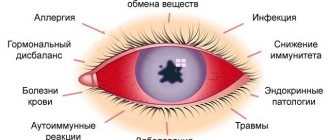
The factors causing the disease are poorly understood and completely unknown. At the same time, there are a number of reasons that could serve as a predisposing factor to the development of the disease. These include:
- Genetic predisposition. According to statistics, children born into a family prone to the formation of vascular moles have a much higher chance of developing hemangiomas.
- Presence of estrogen receptors in tumor tissue. The hormone acts as a growth stimulator.
- Taking certain groups of medications by the mother during pregnancy.
Contributing factors are also unfavorable environmental conditions suffered by the mother during pregnancy, infectious diseases, and multiple pregnancies.
The neoplasm is not prone to malignancy and, if all the recommendations of the attending physician are followed, with positive dynamics of the pathology, it completely disappears by about 5-7 years. They are not dangerous and do not lead to cancer. At the same time, large vascular growths can lead to organ dysfunction or cause improper development of the body as a whole. Hemangiomas are very easily injured, and therefore there is a risk of infection of the child through an open wound. They also help reduce blood clotting.
Main services of Dr. Zavalishin’s clinic:
- consultation with a neurosurgeon
- treatment of spinal hernia
- brain surgery
- spine surgery
Complications with hemangioma
In rare cases, the growth of angioma is accompanied by complications:
- when located on the eyelid – disruption of normal vision development;
- when localized in the area of large arteries, venous angioma can also affect the integrity of the vessel walls;
- a tumor in infants is easily injured, ulcerated and infected;
- Possible deterioration of blood clotting due to excess production and then depletion of platelets;
- angioma of the brain or internal organs can cause internal bleeding;
- vertebral hemangioma is complicated by back pain, pathological fractures, compression of the spinal cord; if the cervical spine is affected, paralysis of all limbs and dysfunction of the pelvic organs may develop.
Classification
Depending on the histological structure, pathology is divided into:
- Capillary.
The appearance of neoplasms depends on the degree of involvement of the dermis in the process, the depth, diameter and location of the lesion. The manifestation of the disease is typical already in the first months of a child’s life, as well as complete recovery by 5-6 years (in approximately 30% of cases).
- Cavernous.
They are represented by a limited subcutaneous nodular formation with a soft-elastic consistency. The basis consists of separate caves (cavities) filled with blood. The skin on top is rarely changed. As the tumor grows, the skin acquires a blue-purple tint.
- Mixed.
According to their localization, all hemangiomas are divided into superficial and subcutaneous forms.
PREVENTION OF RECURRENCE OF HEMANGIOMA IN NEWBORNS
Considering that neonatal hemangioma is a benign tumor with a potentially malignant course due to the absence of a capsule, in my practice I warn the parents of our patients about possible relapses and continued growth of hemangioma if the following recommendations are not followed:
- Exclude all types of vaccinations for the entire period of treatment and observation of the child - up to one and a half years.
- Avoid all types of physiotherapy and massage for up to 1 year.
- Avoid (if possible) the use of vasodilators and biostimulants (L-carnitine), vitamins and iron supplements.
- Avoid visiting baths and saunas.
- Mandatory reduction of temperature during acute respiratory infections.
The above limitations are well known to oncologists. Therapeutic manipulations that accelerate blood circulation in the child’s body immediately lead to recurrence of the hemangioma or its continued growth. Over the 7 years of monitoring our patients, we have been convinced more than once that our recommendations are correct. Typically, tumor growth resumed approximately 2-4 weeks after exposure to drugs that increase blood circulation, and we would put iron supplements first.
Typically, after receiving any vaccine, children may experience a fever for 2-4 days, and this may also be enough time for the hemangioma to re-grow. Although this is only the tip of the iceberg in explaining the reasons for tumor recurrence after vaccination. Large randomized trials are indeed needed to examine this complex, understudied issue. In practice, I have seen the greatest number of hemangioma recurrences after hepatitis B vaccination.
Compliance with all of the above recommendations allows you to avoid relapses of neonatal hemangioma after the end of laser treatment and refuse to use systemic drug therapy with beta blockers.
Features of diagnosis and treatment in children
Examination of tumor growth includes a consultation with a pediatric dermatologist and surgeon, x-ray of the affected area, ultrasound, CT, MRI, angiography and a blood clotting test. Hemostasis indicators are also important.
To treat hemangiomas, the following is carried out:
- electrophoresis with calcium chloride;
- cryotherapy;
- sclerosing therapy;
- radiation therapy;
- surgical intervention.
Depending on the degree of damage and the shape of the hemangioma, treatment tactics are determined. Initially, strict control is established over the tumor, the intensity of growth, the degree of cosmetic defect and the effect on vital organs are assessed.
The use of electrophoresis with calcium chloride in treatment allows the neoplasm to be completely removed without leaving a scar on the body. Treatment takes a long time.
Cryotherapy is carried out using carbon dioxide snow. The use is rational for small diameter tumors. Snow is applied to the affected area, capturing up to 1 cm of healthy tissue. Over time, a bubble forms, covered with a scab.
Sclerosing therapy is carried out with the injection of 70% alcohol and a solution of quinine-urethane. After this, a process similar to cryotherapy occurs. The method is effective when hemangioma is localized on the oral mucosa or upper eyelid.
Radiation therapy is successfully used to treat tumors located on the internal organs of a child. But due to the strong impact on the body, the method has many contraindications, incl. and an age limit - therapy can only be performed on children over 6 months old.
Rapidly progressing tumor growth is a direct indication for surgery. Surgical treatment is carried out only if the operation does not entail a severe cosmetic defect and does not disrupt the function of internal organs.
All about infantile hemangiomas
What it is?
Infantile hemangiomas are the most common vascular tumors that are most often diagnosed in young children. They usually occur in the first weeks after birth, but in some cases the phenomenon can develop during the first months. According to statistics, infantile hemangiomas occur more often in girls than boys and more often in people with light skin than dark skin. Also, according to observations, vascular tumors often occur in premature babies, and the longer the period of prematurity, the higher the risks of its development.
Causes
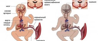
The common cause is hypoxia, i.e. lack of oxygen in the tissues, which can be not only due to pathologies of the placenta, there can also be a hereditary predisposition, but we do not oppose it to hypoxia, it is an additional factor.
Symptoms
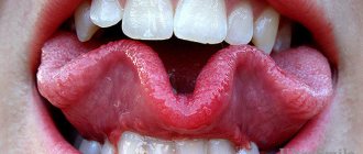
In most cases, the pathology is localized in the face and head, but it can also occur on other parts of the body and even on the mucous membranes and internal organs of the baby. The formations can be located on the surface or inside the skin. Most often, a hemangioma is visually similar to an iceberg - a small part of it is visible on the surface of the skin, and the main part is located in its deeper layers.
The main distinguishing feature of infantile hemangiomas is the way they change over time. So, at birth, the initial hemangioma may look like a bright spot with a vascular point or capillaries in the middle. Then, within 3-4 months, it actively grows and acquires a bright red color. When shouting, physical activity, or taking a warm bath, the tumor may swell and become more pronounced. After 4 months, growth continues, but becomes slow, and closer to a year it stops altogether. At the final stage, involution occurs, that is, the pathology begins to disappear on its own, 90% of tumors regress completely by 4-5 years.
At the stage of involution, the tumor gradually decreases, turns pale and after some time acquires the color of the skin (however, in some cases, a trace may still remain in the form of a light spot, scar or spider veins).
Medical tactics
By their nature, hemagiomas are absolutely benign and are in no way related to the presence of oncology, which must be urgently removed. But there is one “But”! Infant hemangiomas require mandatory medical supervision. The tactics for managing a patient with a vascular tumor will depend on the size, location, number of tumors and the presence of complications.
As a rule, for uncomplicated formations, a wait-and-see approach or “active non-intervention” is used. Its essence is regular monitoring by a doctor with recording of any changes (it is most convenient to take photographs of measurements with a ruler).
The frequency of examinations depends on the age of the child. The following rule is used here: the child's age in months must be equal to the examination interval in weeks. That is, a 3-month-old baby should be shown to the doctor every 3 weeks, a 6-month-old baby every 6 weeks, etc.
Very often parents begin to worry: “How can this be! The tumor is growing, but the doctor is doing nothing.” But specifically for hemangiomas, such tactics are completely justified, because the formation is more likely to regress on its own with minimal cosmetic complications. But surgical intervention, on the contrary, can have significant consequences.
Is treatment necessary?
Treatment of hemangioma (including surgery) is justified in the following cases:
1. The neoplasm is localized on the face, and there is a high risk of severe cosmetic defects.
2. Hemangioma is located in the area of the eyelids, nose, lips and throat, thereby interfering with the normal functioning of these organs.
3. The tumor has reached a large size and is subject to constant friction (for example, with a diaper).
There are several specific syndromes in which hemangiomas are combined with other developmental anomalies. In this case, the pathology requires not only elimination of the tumor, but also the use of other treatment approaches.
In some cases, to determine treatment tactics, the doctor prescribes additional examinations:
- Ultrasound (to assess the structure and depth of the tumor);
- Dopplerography (to study blood flow);
- blood tests (to assess clotting);
- MRI, etc.
Another cause for concern for parents is the bleeding of hemangioma. Since this formation is vascular, bleeding is possible, but it is unlikely that it will occur spontaneously. Bleeding can occur due to injury, cut, or tumor injury. If such a situation occurs, the bleeding must be stopped using a pressure bandage and, if its flow does not stop for a long time, seek medical help.
According to various data, only 10-30% of childhood vascular tumors require treatment.
Let's summarize which hemangiomas require treatment:
- prone to ulceration and bleeding;
- large in size and fast growing;
- localized on the face and other open areas of the body, thereby representing a pronounced cosmetic defect;
- located in the area of the eyes, nose, lips, genitals and interfering with the normal functioning of these organs;
- posing a threat to the child’s life (very large or located on the mucous membranes of the gastrointestinal tract and respiratory tract);
- accompanied by other anomalies;
- located in places subject to frequent friction (for example, in the groin area);
- causing severe psychological discomfort to the child.
In modern medicine, several treatment options for infant hemangiomas are used: medications (drugs for oral administration), external (ointments, gels) and surgical. Let's look at each method in more detail.
Systemic therapy
In evidence-based medicine, two drugs are most often used to treat hemangiomas:
1. Propranolol.
A drug from the group of beta-blockers. The main indication for its use is the treatment of diseases of the cardiovascular system. However, it is also suitable as systemic therapy for complicated hemangiomas, as well as for the treatment of hemangiomas localized on the face and disrupting the functioning of organs.
Pros: speed of achieving the desired effect (a positive result is visible literally the next day).
Cons: the possibility of side effects from the respiratory and cardiac systems of the body; presence of contraindications for use.
2. Prednisolone.
The drug is a systemic glucocorticoid. It is used mainly for hemangiomas that threaten the child's life.
Pros: has an anti-inflammatory and anti-allergic effect, due to which tumor growth quickly stops.
Cons: Due to the need for long-term use, it may cause side effects.
Note that any systemic therapy begins within the hospital, under the strict supervision of specialists. The duration of treatment is up to 6 months.
Local therapy
Two groups of agents can be used in local therapy:
1. External beta-blockers (Timolol, Propranolol).
The use of these drugs is advisable for small hemangiomas. The drug has good antitumor effectiveness combined with low absorption (practically does not penetrate into the blood) and an almost complete absence of side effects.
2. External glucocorticoids and the antitumor drug Imiquimod.
The drugs show positive results on limited lesions, but are not the drugs of choice due to the high risk of complications.
Surgery
The classical surgical method of excision for hemangioma is used extremely rarely. At the moment, the most preferred methods are laser surgery and cryotherapy. Lasers have proven themselves to be effective in the treatment of ulcerated, shallow tumors, as well as for eliminating residual effects after a course of systemic therapy.
Cryotherapy is also used on small, shallow tumors, but this method can be accompanied by pain, burns and scarring.
In conclusion, I would like to remind you once again that in most cases, infant hemangiomas do not require treatment. The most reasonable tactic is “active non-intervention” with regular monitoring by the attending physician.
CONCLUSIONS
- The high penetrating power of neodymium laser radiation with a wavelength of 1064 nm allows sclerosis of vessels located at a depth of 4 mm, which distinguishes it favorably from IPL, CTP, PDL and alexandrite lasers.
- Laser treatment using a neodymium laser with a pulse duration of 650 μs allows sclerosis of not only superficial, but also mixed hemangiomas.
- 650 microsecond technology delivers maximum power at the point of application with minimal thermal impact on surrounding tissue.
- Due to the depth of penetration of neodymium laser radiation, just one procedure is sufficient for complete sclerosis of the hemangioma (subject to the required laser radiation parameters).
- The penetration depth of the neodymium laser with 650 microsecond technology makes it possible to begin treatment already in the proliferative phase of growth without the fear of causing ulceration at the site of sclerosis (as has been reported with PDL lasers). This is a very important point, since timely treatment can accelerate the involutional phase of growth and avoid large cosmetic defects, especially in the facial area.
- A neodymium laser with 650 microsecond technology allows sclerosis of hemangiomas in the neck, eyelids, nose, ear and genitals without the risk of scarring.
- To achieve maximum cosmetic results, you should not sclerose a hemangioma larger than 2 cm in diameter. We noted faster wound healing and crust rejection, in about 2–3 weeks, with this volume of treatment. It is also easier for the child’s parents to care for a small wound and preserve the crust until it is physiologically rejected.
- Repeated laser irradiation in the same place is undesirable, as it can lead to scar formation and hypopigmentation.
- If it is necessary to sclerosis residual foci of hemangioma or tumor recurrence in the same place, it is better to repeat the procedure no earlier than 1 month after the first irradiation. During this time, the biological resistance of surrounding tissues to damage is restored. As a result, the likelihood of scar tissue forming at the site of laser radiation is reduced.
- To prevent recurrence of hemangioma, it is necessary to follow the recommendations during and after treatment for up to 1 year and 6 months.
- Compliance with the recommendations during and after laser treatment allows in most cases (and in our studies - completely) to avoid long-term use of systemic drug therapy with beta blockers. However, this situation does not apply to rapidly progressive, extensive, multifocal hemangiomas, when time is against the patient. In such cases, we also recommend the use of beta blockers according to the standard regimen. Thus, we use a combined treatment method.
Literature
- Bard S., Goldberg DJ Laser Treatment of Vascular Lesions. Karger, 2014.
- Ni N., Langer P., Wagner R., Guo S. Topical timolol for periocular hemangioma: report of further study. Arch Ophthalmol 2011; 129(3): 377–379.
- Léauté-Labrèze C., Hoeger P., Mazereeuw-Hautier J., et al. A randomized, controlled trial of oral propranolol in infantile hemangioma. N Engl J Med 2015; 372(8): 735–746.
- Schiestl C., Neuhaus K., Zoller S., et al. Efficiency and safety of propranolol as fi rst-line treatment for infantile hemangiomas. Eur J Pediatr 2011; 170(4): 493–501.
- Zur KB, Wood RE, Elluru RG Pediatric postcricoid vascular malformation: a diagnostic and treatment challenge. Int J Pediatr Otorhinolaryngol 2005; 69(12): 1697–1701.
- Al Buainian H., Verhaeghe E., Dierckxsens L., Naeyaert JM Early treatment of hemangiomas with lasers. A review. Dermatology 2003; 206(4): 370–373.
- Barlow RJ, Walker NP, Markey AC Treatment of proliferative haemangiomas with the 585 nm pulsed dye laser. Br J Dermatol 1996;134(4):700–704.
- Witman PM, et al. Complications following pulsed dye laser treatment of superficial hemangiomas. Lasers Surd Med 2006; 38: 116–123.
- Geronemus RG, Quintana AT, Lou WW, Kauvar AN High-fluence modifi ed pulsed dye laser dynamic cooling of port-wine stains of infancy. Arch Dermatol 2000; 136:942–943.
Shall we wait?
If the red spot on the baby’s skin does not grow, the doctor may suggest simply observing him, because there is a chance that the ugly blot will disappear completely over time. The fact that “the process has begun” will be hinted at by the appearance of a white spot. But if the hemangioma begins to actively grow or some complications arise, such as ulceration with infection, then treatment should not be delayed. Also, you should not delay the removal of such a vascular tumor, which, although it does not grow, spoils the appearance of the child and threatens to sooner or later lead to psychological trauma. Hemangiomas located on the face (nose, lips, eyelids) are subject to early removal - the risk of complications is too high here. Hemangiomas can be removed at any age, even in babies who are not even one month old.
Dangerous spot. How to detect basalioma in a timely manner
More details
Surgery
At the Northern Clinic, surgical treatment of vascular formations is carried out. Some of them are amenable to drug therapy (capillary, mixed, combined hemangiomas), some of them can be eliminated with a laser (capillary hemangiomas), and a number of vascular formations (cavernous hemangiomas, lymphangioma, vascular malformations) can only be removed surgically.
Without a queue for compulsory medical insurance for residents of Russian regions
More details