Cholangiography is an examination of the bile ducts under X-ray or MRI using a special contrast agent. In fact, cholangiography “paints” a picture of the excretion of bile in a given specific period of time and shows internal obstacles to the normal functioning of the system - stones, tumor, cicatricial narrowing. Cholangiography is an examination process, and the image recorded by x-ray is a cholangiogram.
- Indications
- Types of cholangiography
- Preparation for ERCP
- ERCP technique
- Complications
Indications
Bile is synthesized by liver cells and collected through intrahepatic ducts in the gallbladder, which lies on the lower surface of the liver. Some time after eating, bile from the bladder is supplied to the duodenum, where it interacts with semi-digested food, breaking down its components into its component parts. The gallbladder and duodenum are connected by a common bile duct, into which the pancreatic duct flows just above the intestine.
During cholangiography, an X-ray contrast agent stains the extrahepatic ducts, showing their structure and patency, recording the sites of narrowing and the cause of the narrowing. Therefore, cholangiography is resorted to in the following cases:
- Jaundice of a non-infectious nature, when, due to a mechanical obstruction to the normal flow of bile, bile acids do not enter the intestine, but are absorbed into the blood, which is possible when the main bile duct - the common bile duct - is blocked by a stone, compressed by scars, as well as a tumor blocks the duodenal papilla - the confluence of the duct into the duodenum.
- Pancreatic cancer , when it is necessary to restore the patency of the common bile duct compressed by a cancerous conglomerate with a special drainage or a stent, so that the condition improves, obstructive jaundice regresses and the patient is prepared for a planned operation.
- Inflammation of the pancreas with frequent exacerbations to find the cause of the chronicity of the pathology.
- The presence of pathological purulent tracts - fistulas between the pancreas and duodenum, formed after severe pancreatitis.
After establishing the exact cause of bile excretion pathology, cholangiography from a diagnostic measure can become a therapeutic procedure that eliminates this pathological obstacle.
ERCP technique
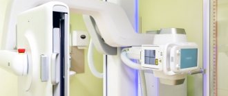
After the preparatory procedures, the patient is sent to a specially equipped room, where the study will be carried out.
Before ERCP, drugs that relax the duodenum (for example, sedatives) are often used.
To reduce discomfort during diagnosis, the oral cavity is irrigated with a local anesthetic (usually containing lidocaine). This reduces sensitivity and reduces discomfort.
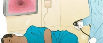
At the beginning of the study, the patient is placed on his left side. After this, the specialist slowly begins to insert an endoscope (a special tube with a camera) through the oral cavity. Gradually, the endoscope is lowered through the esophagus, stomach to the duodenum. At this time, an image from the camera is projected onto the monitor, and the condition of the mucous membrane of the digestive tract is assessed.
Once the endoscope reaches the duodenum, it is inspected and a test injection of contrast agent is performed. A special thin tube (catheter) is then passed through the endoscope into the pancreatic and bile ducts and a contrast agent is injected. After this, a series of photographs are taken, developed and the results are analyzed.
Types of cholangiography
The contrast agent can enter the bile in two ways - directly mixing with bile in the duct or after intravenous administration, initially entering the hepatic bloodstream, and from there being excreted in the bile.
Direct mixing of contrast with bile is possible in several ways:
- retrograde or, in Russian, countercurrent cholangiography (RCCP, ERCP) today is the “gold standard” of examination for pathology of biliary excretion, carried out with simultaneous endoscopy of the duodenum and X-ray examination;
- transhepatic cholangiography involves the introduction of contrast into the large bile duct during puncture through the abdominal wall, the procedure is not free from immediate complications, so it is preferable to carry it out before surgery;
- percutaneous cholangiography is performed during laparoscopy, and a contrast agent is first injected into the gallbladder with a needle, the manipulation is fraught with the subsequent flow of bile along the tract formed by a thick needle;
- Fistulocholangiography is possible in the presence of fistulas, because the fistulous tract is contrasted and the release of the drug by the duct connected to it is then traced.
The release of contrast when introduced into the circulatory system is carried out:
- with intravenous cholangiography , today recognized as not very effective and precise manipulation, as a result of which it was abandoned in favor of a more accurate examination;
- Magnetic resonance cholangiography excludes puncture of the bladder and bile ducts, guaranteeing high quality examination and 90% diagnostic accuracy, but for tiny stones it is inferior to the effectiveness of ERCP, and it is a rather expensive procedure that requires special equipment.
According to the time of execution, cholangiography is divided into the following types:
- preoperative , most often ERCP is performed, less often - percutaneous-transhepatic diagnostic manipulation;
- intraoperative - during surgery by inserting a catheter into the duct;
- postoperative , usually used to monitor the result of surgery, when contrast is supplied through a previously installed drainage catheter.
The undisputed leadership belongs to ERCP, which combines high sensitivity and the same diagnostic accuracy, the possibility of detailed endoscopic examination of the upper gastrointestinal tract and the implementation of a treatment procedure immediately after identifying the cause of the pathology.
Preparation for ERCP
Endoscopic retrograde cholangiopancreatography is not performed on an outpatient basis in a clinic; short-term hospitalization of the patient is necessary, since preliminary drug preparation for the procedure is required. The intervention requires good anesthesia or even anesthesia, and after the manipulation, monitoring of the condition and timely detection of complications, if any, are carried out.
On the eve of the procedure, the patient is given sedatives that have a positive effect on the central and peripheral nervous system, and antispasmodics that relax the muscle layers of the gastrointestinal tract. Very pronounced intestinal peristalsis is stopped by the administration of drugs. Before inserting the gastroscope, the pharynx is anesthetized, but it is also possible to perform diagnostic manipulations under anesthesia, which makes it easier for the patient to tolerate and increases the ability to visualize pathology.
As for the patient’s personal preparation, he must refuse food at least 12 hours in advance.
Anesthesia for ERCP
Share information with your Facebook friends
VK
Doctors' opinions
Dr. Kroh: Basically, I am for this approach. This allows for better airway control and sedation. However, general anesthesia is not always necessary for stent removal without cholangiography and other specific indications.
Dr. Schulman: Although deep sedation should be used in most cases, general anesthesia with endotracheal intubation is not necessary. For ERCP of short duration, the risks and costs associated with general anesthesia must be weighed against the benefits.
Dr. Abu Dayyeh: ERCP remains an advanced endoscopic procedure that carries a significant risk of complications. I don't think we need general anesthesia for simple cases like stent replacement. Keeping the patient calm may help the physician perform the procedure with less collateral damage to the papilla and better perform the fine movements required for ERCP. Therefore, in most cases, general anesthesia is recommended. All anterograde double-balloon enteroscopy should be performed under general anesthesia.
Dr. Erim: Hypoxemia and aspiration are common complications of anterograde tube enteroscopy procedures. A review of adverse complications several years ago prompted us to change practice and since 2013 we have used general anesthesia for all anterograde double balloon enteroscopy procedures. Since then, rates of hypoxemia and aspiration have decreased significantly.
Dr. Schulman: Given the labor-intensive nature of these procedures and the endoscope manipulation that may be required, the best results of these procedures can often be achieved using general anesthesia.
Dr. Abu Dayyeh: The anterograde double-balloon enteroscopy procedure requires a long time; therefore, to minimize the risks associated with longer endoscopic procedures and to promote successful completion of the procedure, general anesthesia is recommended.
Dr. Kroh: It depends on the indications and experience of the endoscopist. General anesthesia often makes the procedure more comfortable for both patients and doctors.
| Barham Abu Dayyeh, MD, MPH Associate Professor of Medicine and Director of the Bariatric and Metabolic Endoscopy Program at the Mayo Clinic in Rochester, Minnesota. |
| Tolga Erim, DO Department of Gastroenterology at the Cleveland Clinic, Florida, in Weston |
| Matthew Kroh, MD, FACS, FASMBS, FASGE Chairman of the Institute of Digestive Diseases at the Cleveland Clinic Abu Dhabi; Assistant Professor of Surgery at the Cleveland Clinic, Lerner College of Medicine |
| Allison R. Schulman, MD, MPH Associate Professor, Department of Gastroenterology and Hepatology; director of bariatric endoscopy at the University of Michigan in Ann Arbor |
print version
Tagsanesthesia Health Anesthesia Endoscopy
ERCP technique
The combination of endoscopy with x-ray examination is optimal in all respects: the highest possible diagnosis and accessibility for inspection of “secluded corners” while recording the entire process of identifying pathology on an x-ray and disk, which will allow subsequent consultation by involved specialists not blindly, but by video.
The first step is sequential endoscopy of the esophagus, stomach, and duodenum. Next, a catheter is inserted through a special channel of the endoscope, the tip of which is directed into the common bile duct, where the contrast agent is supplied. The location of the contrast in the anatomical area is filmed on X-ray film, and the process of movement of the endoscopic equipment is recorded on video. If any neoplasm is detected inside the intestine or duct, a piece of tissue is taken with tweezers - a biopsy; if a stone blocks the duct, it is also crumbled with tweezers and removed. If the duct is narrowed, a stent or drainage catheter is installed.
Complications
ERCP involves intravenous injection, that is, an invasive procedure, plus a contrast agent that is foreign to the body is used. The most common, but in fact very rare complication is inflammatory changes in the delicate tissue of the pancreas - on average three per hundred patients. It manifests itself clinically as pancreatitis - abdominal pain, threefold increase in a specific blood enzyme - amylase. As a rule, the complication develops already on the first day and resolves within a couple of days. Our clinic has developed a method for reducing and preventing this type of ERCP complications.
Bleeding from the site of manipulation is twice as rare when a vessel is damaged during dissection of the duodenal papilla in a patient with blood clotting disorders, which often occurs with prolonged or severe jaundice.
Rupture of the duct is possible in one or two patients out of a hundred, which is due to the looseness of the tissue with reduced extensibility due to scars. The complication can be successfully treated if detected early.
And the most common and common thing is an allergic reaction to a contrast agent. Moreover, repeated examinations using contrast do not guarantee its absence in the future; allergies arise according to the principle: it was not there yesterday, but today it arose.
Cholangiography at Euroonco is performed by experienced medical specialists using modern equipment. Find out more, contact us.
Book a consultation 24 hours a day
+7+7+78
For patients: What is RCP
Full text of the article:
ERCP (endoscopic retrograde cholangiopancreatography).
ERCP is one of the most informative methods for diagnosing pathology of the bile and pancreatic ducts. This method combines the capabilities of endoscopic and x-ray examinations.
Indications for ERCP are: symptoms of mechanical jaundice, increased diameter of the bile duct, frequent exacerbations of chronic pancreatitis, digestive disorders, congenital duct strictures.
Before ERCP, a conventional endoscopy is usually performed. After reaching the duodenum, the place of communication of the common ampulla of the ducts with the duodenum is determined. Then a special thin cannula is inserted into this ampoule, through which an X-ray contrast agent is injected into the bile duct and pancreatic duct.
The second step is to take an X-ray of the corresponding area. The image clearly visualizes all structures containing endoscopically introduced contrast: common ampulla, common bile duct, extra- and intrahepatic bile ducts, gallbladder, pancreatic duct, network of small ducts in the pancreas. The data obtained is analyzed, the level of disruption of the outflow of bile or pancreatic juice is revealed, and the possible cause of this block is determined (gallstones, compression from the outside by a tumor).
After a thorough assessment of the data obtained, a decision is made on the need to proceed to the third stage of ERCP - therapeutic manipulations.
At the junction of the common ampulla and the lumen of the duodenum there is a sphincter, the diameter of which is significantly smaller than the diameter of the ducts, so small gallstones very often collect here and prevent the normal entry of bile and pancreatic secretions into the duodenum. With ERCP, stones in this area are clearly visualized, and to remove them, sphincterotomy is performed (endoscopic dissection of the sphincter, papillosphincterotomy, EPST, EPT). As a result, the diameter of the latter increases, and all stones have the opportunity to exit into the lumen of the duodenum.
If stones are found in other parts of the bile duct system, they can also be removed using special endoscopic tools. Disturbances in the outflow of bile or pancreatic juice are also observed with various narrowings (strictures) of the ducts, which occurs after inflammatory diseases (cholangitis, pancreatitis), with compression by a tumor from the outside, with congenital malformations. In this case, under the control of ERCP, a stent can be placed in the pathologically altered duct - a self-expanding wire frame that eliminates local narrowing and maintains the required diameter.
After sphincterotomy, removal of gallstones or installation of a stent in the biliary tract and pancreatic duct, regular monitoring is recommended and conservative therapy is prescribed to prevent relapse of the disease (re-formation of stones, exacerbation of chronic pancreatitis).