Decompression sickness
(lat. prefix de- extraction, destruction + compressio compression, squeezing) - a pathological condition that develops as a result of the formation of gas bubbles in the blood and tissues of living organisms when external pressure decreases (in a person when leaving a caisson, ascending from depth to the surface, when ascending to height). In the literature there are other names for Decompression sickness: decompression sickness, decompression sickness of divers, high-altitude decompression sickness, or subatmospheric sickness of aviators, dysbarism, aeroembolism, desaturation aeropathy, aerobullesis, “bends”. However, they are less successful, because they do not reflect the essence of the disease or emphasize only certain forms of its manifestation. Decompression sickness often includes other disorders that develop with a decrease in external pressure, such as flatulence, barootitis, barosinusitis, barodontalgia, high-altitude tissue emphysema (see Atmosphere, the influence of high and low pressure; Altitude, the main forms of decompression disorders) , barotrauma of the lungs (see Barotrauma). Since it is possible to clearly differentiate the etiology and pathogenesis of disorders associated with decompression, it is more correct to consider them, including D. b. itself, as independent nosological forms of the general group of decompression disorders.
Experimental study of the biological effect of lowering pressure was begun in 1660 by R. Boyle. In the “pneumatic machine” he designed (a bell from which air was pumped out), he observed the appearance of gas bubbles in the fluid of the snake’s eye. Boyle pointed out that the appearance of gas bubbles in the liquid media of an animal body can be the cause of deadly functional disorders. However, the problem of functional disorders during decompression acquired practical significance only in the mid-19th century. in connection with the development of caisson work and diving. As soon as the depth of work began to reach 20-25 m (pressure 3-3.5 atm), widespread cases of severe functional disorders appeared in caisson workers (caisson disease) and divers (decompression sickness), which occurred when returning to normal pressure and manifested in the form sharp muscle and joint pain, damage to c. n. pp., severe disorders of the cardiovascular and respiratory systems, often fatal.
Hoppe-Seyler (F. Hoppe-Seyler, 1857) and Bucquoy (1862) showed a direct connection between the development of the disease and decompression. Pol and Watelle (B. Pol, T. Watelle, 1854) figuratively expressed this connection as “payback for getting out of pressure.” Ber (P. Bert, 1878) put forward and experimentally substantiated the theory of “gas embolism” D. b. Fundamental research by J. Haldane, as well as A. Hill, confirmed the correctness of this theory, which made it possible not only to explain the mechanism of the disease, but also to develop scientifically based methods of treatment and prevention.
When studying Decompression sickness, which occurs during underwater work, a hypothesis was put forward about the possibility of developing a similar disease in high altitude conditions [A. Boycott et al., 1908; J. Haldane, 1908 and others; Henderson (I. Henderson), 1917]. Later cases of D. b. were indeed noted initially during “ascents” in the pressure chamber [J. Jongbloed, 1930; V. V. Streltsov, 1932; V. G. Mirolyubov, A. P. Apollonov, 1938, P. K. Isakov et al., 1939, etc.], and then in high-altitude flights on airplanes [Carmichael (E. Carmichael), 1939]. A need arose to study the characteristics of high-altitude air defense and to develop preventative measures and means of protecting aircraft crew members from the adverse effects of the rarefied atmosphere.
In the 40-50s. 20th century Many studies have been carried out concerning theoretical and practical issues of D. b. in aviation: the clinical picture of the disease was studied in detail, the etiological relationship between the high-altitude and caisson forms of D. was substantiated, the physical characteristics were studied in detail. patterns of development of gas bubbles in the body have been identified. and fiziol, factors influencing the development of D. b. at altitude, and effective means of preventing and treating the disease have been found.
Certain features of space flight conditions (intense muscular work during spacewalk, long duration of stay in a rarefied atmosphere, the possibility of repeated decompressions when leaving the spacecraft) increased the likelihood of the occurrence of D. b. and demanded the development of new methods for its prevention and treatment. Much has been done in this direction by domestic and foreign scientists: A. G. Kuznetsov, A. M. Genin, P. M. Gramenitsky, V. P. Nikolaev, D. Busby, E. Roth, Allen ( T. N. Allen), Lambertsen (S. Lambertsen).
Comparative analysis of the number of diseases D. b. convincingly shows that by the 70s. the incidence of D. b., especially severe forms, has decreased significantly. If in the initial period of caisson work (1840–1860) there was heavy drilling. (caisson disease) developed in almost every case of decompression (see Caisson work), then by the middle of the 20th century. D. b. began to be observed in less than 1% of divers and 0.6% of caisson workers.
Etiology and pathogenesis
Decompression sickness is a consequence of the transition of blood and tissue gases from a dissolved state to a free gaseous state as a result of a decrease in ambient atmospheric pressure. The resulting gas bubbles disrupt normal blood circulation, irritate nerve endings, and deform and damage body tissues. At normal atmospheric pressure, there is a dynamic equilibrium between the partial pressure of gases in the lungs and their tension in the blood and tissues of the body. The main part of the total gas pressure in the lungs, and therefore in the blood and tissues (approx. 570 out of 760 mm Hg) is accounted for by nitrogen, a physiological inert gas that does not participate in gas exchange. The high partial pressure of nitrogen in the lungs (and, accordingly, in the blood and tissues), its physiological and chemical “inertness” determine its leading role in the formation of gas bubbles during decompression.
The nitrogen content in the body as a whole is determined by the level of its partial pressure, the duration of exposure under given conditions and solubility in fluids and tissues. The solubility coefficient of nitrogen is different for different tissues of the body. It is highest for adipose tissue.
At a pressure of 760 mm Hg. Art. 100 ml of blood contains approx. 1 ml of nitrogen, and 100 ml of adipose tissue contains 5 times more. The adult human body contains approx. 1 liter of nitrogen, adipose tissue accounts for approximately 350 ml.
When the partial pressure of nitrogen in the external and alveolar air changes, the time for establishing dynamic equilibrium (for nitrogen) is different for different tissues of the body. The blood, lymph, and well-perfused tissues are most quickly saturated with nitrogen and lose it. Due to poor vascularization, adipose tissue is saturated with nitrogen much more slowly. However, with prolonged exposure at elevated pressure, due to the high solubility of nitrogen in fats, its content in adipose tissue can be significant.
When the ambient pressure decreases (a diver rises from depth to the surface, a worker exits the caisson, a pilot rises to altitude), the gas dynamic equilibrium is disrupted, the tissues and fluids of the body become supersaturated with gases, and primarily with nitrogen. A process of desaturation occurs (see Saturation). With sufficiently slow decompression, the process of removing excess nitrogen from tissues until a new gas equilibrium is established usually occurs without the formation of gas bubbles. In the case of sufficiently rapid decompression, tissue oversaturation with gases reaches critical levels. Conditions are created for the formation of gas bubbles in tissues and liquids. The diffusion of gases from tissues into the initially formed bubble will cause its growth until gas equilibrium is established. The dimensions of the gas bubble also significantly depend on the elastic elastic properties of the tissue (deforming pressure, according to Fulton).
According to J. Haldane (1908), the permissible nitrogen supersaturation coefficient (i.e., the ratio of the initial pressure to the final pressure after decompression, at which gas bubbles begin to form) for the human body is approx. 2.25. This means that with a 2.25-fold decompression (after ascent from a depth of 12.5 m to the surface and a transition from a pressure of 2.25 am to normal or from normal to reduced - less than 300 mm Hg. Art.), a person can gas bubbles will form and decompression sickness will develop. For divers decompressing from very high pressures, which cause high tissue saturation with nitrogen, the permissible coefficient may be below 2.25. Therefore, in such cases, great care is required during decompression.
The process of formation of gas bubbles in tissues and liquids supersaturated with gases is significantly facilitated by the existence in the body of the so-called. gas "seeds". Their role in the body can be played by gas micro-inclusions measuring 10-7 - 10-6 cm that are relatively stable at normal pressure, cone-shaped and wedge-shaped niches on the surface of hydrophobic tissues, “funnels” of turbulent zones of blood flow, and especially microareas with negative hydrostatic pressure that arises when muscle contraction. All these gas “embryos”, under conditions of oversaturation of the body with nitrogen, act as primary centers for the formation of gas bubbles. Gas bubbles that appear in the body during decompression interact with tissues and liquid media and undergo a certain evolution.
There are two main types of post-decompression gas bubbles. The first type is autochthonous (extravascular) bubbles, the formation and reverse development of which is entirely determined by the process of diffusion—the exchange of gases between the bubble and its environment. Interstitial vesicles appear to be of this type. When there is an excess of gases dissolved in the tissue, they grow and put pressure on the surrounding tissues, causing them to deform, which can lead to pain. This is, apparently, the mechanism of development of muscle-articular decompression pain. It is believed that autochthonous vesicles also form inside cells (in parenchymal organs and adipose tissue), causing their destruction. Getting into the bloodstream with lymph or through damaged vessels, gas bubbles and cell destruction products can form gas and fat emboli.
Rice. 1. The brain of a dog with vascular aeroembolism (gas bubbles - light spots on a dark background of the vessel) after decompression. Rice. 2. Microscopic specimen of a hamster’s buccal pouch with vascular aeroembolism (gas bubbles are indicated by arrows) after decompression.
The second type is gas bubbles, the evolution of which is determined not only by diffusion processes, but also by merging with each other or, conversely, breaking into smaller ones. Most intravascular gas bubbles should be classified as this type. Originating in the venous bed (in tissue capillaries), they merge with each other, causing the possibility of developing acute aeroembolism in the circulatory system (Fig. 1 and 2). Gas bubbles are enveloped in blood colloids, proteins, lipoids, fats and other substances suspended in the blood are deposited on their surface, making their resorption difficult.
The hemodynamic disorder that occurs during aeroembolism (see) is aggravated by an increase in blood viscosity (see Viscosity). Sedimentation of blood cells on the surface of the vesicles, increased blood viscosity, as well as disruption of the normal relationship between the vessel wall and blood lead to the formation of aerothrombi, the basis of which is gas, and the capsule is a thrombotic mass. Wedge, the consequences of aerothrombosis, which causes persistent circulatory disorders, are more serious and lasting than with aeroembolism.
Disruption of blood circulation by gas and fat emboli or extravascular gas bubbles causes the development of protective reflex reactions in the form of dilation of arterioles and capillaries, increased blood pressure and increased blood flow. These reactions promote the movement of the embolus into larger venous vessels. Collateral circulation becomes more intense (see Vascular collaterals), aimed at eliminating local ischemia (see). Aeroemboli that enter the right ventricle of the heart with the bloodstream are crushed into small gas bubbles. The latter, due to an increase in capillary pressure as their diameter and gas pressure inside the bubbles decrease, are more easily absorbed. It was noticed that in repeated experiments with decompression, the effectiveness of protective reactions increases: the animals tolerated decompression more easily. This suggested the possibility of targeted training of the body to a relatively rapid decrease in pressure.
Development of D. b. contribute to severe physical stress, hypothermia (see Cooling the body), hypercapnia (see), hyperoxia (see), violations of the established regime and rest.
For experimental reproduction of D. b. Usually, recompression chambers and pressure chambers are used (see Pressure chamber), which make it possible to create gas environments of a certain composition and pressure in relation to caisson (see Caisson work), diving (see Diving work) and aerospace practice (see High-altitude flights). Aeroembolism can be simulated by introducing inert gases into the venous system of animals.
New publications
- Middle ear cholesteatoma
- Rinsing ears from wax plugs
- Spinal shock in humans
Alexey Portnov, medical editor Last edited: 06/24/2018
X
All iLive content is reviewed by medical experts to ensure it is as accurate and factual as possible.
We have strict sourcing guidelines and only link to reputable sites, academic research institutions and, where possible, proven medical research. Please note that the numbers in parentheses (, etc.) are clickable links to such studies.
Caisson sickness occurs when there is a rapid decrease in pressure (for example, when ascending from depth, leaving a caisson or pressure chamber, or ascending to a height).
In this case, gas previously dissolved in the blood or tissues forms gas bubbles in the blood vessels. Characteristic symptoms include pain and/or neurological impairment. Severe cases can be fatal. Diagnosis is based on clinical findings. The main treatment for decompression sickness is recompression. Diver compliance with safety rules is vital to the prevention of decompression sickness.
Henry's law states that the solubility of a gas in a liquid is directly proportional to the pressure exerted on the gas and liquid. Thus, the amount of inert gases (eg nitrogen, helium) in the blood and tissues increases at higher pressures. During ascent, when the ambient pressure decreases, gas bubbles may form. Free gas bubbles can occur in any tissue and cause local symptoms, or they can travel through the bloodstream to distant organs. Blisters cause symptoms by blocking a vessel, rupturing or compressing tissue, or activating the coagulation and inflammatory cascades. Because N is readily soluble in fat, lipid-rich tissues (eg, the central nervous system) are particularly sensitive to rapid decreases in pressure.
Decompression sickness occurs in approximately 2 to 4 cases per 10,000 dives. Risk factors include cold water diving, stress, fatigue, asthma, dehydration, obesity, age, exercise, flying after diving, rapid ascents, and long and/or deep sea dives. Because excess N remains dissolved in body tissue for at least 12 hours after a dive, repeated dives on the same day require special techniques to determine adequate decompression, and the development of decompression sickness is more likely.
, ,
Pathological anatomy
The most pronounced and specific morphological changes in rapid death from severe decompression sickness are the presence of numerous gas bubbles in the venous system, the right half of the heart overfilled and distended with gas bubbles, the phenomena of edema and emphysema of the lungs, multiple foci of hemorrhages in various organs and tissues. Sometimes foamy blood is found in the pulmonary artery. The corpses of those killed by D. b. undergo rapid and severe rigor mortis. The skin and mucous membranes are cyanotic.
When is oxygen harmful?
Oxygen is of greatest importance for humans, thanks to which oxidative processes occur in the tissues of the body, in particular the oxidation of nutrients, accompanied by the release of energy and the formation of toxins.
An increased oxygen content in the inhaled air, up to breathing pure oxygen, does not lead to health problems. However, pure oxygen has a drying effect on the mucous membranes of the respiratory tract; with prolonged inhalation of it, phenomena of irritation may occur. Therefore, when breathing pure oxygen, it is recommended to humidify it.
It should be borne in mind, however, that inhaling oxygen under pressure (over 3 atm) is not safe. Oxygen under pressure has a direct toxic effect on the central nervous system, causing convulsions and other intoxication phenomena. Such situations can arise when using oxygen equipment when diving under water (for scuba divers). This influence of oxygen should also be taken into account when conducting hyperbaric oxygenation.
A decrease in the amount of oxygen in the inhaled air causes a decrease in the intensity of metabolic processes in the body, leads to the accumulation of under-oxidized metabolic products and disruption of the vital functions of the body. To some extent, the body is able to compensate for oxygen deficiency in the inhaled air by turning on compensatory mechanisms: increasing pulmonary ventilation and blood circulation rate, the amount of hemoglobin and red blood cells in the blood, increasing the absorption of oxygen by tissues, etc. In this case, respiratory movements become more frequent and deep, and the heart rate increases.
However, when the oxygen content in the air decreases to less than 17-18%, the body is not able to cope with oxygen deficiency, and it begins to affect its functions.
Shortness of breath and palpitations increase, the skin turns pale, acrocyanosis appears, dizziness and headaches may occur, sweating increases, the functional abilities of the central nervous system decrease, etc. Tolerance to oxygen deficiency depends on the state of health and fitness of people.
Healthy and well-trained people (pilots) can briefly stay in an atmosphere with an oxygen content of 10% or even slightly less. However, the time spent in such an atmosphere is limited to a few minutes.
Oxygen deficiency usually occurs when climbing mountains or flying. The maximum achievable height to which a person can rise without the use of breathing equipment is considered to be 8 km. In addition, air with an insufficient amount of oxygen may be contained in any closed spaces (cisterns, vats) or in places with very limited air exchange, where oxidative processes occur, accompanied by the absorption of oxygen and the release of carbon dioxide (rotting, smoldering, fermentation).
The most dangerous in this regard are fermentation tanks, abandoned wells and mines, in the air of which there is a lot of carbon dioxide and little oxygen. It is believed that when a person enters such an atmosphere, the greatest danger is created by carbon dioxide, whose concentrations can reach 15-20%.
At such concentrations, death occurs very quickly as a result of paralysis of the respiratory center. At lower concentrations of this gas, human existence is possible for a longer time, but a level of carbon dioxide in the air of about 10% is already considered life-threatening. Because carbon dioxide is heavier than air, it tends to accumulate at the bottom of these containers. Therefore, going down into tanks, wells, and mines is very dangerous.
If it is necessary for a person to stay in such places (cleaning wells, vats, etc.), a fairly simple test is used. The fact is that air containing more than 6% carbon dioxide does not support combustion. Therefore, it is enough to lower some burning object (candle, torch) into the container under study to assess the state of the atmosphere in it. If it continues to burn, then it is safe to work in such an atmosphere (although there may be shortness of breath, palpitations and other unpleasant sensations). If the burning object in such a container goes out, then the concentration of carbon dioxide in it is high and it is possible to work there only after intensive ventilation or in an insulating gas mask (a filtering gas mask is useless). It should be taken into account that carbon dioxide in concentrations of about 2-5% also has a toxic effect on the body, but short-term exposure to an atmosphere with such a carbon dioxide content is not dangerous to life.
Clinical picture
The course, symptoms and severity of the disease are determined by the size, quantity and localization of gas bubbles in the body, the presence of provoking factors and the timeliness of treatment. Based on the severity of the course, three forms of decompression sickness are conventionally distinguished: mild, moderate and severe. The mild form is characterized by skin itching and rash, mild pain in the muscles, bones, joints and along the nerve trunks. With D. b. of moderate severity, there is a sharp deterioration in the general condition, cold sweat appears, severe pain in the muscles, bones and joints is noted, sometimes accompanied by bloating, nausea, vomiting, as well as short-term loss of vision. In severe forms, patients develop symptoms of c. n. With. (paresis and paralysis of the limbs), cardiovascular and respiratory systems. There are chest pains, suffocation, cyanosis, collapse (see).
Rice. 4. Radiographs of the hips with diaphyseal lesions (foci of necrosis are indicated by arrows) of the femurs in the chronic form of decompression sickness: 1 - lesion without calcification; 2 - complete calcification of the lesion.
Some authors also distinguish fatal and chronic forms of D. b. (M. I. Jacobson, 1950; Miles, 1971). In the fatal form, massive air embolism occurs, leading to blockage of blood circulation and damage to the lungs, heart, and brain that are incompatible with life. The chronic form is characterized by late consequences of aeroembolism and aerothrombosis in the form of aseptic bone necrosis (Fig. 4), deforming osteoarthrosis, etc.
When returning from high blood pressure to normal, symptoms of the disease in most cases develop within the first hour after decompression. Sometimes they are noted already during the process of stepwise decompression and, as an exception, after 6-12 hours. after decompression. It is important to remember that relatively mild symptoms at the onset of the disease can quickly develop into severe ones with a sharp deterioration in cardiac activity, pulmonary edema (see) and paralysis of the respiratory center.
The most common symptoms of D. b. are slowly developing dull pains, usually in or near one joint, often arising when moving. The pain is preceded by a feeling of numbness or “unease” in the joint. The skin over the affected area may become pale and sometimes become swollen. The patient often experiences general weakness, chills or a feeling of heat. With severe pain, tachycardia and increased blood pressure are observed. The nature of the pain can be aching, tearing, drilling, gnawing, shooting. They develop due to irritation of interoceptors by gas bubbles or tissue hypoxia when blood vessels are blocked by gas bubbles, as well as when nerve fibers are irritated by bubbles formed in the myelin sheath.
Often D. b. manifested by skin itching, burning in limited or extensive areas of the body, where polymorphic focal redness appears. Skin lesions are caused by the formation of gas bubbles in the blood vessels of the skin and sweat glands, which disrupt blood circulation and irritate nerve endings.
After deep-sea descents in patients with D. b. in approximately 5% of cases various symptoms of damage to the c are noted. n. pp.: dizziness, temporary deafness, visual impairment, aphasia, loss of sensitivity, paresis, spastic paralysis of one or both legs, convulsions. These symptoms develop due to the formation of gas bubbles in the myelin sheaths of motor nerve fibers or the white matter of the spinal cord and brain. Most often, gas bubbles form in the lumbosacral segments of the spinal cord.
With multiple aeroembolism, gas bubbles of various sizes can accumulate in significant quantities in the cavities of the right heart and the vessels of the lungs, causing disturbances in cardiovascular activity - pallor, severe weakness, frequent and shallow breathing appear, and blood pressure drops. There are chest pains, especially when inhaling, and coughing attacks. The pulse is rapid at first, then slows down, the skin becomes pale gray or bluish. With severe symptoms of hypoxia (see) and hypotension, the patient loses consciousness.
Damage to the inner ear is a consequence of the accumulation of gas bubbles in the fluids and tissues of the vestibulocochlear organ; a pathological condition of the Meniere syndrome type develops (dizziness, nausea, vomiting, nystagmus, weakness). Prodromal signs may include complaints of tiredness and tiredness. This syndrome usually develops after divers stay at great depths and perform heavy physical activity there. work, as well as in case of violations of the decompression regime.
Complications of DCS
Most often, patients suffer from chronic Meniere's syndrome, in which pathology affects the middle ear. The person experiences dizziness and hearing gradually deteriorates. Another possible disorder is aeropathic myelosis, which is a lesion of bone marrow cells.
With moderate and severe variants of the disease, all kinds of cardiac pathologies of an inflammatory and degenerative nature occur. Among the most common are endocarditis and myocarditis, cardiosclerosis. Pneumonia may develop from the respiratory system. The most common neurological manifestations of the disease are paresis, muscle paralysis, and loss of sensitivity.
Diagnosis
The diagnosis is made on the basis of characteristic complaints and clinical symptoms that appear after decompression. In this case, both the decompression regime and the conditions of being under high pressure should be strictly taken into account. The appearance of skin itching, pain, Meniere's syndrome, paralysis, the sudden development of collapse - all this, taking into account the previous decompression, serves as direct evidence of D. b. The correctness of the diagnosis is verified by repeatedly placing the victim under conditions of increased pressure (re-compression). If the symptoms of the disease stop, the diagnosis is correct; if during recompression the symptoms do not disappear or even weaken, then the diagnosis is D. b. becomes very doubtful.
Rice. 3. X-ray of the knee joint after decompression (the arrow indicates the gas bubble).
During X-ray examination, in some cases of Decompression sickness, gas bubbles are found in the joint cavity (Fig. 3), bubbles in the synovial tendon sheaths, muscle fascia, and perivascular formations. However, the diagnostic value of such findings is relative: they can be detected without a wedge, manifestations of the disease and absent when its symptoms are severe.
Sometimes radiography reveals characteristic aseptic necrosis of bones (Fig. 4), deforming osteoarthritis - the consequences of undergoing D. b. Necroses are foci of destruction, involving 1/3-1/2 of the epiphysis of the bone, but are sometimes observed in the diaphysis. Typically, necrosis develops in the femurs - in their proximal epimetaphyseal sections. Tissue defects form on the articular surface of the affected bone. In the head, neck and trochanters of the femur, the structure of bone tissue, as well as articular cartilage, is disrupted, which leads to the development of deforming osteoarthritis (see Arthrosis). Bone lesions are often bilateral, symmetrical and develop within several years after undergoing D. b. Sometimes deformation and flattening of the thoracic vertebral bodies (caisson platyspondyly) are found.
In experimental practice, attempts are being made to use ultrasound to locate gas bubbles in the body of animals and humans during decompression.
Differentiate
D. b. follows from barotrauma of the lungs (see Barotrauma;), nitrogen intoxication (see), oxygen poisoning (see Hyperoxia), hypercapnia (see), acute hypoxia (see).
Carbon dioxide
Since carbon dioxide has the ability to trap long-wave infrared radiation emitted by the earth’s surface into space, as a result of the gradual accumulation of this gas in the earth’s atmosphere, the so-called “greenhouse effect” can occur, i.e., the climate on earth will warm.
An increase in average annual air temperature of several degrees causes polar ice to melt, and the resulting water will raise the level of the world's oceans. As a result, water will flood a significant part of the land, and its most fertile part.
A decrease in the area of fertile lands with a simultaneous increase in the world population will lead to a sharp decrease in mankind’s food supply. In this regard, the struggle to reduce air pollution with carbon dioxide, to reduce the intensity of oxidative processes (mainly combustion processes), and to increase the area of green spaces is of particular importance for the existence of mankind.
This problem is one of the reasons that encourages humanity to take care of green spaces and strive to increase their area, as well as to find new ways to obtain energy to replace the energy of burned fuel (the development of nuclear energy, the widespread use of solar, wind, water, etc.) . In our country, issues of protecting green vegetation are of a state nature; they are legislatively enshrined in a number of government decrees. However, to successfully solve this problem, it is necessary to understand its significance among broad sections of the population.
Treatment
A radical method of treating decompression sickness is recompression - exposing the patient to increased pressure in a recompression chamber. The method is based on the fact that when pressure increases, gas bubbles in the patient’s body decrease in volume and dissolve. Recompression promotes the resorption of vesicles, i.e., elimination of the etiological factor of the disease. Subsequent decompression to normal pressure is carried out slowly, taking into account the laws of desaturation of the body from excess inert gas without the formation of gas bubbles. Therapeutic recompression is carried out according to special regimes. Depending on the patient’s condition, symptomatic treatment is also used: stimulation of the cardiovascular system, warming, oxygen, painkillers, measures aimed at preventing or eliminating pulmonary edema. New methods of treating D. are being developed, in particular hyperbaric oxygenation (see Hyperbaric oxygenation, Hyperoxia).
Varieties
Depending on the intensity of symptoms, there are several stages of decompression sickness:
- mild – manifested by slight pain in the joints and muscles;
- moderate severity – symptoms include dizziness, nausea, temporary loss of vision;
- severe – convulsions (a substance located in the spinal cord is involved in the pathogenic process), systemic speech impairment;
- lethal - symptoms develop against the background of acute heart failure or cerebral circulatory disorders.
In addition, there are two types of disease:
- first - lymph nodes, skin, muscles, joints are involved in the process;
- the second – there is damage to the brain and spinal cord, respiratory and cardiovascular systems.
High altitude decompression sickness
High-altitude decompression sickness develops during decompression from normal pressure to low pressure - in high-altitude flights, during “ascents” in a pressure chamber, spacewalks, ascents without prior desaturation; usually occurs in the first 15-60 minutes. stay at a pressure less than 300 mm Hg. Art. (altitudes over 7000 m). At lower altitudes D. b. may develop 2-4 hours after decompression. The most common symptoms are muscle and joint pain, which is why the disease is often called “high altitude pain.” However, severe forms (suffocation, paralysis, collapse) and even death are possible. Without the use of preventive measures, the frequency and severity of the disease are directly dependent on the level of altitude (multiplicity and magnitude of decompression), time spent at altitude, speed of decompression and the action of provoking factors (muscular work, cold, hypercapnia, obesity). The disease is controlled by descending to altitudes less than 7000 m or increasing the pressure in the cabin and high-altitude equipment (see) to the highest possible level. In severe cases, therapeutic recompression of up to 3-5 atm is used.
Prevention of high-altitude decompression sickness is ensured by the use of pressurized cabins (see Aircraft cabins), spacesuits, and preliminary desaturation of the body from nitrogen through oxygen breathing (see Oxygen therapy). See also Altitude sickness.
General information
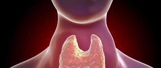
Decompression sickness (decompression sickness, DCS) is a complex of changes that develop during the transition from high atmospheric pressure to normal, and less often - from normal to low. The pathology got its name from the word “caisson”, meaning a chamber created in the 40s of the 19th century and intended for work under water or in water-saturated soil conditions. DCS is considered an occupational disease of submariners and specialists working in caisson chambers; in some cases it is diagnosed in pilots. In recent years, due to the widespread prevalence of diving, it has been identified in other groups of the population. According to statistics, the incidence of the disease is 2-4 cases per 10,000 dives.
Caisson disease
Carbon dioxide indoors
Carbon dioxide is also an indicator of the degree of cleanliness of indoor air. During the life of the body, various metabolic products and foreign impurities are released into the air: the concentration of carbon dioxide, air humidity, dust and bacterial contamination increase; decomposition products of sweat and sebum, ammonia and other foul-smelling gases enter the air. With a significant concentration of people in the room, all this creates an atmosphere of stuffy, “living” air. All polluting components accumulate in the air in proportional quantities. Therefore, it turned out to be very convenient to assess the degree of air pollution by the concentration of carbon dioxide, the determination of which is very accessible.
It has been established that at carbon dioxide concentrations of up to 0.1%, overall air pollution is insignificant and does not significantly affect human well-being and performance. Therefore, this concentration is accepted as the maximum permissible for the air in residential and public premises.