What is urea
Urea is a product that is formed in the liver as a result of the synthesis of ammonia and carbon dioxide. The resulting compound is delivered by blood to the kidneys, where the filtering organ separates it from the general bloodstream and sends it into the urine.
Urea is a product unnecessary by the body, which must be completely eliminated, since its accumulation gives an osmotic effect, which can cause swelling of the internal organs in which it accumulates:
- liver;
- kidney;
- thyroid gland;
- spleen;
- pancreas.
Normally, the final product - urea - should be excreted from the body, but if there are any abnormalities in the functioning of the system, it is detected in the blood.
What does urea in the blood mean?
Based on the state of the blood, namely the presence of high concentrations of urea in it, one can judge:
- the functioning of the kidneys, which are required to remove waste substances;
- about the function of the liver, which synthesizes urea from ammonia;
- about the health of muscle tissue, since the breakdown of protein in the muscles also produces urea.
Indications for testing may include:
- cardiac ischemia;
- rheumatoid arthritis;
- systemic scleroderma;
- liver diseases;
- screening results that do not fit within the normal range;
- suspected kidney infections;
- preparation for drug therapy;
- studies before hospitalization;
- assessment of the dialysis procedure performed.
Most often, an increase in urea indicates chronic or acute kidney disease. Separately, a comparative analysis is carried out for the urea content in urine and blood. If the level of the substance in the urine is lower than what is found in the blood, they speak of a disease of the filtration system - renal failure.
Indications for the purpose of the study
Urea in the blood is synthesized in the liver from ammonia and carbon dioxide, transported by the blood to the kidneys, there it is filtered through the glomerulus, and then excreted in the urine. Urea is an osmotically active substance, so its accumulation leads to swelling of the tissues of parenchymal organs (liver, kidneys, lungs, spleen, pancreas, thyroid gland), myocardium, central nervous system, subcutaneous tissue. An increase in urea concentration several times relative to the norm, accompanied, as a rule, by a pronounced clinical syndrome of intoxication, is called uremia.
Very often, against the background of kidney disease, simultaneously with an increase in the concentration of urea in the blood, its content in the urine decreases (a decrease in kidney function leads to an increase in urea in the blood).
The study is prescribed for coronary heart disease, systemic connective tissue diseases (rheumatoid arthritis, rheumatism, systemic scleroderma, etc.), arterial hypertension (regardless of the duration of its existence), when detecting abnormalities in a general urine test during a screening study, liver disease accompanied by dysfunction (hepatitis, cirrhosis), if inflammatory or infectious diseases of the kidneys are suspected, diseases of the gastrointestinal tract, as well as before and during drug therapy, before hospitalization of the patient due to an acute illness, after dialysis sessions for evaluation their effectiveness.
The concentration of urea in the blood characterizes:
- the state of the excretory function of the kidneys, that is, their ability to excrete substances unnecessary to the body with urine;
- the condition of muscle tissue (as a result of the breakdown of protein in the muscles, urea is formed);
- liver function, where ammonia is converted to urea.
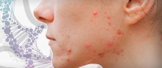
Why might urea be elevated?
To determine the level of urea in the blood, a laboratory method called a biochemical blood test is used. It involves taking material from a vein. For each age category of patients there is its own norm for urea content in the blood:
- in newborns from 1.7 to 5;
- in children of the first year of life from 1.4 to 5.4;
- from one to 15 years old can have 1.8-6.7;
- women over 18 years old - from 2 to 6.7;
- men over 18 years old - from 2.8 to 8.
Several factors influence the urea content. One of which is the level of amino acids in the body, since ammonia is formed from them during metabolism, which becomes one of the components of urea. On the other hand, with a sufficient amount of amino acids, the diseased liver will not be able to synthesize urea, which will be the reason for the detection of its negligible amount. The third important factor is the performance of the kidneys, which must, by filtering the blood, extract urea from it and send it out of the body with urine.
If we exclude all possible pathologies, then we can say that the following factors not related to the disease can increase the level of urea in the blood:
- on the eve of the collection of material, severe emotional stress was experienced;
- the patient had to buy and take a drug from the list: Tetracycline, Euthyrox, Neomycin, Lasix, any drug from the group of corticosteroids or sulfonamides, anabolic steroids, steroids, salicylates;
- the patient fasted for a long time or, on the contrary, ate a lot of protein foods;
- the patient on the eve of the delivery of the material or daily subjected the body to heavy physical activity.
When a doctor tries to determine the quality of kidney function, the presence of urea in the blood may indicate:
- blockage of the urinary ducts;
- chronic. renal failure;
- glomerulonephritis;
- pyelonephritis;
- dehydration, which could be preceded by myocardial infarction, shock or heart failure.
Separately, we should highlight the reasons why urea is formed in the human body in huge quantities:
- prostate neoplasms;
- burn disease;
- severe intestinal infections;
- hematological diseases;
- state of shock.
To accurately determine the patient’s condition, it is necessary to conduct several laboratory tests in a row to see the picture in dynamics; in addition, it is necessary to measure the amount of urea in the urine in order to be able to compare this indicator with blood data.
Topical use of urea in skin care: a review
Leonardo Celleno, Catholic University of the Sacred Heart, Rome, Italy, WILEY Dermatological Therapy, July 19, 2021, DOI: 10.1111/dth.12690
Impaired barrier function has been associated with a number of skin diseases, including xerosis, atopic dermatitis and psoriasis. Urea, a component of the skin's natural moisturizing factor, plays an important role in maintaining skin hydration and integrity. Several studies have examined the effects of urea in clinical settings. In this article, we summarize the available clinical evidence on the effects of urea in maintaining skin health and treating skin diseases. At lower doses (≤10%), topical urea preparations act as a skin moisturizer, while at higher concentrations (>10% urea), urea-based preparations have a keratolytic effect. (eg, Aquapiling (aquapiling.net)) Urea is also useful in combination therapy with anti-inflammatory and antifungal drugs due to its activity as a penetration enhancer.
KEYWORDS
atopic dermatitis, ichthyosis vulgaris, keratolytic agent, moisturizer, onychomycosis, psoriasis, topical urea, xerosis
The role of the skin is to protect the body from harmful environmental influences while maintaining appropriate mechanical properties, including elasticity. Healthy skin is characterized by effective control of water loss, allowing it to maintain a good level of hydration and therefore a strong physical and chemical barrier (Verdier-Sévrain & Bonté, 2007).
Urea is a polar, hygroscopic molecule produced endogenously by the human body and found naturally in the skin. Urea is formed from the metabolism of proteins and other organic nitrogen compounds and is excreted in urine and sweat (Kapuscinska & Nowak, 2014; Mosher, 1933). As one of the components of natural moisturizing factor (NMF), urea helps maintain healthy skin hydration levels. Despite the constant discovery of new ingredients and new skin care formulations, urea still remains one of the most useful molecules available to dermatologists due to its molecular and functional characteristics. In this article, we present a review of the clinical evidence supporting the use of urea to maintain skin integrity and treat diseases associated with skin barrier dysfunction.
The stratum corneum (SC), the outermost layer of the skin, protects the body from external agents and controls exchanges with the environment, especially transepithelial water loss (TEWL). The structural organization of the stratum corneum and the presence of hygroscopic molecules, collectively called NMF, allow the stratum corneum to retain water, thus keeping the epidermis hydrated and elastic. The stratum corneum consists of terminally differentiated keratinocytes called corneocytes, which are arranged in dense layers and are united by corneodesmosomes. Corneocytes are embedded in a hydrophobic lipid extracellular matrix, the keratinized cell envelope (Pouillot, Dayan, Polla, Polla, & Polla, 2008). Although the barrier created by the stratum corneum is similar in structure to a brick wall, it is more dynamic and complex in nature (Elias, 1983). In fact, the keratinized cell membrane surrounding the nondividing keratin-filled corneocytes represents an active environment consisting of enzymes involved in epidermal differentiation and NMF formation (Pouillot et al., 2008).
Adequate levels of hydration are necessary to maintain skin mechanical properties such as strength, flexibility, and elasticity (Mojumdar, Pham, Topgaard, & Sparr, 2017). In vivo, normal hydration levels range from 30–50% of the dry weight of the stratum corneum (Caspers, Lucassen, Carter, Bruining, & Puppels, 2001). Water found in the stratum corneum is mainly absorbed by corneocytes, which can increase their weight when swollen by up to 50% (Richter, Müller, Schwarz, Wepf, & Wiesendanger, 2001; Mojumdar et al., 2017). Under low relative humidity (RH) conditions, the stratum corneum is brittle and brittle, becoming more elastic as relative humidity increases (Mojumdar et al., 2017). Changes in relative humidity also alter the mobility of keratin filaments present in corneocytes, thereby supporting the role of water in keratin plasticization (Mojumdar et al., 2017).
NMF is critical to maintaining a healthy stratum corneum as it plays an important role in skin hydration (Robinson, Visscher, Laruffa, & Wickett, 2010). A decrease in NMF levels causes water loss in the stratum corneum and reduces the elasticity of the epidermis (Verdier-Sévrain & Bonté, 2007). NMF consists of molecules that are formed from the breakdown of proteins or are secreted by the sebaceous and sweat glands. The detailed composition of NMF is depicted in Figure 1. Degradation of filaggrin, a keratin-aggregating protein of the keratinized cell membrane that is formed during keratinocyte differentiation (Simon et al., 1996; Kezic, Kammeyer, Calkoen, Fluhr, & Bos, 2009), results in the formation of hygroscopic amino acids and other by-products including urea (Björklund et al., 2014). In a healthy stratum corneum, urea corresponds to 7% NMF, a percentage that decreases with age (Verdier-Sévrain & Bonté, 2007).
NMF components (%)
Clockwise: free amino acids; pyrrolidonecarboxylic acid; lactate; Sahara; urea; chlorides; sodium; potassium; ammonia, uric acid, glucosamine, creatinine; calcium; phosphates; citrate and formate.
FIGURE 1 Chemical composition of NMF. Data from (Verdier-Sévrain & Bonté, 2007)
The moisturizing effects of urea have been studied in vivo for many years (Serup, 1992; Bettinger, Gloor, Gehring, & Wolf, 1995; Treffel & Gabard, 1995; Loden, 1996, 1997; Kuzmina, Hagströmer, & Emtestam, 2002); Grether-Beck, Mühlberg, Brenden, & Krutmann, 2008; Borelli, Bielfeldt, Borelli, Schaller, & Korting, 2011). Pan and colleagues analyzed these clinical data in 2013 (Pan, Heinecke, Bernardo, Tsui, & Levitt, 2013). Urea has been noted to reduce transepidermal water loss (Loden, 1996, 1997; Borelli et al., 2011), improve hydration (Borelli et al., 2011) and water retention (Treffel & Gabard, 1995). In addition, urea can increase the amount of free water under high humidity conditions (Bettinger et al., 1995) and act as a powerful skin moisturizer and exfoliant (Serup, 1992).
Of note is the fact that all of these clinical studies used topical cream, emulsion or foam formulations with a urea concentration of 10% or less. No adverse events were reported, confirming the safety of topical urea-based products.
Urea increases the water content of the stratum corneum, acting as a humectant but also by maintaining the fluidity of the stratum corneum (Albèr et al., 2014; Mojumdar et al., 2017). By measuring corneometry, Albèr et al. found that urea promoted skin hydration even when used in a formulation with reduced water activity (Albèr et al., 2014). A subsequent study examined the molecular characteristics of keratin and macroscopic properties of the stratum corneum (SC) after the addition of urea to dehydrated SCs and corneocytes and found that the changes were similar to those occurring with increasing relative humidity in the absence of urea (Mojumdar et al., 2017) . These data support the hypothesis that urea acts as a natural endogenous humectant, replacing water under low-humidity conditions and maintaining fluid levels within the stratum corneum (Mojumdar et al., 2017). At higher concentrations (>10%) (eg Aquapiling (aquapiling.net)) urea has an emollient/keratolytic effect. The first evidence came from Swanbeck's studies in the 60s. These studies showed that preparations with high concentrations of urea could be used to treat ichthyosis and other hyperkeratotic conditions (Swanbeck, 1968a, 1968b; Swanbeck & Rajka, 1970). Swanbeck suggested that at high concentrations, urea is able to dissolve keratin, promoting the destruction of hydrogen bonds. Further studies showed that urea can cause conformational changes in keratin, causing denaturation of the protein structure (Pan et al., 2013).
In addition to its moisturizing properties, preserving the fluidity of SK and promoting keratin denaturation, urea is also able to participate in the regulation of gene expression (Friedman, von Grote, & Meckfessel, 2016). In a study by Grether-Beck et al. indicates that urea induces the expression of epidermal genes (Grether-Beck et al., 2012). Although more research is needed to better understand the involvement of urea in the regulation of gene expression in SK, the active role of urea as an inducer of epidermal gene expression may explain the beneficial effects of urea in preserving skin barrier function.
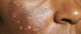
Keratinization disorders are characterized by qualitative or quantitative changes in the structure of the stratum corneum. Urea, used alone or in combination therapy, improves the treatment of pathological conditions including xerosis, atopic dermatitis, ichthyosis vulgaris, psoriasis and onychomycosis (Pan et al., 2013).
Xerosis or dry skin may be the result of dehydration or changes in lipid production. Dehydrated skin, typical of older people, whose tissues lack water - thin, weak and fragile. Low sebum levels and abnormal lipid levels in the epidermis (as in atopic dermatitis) make the skin tender, opaque, red, and hypersensitive to external agents. In both cases, topical formulations containing urea play an important role in the treatment of diseases. When used in low doses (mostly <10%, one study used 40% urea), urea regulates transepidermal water loss (TEWL) and restores the ability of the stratum corneum to mobilize and maintain hydration (Pan et al., 2013; Danby et al., 2016). Moisturizers containing urea improve fissure healing in diabetic patients (Federici, Federici, & Milani, 2015; Gin et al., 2017). Open heel cracks allow bacteria to enter and can lead to infections and further complications. Additionally, a skin care routine that includes a moisturizing step stops the progression of dry skin diseases, including epidermal hyperproliferation and inflammation (Friedman, von Grote, & Meckfessel, 2016).
From top to bottom, left to right: topical urea formulations; ≤10% urea, moisturizing effect, positive effect on xerosis, AD, IV, psoriasis; ≥10% urea, keratolytic effect, positive effect on psoriasis, onychomycosis; useful in quality; monotherapy; combination therapy, urea – enhances the penetration of corticosteroids, salicylic acid and antifungal drugs.
FIGURE 2 Overview of known effects of topical urea formulations in clinical settings. AD, topical dermatitis; IV, ichthyosis vulgaris
Skin barrier dysfunction has been associated with an increased risk of developing atopic dermatitis (AD) (Egawa & Kabashima, 2018). A weakened skin barrier makes it easier for allergens and other external agents to penetrate and predisposes the body to inflammation. If skin inflammation persists, barrier function is further compromised (Egawa & Kabashima, 2018). AD is the most common inflammatory skin disease, affecting up to 30% of children worldwide (Bieber, 2008). AD is a cutaneous manifestation of a genetic predisposition to allergies and is characterized by highly itchy and recurrent skin lesions on dry skin (Egawa & Kabashima, 2018). Adequate hydration of the stratum corneum appears to be critical in preventing AD, as the use of moisturizers in neonates reduces the prevalence of the disease (Horimukai et al., 2014; Simpson et al., 2014).
Several clinical trials have examined the clinical effects of topical urea formulations in AD (Pan et al., 2013). At a concentration of 10%, urea has been found to improve skin hydration and reduce transepidermal water loss (TEWL) in patients with AD (Sasaki, Tadaki, & Tagami, 1989). However, two recent studies suggest that urea increases skin hydration in AD without reducing TEWL (Hoppe et al., 2015; Ahmad Nasrollahi et al., 2018). These conflicting results can be explained by the low concentration of urea used in the formulations used (only 5%).
Clinical benefit of urea in AD has been observed when used alone and in combination with hydrocortisone or betamethasone-17 valerate (Bohnsack, Tausch, Gassmuller, & Rippke, 1997; Wilhelm, Scholermann, & Bohnsack, 1998; Loden et al., 2002; Wirén et al. al., 2009; Pan et al., 2013). It should be noted that combination therapy with urea was more effective than treatment with hydrocortisone or betamethasone-17-valerate alone (Pan et al., 2013). A recent Cochrane review confirmed that for AD, treatment with moisturizers and topical corticosteroids is more effective than treatment with topical corticosteroids alone (Van Zuuren, Fedorowicz, & Arents, 2017). A systematic review confirmed the beneficial effects of moisturizers on AD but warned of the need for more focused studies that would allow direct comparisons of moisturizers (Lindh & Bradley, 2015). Meanwhile, available evidence supports the use of urea-containing moisturizers as first-line therapy for AD (Lindh & Bradley, 2015).
Ichthyosis vulgaris (IV) is a genetic disorder with a prevalence of 1:300 caused by loss-of-function mutations in the filaggrin gene (FLG) (Brown & McLean, 2012). IV is characterized by flaky, dry skin with symmetrical white or gray scales. Peeling occurs due to the absence of filaggrin protein in the stratum corneum, which causes low cellular regeneration of the stratum corneum and chronic accumulation of keratin. A recent systematic review concluded that urea-containing drugs should be used as first-line therapy for ichthyosis vulgaris (Lindh & Bradley, 2015). Maintenance therapy using 10% urea is noninferior to therapy containing 1% hydrocortisone, 2% salicylic acid, or paraffin moisturizers (Pan et al., 2013).
Psoriasis is a chronic inflammatory skin disease with a worldwide prevalence of 2% (Parisi, Symmons, Griffiths, & Ashcroft, 2013). Psoriasis is characterized by the development of erythematous scaly plaques that result from increased keratinocyte proliferation and altered epidermal differentiation (Zhang et al., 2009). Clinical trials have been conducted to study the effects of urea on psoriatic skin (Pan et al., 2013). One study found that, when used at a concentration of 10%, urea reduced transepidermal water loss (TEWL) and increased stratum corneum hydration (Sasaki, Tadaki, & Tagami, 1989). Subsequently, Hagemann and Proksch also found that urea induces epidermal differentiation (Hagemann & Proksch, 1996). When used at concentrations ranging from 10 to 40%, alone or in combination with bifonazole or dithranol, most studies reported improvement in desquamation and clinical status in patients with psoriasis (Pan et al., 2013). (for example, Aquapiling (aquapiling.net))
At a concentration of 40%, urea shows clinical effects in the treatment of fungal nail infections such as onychomycosis (Pan et al., 2013). Topical treatment with fluconazole 1% and urea 40% is more effective than fluconazole alone in the treatment of onychomycosis (Bassiri-Jahromi, Ehsani, Mirshams-Shahshahani, & Jamshidi, 2012). Urea enhances the penetration of the antifungal agent and thus enhances the therapeutic effect (Pan et al., 2013). (for example, Cream-paste for corns Aquapiling (aquapiling.net))
Urea can be considered a penetration enhancer due to its ability to facilitate the transport of molecular flow through the nail and skin (Trommer & Neubert, 2006). These molecules include the aforementioned antifungal drugs, as well as other compounds used in the treatment of skin diseases, such as corticosteroids (Trommer & Neubert, 2006). An earlier study by Ayres and Hooper examined the potentiating effects of urea on skin penetration of cortisol (Ayres & Hooper, 1978). The authors observed that a topical formulation containing 1.0% cortisol and 10% urea delivered eight times more cortisol to the skin than a topical cream used as a control (1.0% cortisol cream mixed with water). Subsequent studies showed that urea enhances the penetration of hydrocortisone, progesterone and leuprolide through the subcutaneous tissue (Trommer & Neubert, 2006). It is noteworthy that in an in vitro study examining transcutaneous diffusion of progesterone, urea increased progesterone penetration by 2.5-fold (Valenta & Wedenig, 1997).
The mechanism by which urea increases the permeability of the stratum corneum is not fully understood, but it is thought to be related to the ability to increase the water content absorbed by corneocytes, which is a direct consequence of its high water-binding capacity (Mueller et al., 2016). At higher doses, the keratolytic action of urea may also play a role in enhancing drug penetration (Trommer & Neubert, 2006). Because of its recognized action as a chemical penetration enhancer, urea should be used in conjunction with topical corticosteroids, salicylic acid, or antifungals to improve drug delivery and treatment outcomes.
Urea is a well-known moisturizer and keratolytic agent. Urea has been used safely for over a century to support skin health and treat a range of skin conditions. Clinical studies have confirmed the positive effect of urea in diseases associated with skin barrier dysfunction. When used in different concentrations, urea has different effects (Fig. 2). Lower concentrations of urea (≤10%) are associated with a moisturizing effect, whereas higher concentrations (>10%) have an emollient/keratolytic effect. As a skin penetration enhancer, urea facilitates the crossing of the skin barrier by other molecules and thus improves the action of drugs used in combination therapy. Topical formulations containing urea are available in a variety of concentrations and provide clinicians with a wide range of alternatives to enhance the treatment of skin conditions and improve patient well-being.
Medical writing and editing support was provided by Raquel Carvalhosa, Ph.D., and . Financial support for the compilation and editing of medical texts was provided by.
CONFLICT OF INTEREST
The authors declare no conflict of interest.
ORCID Leonardo Celleno https://orcid.org/0000-0002-7129-198X
REFERENCES
Ahmad Nasrollahi, S., Ayatollahi, A., Yazdanparast, T., Samadi, A., Hosseini, H., Shamsipour, M., … Firooz, A. (2018). Comparison of linoleic acid-containing water-in-oil emulsion with urea-containing water-in-oil emulsion in the treatment of atopic dermatitis: A randomized clinical trial. Clinical Cosmetic and Investigational Dermatology, 11, 21–28. https://doi.org/10.2147/CCID.S145561 Albèr, C., Buraczewska-Norin, I., Kocherbitov, V., Saleem, S., Lodén, M., & Engblom, J. (2014). Effects of water activity and low molecular weight humectants on skin permeability and hydration dynamics—a double-blind, randomized and controlled study. International Journal of Cosmetic Science, 36(5), 412–418. https://doi.org/10.1111/ics.12136 Ayres, P. J., & Hooper, G. (1978). Assessment of the skin penetration properties of different carrier vehicles for topically applied cortisol. British Journal of Dermatology, 99(3), 307–317. https://doi.org/10.1111/j. 1365-2133.1978.tb02002.x Bassiri-Jahromi, S., Ehsani, A.H., Mirshams-Shahshahani, M., & Jamshidi, B. (2012). A evaluation comparative of combination therapy of fluconazole 1% and urea 40% compared with fluconazole 1% alone in a nail lacquer for treatment of onychomycosis: Therapeutic trial. Journal of Dermatological Treatment, 23(6), 453–456. https://doi. org/10.3109/09546634.2011.588191 Bettinger, J., Gloor, M., Gehring, W., & Wolf, W. (1995). Influence of emulsions with and without urea on water-binding capacity of the stratum corneum. Journal of the Society of Cosmetic Chemists, 46(5), 247–254. Bieber, T. (2008). Atopic dermatitis. New England Journal of Medicine, 358(14), 1483–1494. https://doi.org/10.1056/NEJMra074081 Björklund, S., Andersson, J. M., Pham, Q. D., Nowacka, A., Topgaard, D., & Sparr, E. (2014). Stratum corneum molecular mobility in the presence of natural moisturizers. Soft Matter, 10(25), 4535–4546. https://doi. org/10.1039/C4SM00137K Bohnsack, K., Tausch, I., Gassmuller, J., & Rippke, F. (1997). Efficacy on the symptom “dry skin” and long-term dermal tolerance of Laceran lotion 10% urea in patients with atopic dermatitis. Zeitschrift für Hautkrankheiten, 72, 34–39. Borelli, C., Bielfeldt, S., Borelli, S., Schaller, M., & Korting, H. C. (2011). Cream or foam in pedal skin care: Towards the ideal vehicle for urea used against dry skin. International Journal of Cosmetic Science, 33(1), 37–43. https://doi.org/10.1111/j.1468-2494.2010.00576.x Brown, S. J., & McLean, W. H. I. (2012). One remarkable molecule: filaggrin. The Journal of Investigative Dermatology, 132(3 Pt 2), 751–762. https://doi.org/10.1038/jid.2011.393 Caspers, P. J., Lucassen, G. W., Carter, E. A., Bruining, H. A., & Puppels, G. J. (2001). In vivo confocal Raman microspectroscopy of the skin: Noninvasive determination of molecular concentration profiles. The Journal of Investigative Dermatology, 116(3), 434–442. https://doi. org/10.1046/j.1523-1747.2001.01258.x Danby, S. G., Brown, K., Higgs-Bayliss, T., Chittock, J., Albenali, L., & Cork, M. J. (2016). The effect of an emollient containing urea, ceramide NP, and lactate on skin barrier structure and function in older people with dry skin. Skin Pharmacology and Physiology, 29(3), 135–147. https://doi.org/10.1159/000445955 Egawa, G., & Kabashima, K. (2018). Barrier dysfunction in the skin allergy. Allergology International, 67(1), 3–11. https://doi.org/10.1016/j.alit. 2017.10.002 Elias, P. M. (1983). Epidermal lipids, barrier function, and desquamation. The Journal of Investigative Dermatology, 80(Suppl), 44s–49s. Federici, A., Federici, G., & Milani, M. (2015). Use of a urea, arginine and carnosine cream versus a standard emollient glycerol cream for treatment of severe xerosis of the feet in patients with type 2 diabetes: A randomized, 8 month, assessor-blinded, controlled trial. Current Medical Research and Opinion, 31(6), 1063–1069. https://doi.org/10. 1185/03007995.2015.1037731 Friedman, A. J., von Grote, E. C., & Meckfessel, M. H. (2016). Urea: A clinically oriented overview from bench to bedside. Journal of Drugs in Dermatology: JDD, 15(5), 633–639. Gin, H., Rorive, M., Gautier, S., Condomines, M., Saint Aroman, M., & Garrigue, E. (2017). Treatment by a moisturizer of xerosis and cracks of the feet in men and women with diabetes: A randomized, double-blind, placebo-controlled study. Diabetic Medicine, 34(9), 1309–1317. https://doi.org/10.1111/dme.13402 Grether-Beck, S., Felsner, I., Brenden, H., Kohne, Z., Majora, M., Marini, A., … Krutmann, J. (2012). Urea uptake enhances barrier function and antimicrobial defense in humans by regulating epidermal gene expression. Journal of Investigative Dermatology, 132(6), 1561–1572. https://doi. org/10.1038/jid.2012.42 Grether-Beck, S., Mühlberg, K., Brenden, H., & Krutmann, J. (2008). Urea plus ceramides and vitamins: Improving the efficacy of a topical urea preparation by addition of ceramides and vitamins. Der Hautarzt; Zeitschrift Fur Dermatologie, Venerologie, Und Verwandte Gebiete, 59(9), 717–718, 720–723. https://doi.org/10.1007/s00105-008-1594-z Hagemann, I., & Proksch, E. (1996). Topical treatment by urea reduces epidermal hyperproliferation and induces differentiation in psoriasis. Acta Dermato-Venereologica, 76(5), 353–356. Hoppe, T., Winge, M. C. G., Bradley, M., Nordenskjöld, M., Vahlquist, A., Törmä, H., & Berne, B. (2015). Moisturizing treatment of patients with atopic dermatitis and ichthyosis vulgaris improves dry skin, but has a modest effect on gene expression regardless of FLG genotype. Journal of the European Academy of Dermatology and Venereology, 29(1), 174–177. https://doi.org/10.1111/jdv.12333 Horimukai, K., Morita, K., Narita, M., Kondo, M., Kitazawa, H., Nozaki, M., … Ohya, Y. (2014 ). Application of moisturizer to neonates prevents development of atopic dermatitis. The Journal of Allergy and Clinical Immunology, 134(4), 824–830.e6. https://doi.org/10.1016/j.jaci.2014.07.060 Kapuscinska, A., & Nowak, I. (2014). The use of urea and its derivatives in the cosmetics industry. CHEMIK, 68(2), 91–98. Kezic, S., Kammeyer, A., Calkoen, F., Fluhr, J. W., & Bos, J. D. (2009). Natural moisturizing factor components in the stratum corneum as biomarkers of filaggrin genotype: Evaluation of minimally invasive methods. British Journal of Dermatology, 161(5), 1098–1104. https:// doi.org/10.1111/j.1365-2133.2009.09342.x Kuzmina, N., Hagströmer, L., & Emtestam, L. (2002). Urea and sodium chloride in moisturisers for skin of the elderly—a comparative, double-blind, randomized study. Skin Pharmacology and Applied Skin Physiology, 15(3), 166–174. https://doi.org/10.1159/000063545 Lindh, J. D., & Bradley, M. (2015). Clinical effectiveness of moisturizers in atopic dermatitis and related disorders: A systematic review. American Journal of Clinical Dermatology, 16(5), 341–359. https://doi.org/10. 1007/s40257-015-0146-4 Loden, M. (1996). Urea-containing moisturizers influence barrier properties of normal skin. Archives of Dermatological Research, 288(2), 103–107. Loden, M. (1997). Barrier recovery and influence of irritant stimuli in skin treated with a moisturizing cream. Contact Dermatitis, 36(5), 256–260. Loden, M., Andersson, A.-C., Anderson, C., Bergbrant, I.-M., Frödin, T., Ohman, H., … Lindberg, M. (2002). A double-blind study comparing the effect of glycerin and urea on dry, eczematous skin in atopic patients. Acta Dermato-Venereologica, 82(1), 45–47. Mojumdar, E. H., Pham, Q. D., Topgaard, D., & Sparr, E. (2017). Skin hydration: Interplay between molecular dynamics, structure and water uptake in the stratum corneum. Scientific Reports, 7(1), 15712. https:// doi.org/10.1038/s41598-017-15921-5 Mosher, H. H. (1933). Simultaneous study of constituents of urine and perspiration. Journal of Biological Chemistry, 99, 781. Mueller, J., Oliveira, J. S. L., Barker, R., Trapp, M., Schroeter, A., Brezesinski, G., & Neubert, R. H. H. (2016). The effect of urea and taurine as hydrophilic penetration enhancers on stratum corneum lipid models. Biochimica et Biophysica Acta (BBA) - Biomembranes, 1858(9), 2006–2018. https://doi.org/10.1016/j.bbamem.2016.05.010 Pan, M., Heinecke, G., Bernardo, S., Tsui, C., & Levitt, J. (2013). Urea: A comprehensive review of the clinical literature. Dermatology Online Journal, 19(11), 20392. Parisi, R., Symmons, D. P. M., Griffiths, C. E. M., & Ashcroft, D. M. (2013). Global epidemiology of psoriasis: A systematic review of incidence and prevalence. Journal of Investigative Dermatology, 133(2), 377–385. https://doi.org/10.1038/jid.2012.339 Pouillot, A., Dayan, N., Polla, A. S., Polla, L. L., & Polla, B. S. (2008). The stratum corneum: A double paradox. Journal of Cosmetic Dermatology, 7(2), 143–148. https://doi.org/10.1111/j.1473-2165.2008.00379.x Richter, T., Müller, J. H., Schwarz, U. D., Wepf, R., & Wiesendanger, R. (2001). Investigation of the swelling of human skin cells in liquid media by tapping mode scanning force microscopy. Applied Physics A, 72(S1), S125–S128. https://doi.org/10.1007/s003390100750 Robinson, M., Visscher, M., Laruffa, A., & Wickett, R. (2010). Natural moisturizing factors (NMF) in the stratum corneum (SC). I. Effects of lipid extraction and soaking. Journal of Cosmetic Science, 61(1), 13–22. Sasaki, Y., Tadaki, T., & Tagami, H. (1989). The effects of topical application of urea cream on the function of pathological stratum corneum. Acta Dermatol-Kyoto, 84(4), 581–586. Serup, J. (1992). A three-hour test for rapid comparison of effects of moisturizers and active constituents (urea). Measurement of hydration, scaling and skin surface lipidization by noninvasive techniques. Acta Dermato-Venereologica. Supplementum, 177, 29–33. Simon, M., Haftek, M., Sebbag, M., MontéZin, M., Girbal-Neuhauser, E., Schmitt, D., & Serre, G. (1996). Evidence that filaggrin is a component of cornified cell envelopes in human plantar epidermis. Biochemical Journal, 317(1), 173–177. https://doi.org/10.1042/bj3170173 Simpson, E. L., Chalmers, J. R., Hanifin, J. M., Thomas, K. S., Cork, M. J., McLean, W. H. I., … Williams, H. C. (2014). Emollient enhancement of the skin barrier from birth offers effective atopic dermatitis prevention. Journal of Allergy and Clinical Immunology, 134(4), 818–823. https:// doi.org/10.1016/j.jaci.2014.08.005 Swanbeck, G. (1968a). A new treatment of ichthyosis and other hyperkeratotic conditions. Acta Dermato-Venereologica, 48(2), 123–127. Swanbeck, G. (1968b). Carbamide cream for dry and hyperkeratotic skin. Läkartidningen, 65(22), 2286–2290. Swanbeck, G., & Rajka, G. (1970). Antipruritic effect of urea solutions. An experimental and clinical study. Acta Dermato-Venereologica, 50(3), 225–227. Treffel, P., & Gabard, B. (1995). Stratum corneum dynamic function measurements after moisturizer or irritant application. Archives of Dermatological Research, 287(5), 474–479. Trommer, H., & Neubert, R. H. (2006). Overcoming the stratum corneum: The modulation of skin penetration. A review. Skin Pharmacology and Physiology, 19(2), 106–121. https://doi.org/10.1159/000091978 Valenta, C., & Wedenig, S. (1997). Effects of penetration enhancers on the in-vitro percutaneous absorption of progesterone. Journal of Pharmacy and Pharmacology, 49(10), 955–959. https://doi.org/10.1111/j.2042-7158. 1997.tb06023.x Van Zuuren, E. J., Fedorowicz, Z., & Arents, B. W. M. (2017). Emollients and moisturizers for eczema: Abridged Cochrane systematic review including GRADE assessments. British Journal of Dermatology, 177(5), 1256–1271. https://doi.org/10.1111/bjd.15602 Verdier-Sévrain, S., & Bonté, F. (2007). Skin hydration: A review on its molecular mechanisms. Journal of Cosmetic Dermatology, 6(2), 75–82. https://doi.org/10.1111/j.1473-2165.2007.00300.x Wilhelm, K. P., Scholermann, A., & Bohnsack, K. (1998). Efficacy and tolerance of a topical preparation containing 10% urea in patients with atopic dermatitis. Aktuelle Dermatologie, 24, 26–30. Wirén, K., Nohlgård, C., Nyberg, F., Holm, L., Svensson, M., Johannesson, A., … Lodén, M. (2009). Treatment with a barrier-strengthening moisturizing cream delays relapse of atopic dermatitis: A prospective and randomized controlled clinical trial. Journal of the European Academy of Dermatology and Venereology, 23(11), 1267–1272. https://doi.org/10. 1111/j.1468-3083.2009.03303.x Zhang, X.-J., Huang, W., Yang, S., Sun, L.-D., Zhang, F.-Y., Zhu, Q.- X., ... Liu, J.-J. (2009). Psoriasis genome-wide association study identifies susceptibility variants within LCE gene cluster at 1q21. Nature Genetics, 41(2), 205–210. https://doi.org/10.1038/ng.31
Symptoms indicating increased urea
Based on his feelings, the patient himself can often tell that the level of urea in the blood may be elevated. This is primarily indicated by pain in the lower back in both men and women. It indicates kidney disease.
Separately distinguished:
- swelling;
- increased amount of urine produced;
- scanty urine production;
- the presence of protein in the urine.
However, indications for donating blood for urea may be:
- ischemic diseases;
- hepatitis;
- cirrhosis;
- decreased absorption of foods;
- connective tissue diseases.
The level of urea indicates several possible pathologies at once, so this analysis is often used to monitor the patient’s condition.
Urea
Urea is the end product of protein and amino acid metabolism. Nitrogen obtained from the breakdown of proteins is converted into urea. Urea is easily filtered by the kidneys and is not reabsorbed by the kidneys. The formation of urea is a constant process associated with metabolism, so there is usually a small but stable amount of urea in the blood.
The level of urea in the blood increases with increased protein breakdown: in the early period after serious injuries, operations, during fever, after extensive burns, after intense physical activity.
Serum urea is currently the most widely used screening test for assessing renal function. A blood urea test along with creatinine is usually prescribed.
In what cases is research usually prescribed?
Analysis for urea and creatinine is included in the basic biochemical screening and is required if kidney pathology is suspected. It is also advisable to examine the level of urea and creatinine in the blood under the following conditions indicating possible kidney pathology:
- Fatigue, lack of concentration, poor appetite or sleep disturbances;
- Swelling of the face, wrists, or ankles
- Decreased urine output (oliguria);
- Pain in the lower back, side, under the ribs, near the location of the kidneys;
- High blood pressure;
What do the test results mean?
An increase in serum urea levels suggests renal dysfunction. This may be due to acute or chronic kidney disease, or may also indicate extrarenal causes leading to decreased blood flow to the kidneys. (congestive heart failure, extensive blood loss, dehydration).
Low urea concentrations are not usually a cause for concern. However, this result can be observed with prolonged fasting or a long-term protein-free diet, in which case additional examination is required.
The serum urea test for renal impairment is less sensitive than the Rehberg test, and serum urea may be normal until renal filtration capacity (determined by creatinine clearance) decreases to 50% of normal. Therefore, doctors more often evaluate these indicators together, which helps them diagnose the disease earlier and more accurately.
Test deadlines.
You can usually get your urea test result the next day.
How to prepare for the analysis?
No special preparation is needed. You should adhere to the general rules of preparation for taking blood from a vein. Detailed information can be found in the corresponding section.
How to take a urea test correctly
You need to prepare for the analysis so as not to receive distorted data. First of all, you need to remember that blood is donated strictly on an empty stomach. In addition, since urea production is affected by the breakdown of muscle fibers, it is important to avoid physical activity on the eve of the test. If you can’t get to the laboratory without excessive physical activity, you need to arrive at the door of the office no later than half an hour before the appointed time in order to have time to calm down and put your nervous system in order.
If the patient is taking any medications, it is better to postpone the morning dose until the blood has already been donated. Immediately before the donation, you should not do massages, biopsies, or x-rays, especially those using contrast.