The urethra (urethra) is part of a woman's urinary system and a man's urinary and reproductive systems.
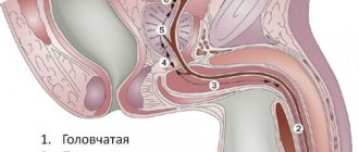
In men, the 20 cm long urethra is located both in the pelvis and inside the penis, and opens into an external opening on the glans. Anatomically, the following sections of the male urethra are distinguished: (1) external opening; (2) scaphoid fossa; (3) penile; (4) bulbous; (5) membranous; (6) prostatic (proximal and distal areas).
Figure taken from www.urologyhealth.org
The prostatic urethra passes through the prostate and is divided into proximal and distal parts at the level of the seminal tubercle. In the proximal part of the prostatic urethra, the excretory ducts of the prostatic glands open at the orifices along the posterolateral surfaces. On the sides of the seminal tubercle are the mouths of the right and left ejaculatory ducts, through which sperm enters the lumen of the urethra from the seminal vesicles and vas deferens. In the distal part of the prostatic part and in the membranous part of the urethra there are elements of the urethral sphincter. Starting from the bulbar region, the urethra passes inside the corpus spongiosum of the penis. The bulbar region is located inside the bulb of the corpus spongiosum. In the membranous and bulbar sections, the urethra bends anteriorly upward. In the penile region, the urethra is located medially along the ventral surface of the penis downward from the cavernous bodies. The capitate part of the urethra is located inside the head of the penis. The inner surface of the male and female urethra is covered with mucous membrane (transitional epithelium, with the exception of a short area near the external opening, where there is flat non-keratinizing epithelium).
Questions about the article
Hope
November 4, 2021 at 08:08 pm
Tell me, is it better to have surgery before or after pregnancy? Or maybe right after giving birth? While recovery is going on? Third pregnancy, cystitis began. I scold myself for not having surgery before pregnancy. But I reassure you that often after childbirth the urethra dropped again
Anton Evgenievich Rotov
November 4, 2021 at 08:11 pm
It is better to do the operation definitely after childbirth, after complete recovery (I advise no earlier than 6 months). Now I advise you to gradually train the muscles of the perineum. And also monitor the microflora of the vagina. If done before childbirth, then no later than a year.
How to avoid complications of urethritis? How not to miss the disease?
To avoid complications, more frequent preventive examinations should be carried out, especially after unprotected sexual intercourse, even in the absence of complaints. The earlier the diagnosis is made, the easier the treatment will be.
In fact, sex, no matter what it is (vaginal, oral or anal), can be equally unsafe, since all body fluids can contain a variety of pathogens. Unsafe sexual intercourse means promiscuity and frequent changes of sexual partners.
Frequent change of sexual partners
But even one woman is not a guarantee of health, since she can have an intimate relationship with another man. In this sense, a condom is a reliable barrier to the spread of infection.
From the data provided, it is clear that only a urologist who conducts special and serological tests can determine the cause of urethritis and what infection caused the urethritis - a possible option, even several infections at once. Some of them may have an incubation period and may not appear immediately. Therefore, having noticed unpleasant signs, there is no need to immediately blame the last partner.
What is the urinary system and how does it work?
The urinary tract is a continuous system of hollow organs whose main function is the production, collection, transportation, storage and excretion of urine.
The urinary system is divided into upper and lower sections. The upper urinary system consists of the kidneys and a tube called the ureter, which transports urine from the kidney to the bladder.
The lower urinary system consists of the bladder and another tube called the urethra, which ends the urinary system and transports urine from the bladder to the outside.
The function of the urinary system is to ensure the removal of metabolic products from the human body, regulate water-salt balance, and also store and transport urine.
The urinary system works in conjunction with the lungs, skin and intestines to maintain the balance of chemicals and water in the body. Adults excrete from 800 to 2000 milliliters of urine per day with normal drinking water consumption per day, which is 1.5-2 liters. Several factors influence the increase in urine production in the body. For example, certain types of medications, such as diuretics (water medications), are sometimes used to treat high blood pressure. Drinks such as coffee and alcohol can also cause an increase in the amount of urine produced in some people.
Features of the disease in men
In men and women, the disease has different manifestations, which is associated with the characteristics of their genitourinary system. And if female urethritis passes with virtually no symptoms, then inflammation of the urethra in men is accompanied by various unpleasant sensations.
General practitioner Elena Vasilyevna Malysheva and cardiologist German Shaevich Gandelman will talk about the features and symptoms of the disease:
The cause of activation of the inflammatory process, according to doctors, is mainly genital infections. The first manifestation of the disease is weakness of the body. A man can feel it just a few hours after the pathogen enters the body. There are also non-infectious causes of the disease (injuries, allergic reactions, poor nutrition or personal hygiene, etc.), but they are quite rare.
It is very important to begin treating inflammation at the initial stage of its development, since urethritis can lead to the development of pathologies of the genitourinary system and reproductive dysfunction.
Diagnostics
The following tests and procedures may be used to diagnose urethral diseases:
- Anamnesis collection.
- Digital rectal examination: examination of the rectum and prostate.
- General urine analysis.
- Urine cytology: A laboratory test in which a urine sample is examined under a microscope for abnormal cells.
- Culture of urethral discharge.
- CT scan.
- Urethroscopy: a procedure for examining the urethra with a special thin instrument.
Treatment of urethritis
It is possible to prescribe the correct treatment only after receiving test results, since each pathogen is treated with specific drugs, and there are no universal magic pills.
It is very important to complete the full course of antibiotic therapy and monitor disease prevention:
- follow the rules of safe sex (condoms);
- avoid sexual intercourse until the infection is completely cured;
- partners should also be tested and treated;
- personal hygiene must be maintained.
A course of antibiotic therapy in the treatment of urethritis
If urethritis is asymptomatic, sexual contacts should be monitored from the last four weeks to six months. All partners who were in a relationship at this time must get tested. Treatment is carried out while maintaining the confidentiality of each patient, so there is no point in being afraid to go to the clinic.
If a man feels well and has stopped treatment, but he is not yet completely cured (recovery has not been confirmed in the laboratory), then the disease may return. Symptoms of urethritis may reappear even after several months, again spontaneously. Such a man, without feeling anything, all this time infects his sexual partners with a sexually transmitted infection. In this case, repeated treatment will be longer and more severe, and the consequences will be much more dangerous, including the development of chronic urethritis, which cannot be completely cured.
ONLINE REGISTRATION at the DIANA clinic
You can sign up by calling the toll-free phone number 8-800-707-15-60 or filling out the contact form. In this case, we will contact you ourselves.
If you find an error, please select a piece of text and press Ctrl+Enter
What complications can you encounter?
The danger, first of all, is acute urinary retention. A person feels complete exhaustion and weakness due to a painful, unbearable desire to urinate. Irritability and sometimes even panic attacks develop. However, intoxication from accumulated waste biological fluid is more dangerous. Stagnant urine is a favorable environment for the spread of bacteria.
More often, against the background of penetration of a foreign body into the urethra, inflammatory processes develop: urethritis. The pathology then spreads upward. Sometimes the coating of handles or nails enters into a chemical reaction with mucous membranes and urine, causing additional intoxication.
Due to the entry of a foreign object into the urethra, severe trauma and disruption of its integrity are possible.
General methods of treating urethral diseases
Treatment of urethral diseases depends on the type of disease. It can be either conservative (various medications and physical procedures) or surgical.
The Department of Urology of the Moscow State University named after M.V. Lomonosov, when choosing surgery as a treatment method, always gives preference to minimally invasive methods - the most progressive method of surgical interventions. This ensures minimal impact on surrounding tissue and rapid recovery.
More information about urethral diseases:
Upper urinary system
Kidneys
The kidneys are a paired organ, they are located on both sides of the spine in the retroperitoneal space just above the lumbar region and have the appearance of a large bean and are approximately the size of a fist. The right kidney is located slightly lower than the left due to the position of the liver. In an adult, the average kidney is 10 cm long, 6 cm wide and 3 cm thick, and weighs about 120-200 g. The left one is usually slightly larger than the right kidney.
Each kidney is covered by a fibrous capsule that protects the kidney from injury. All pain sensations are associated with this capsule: the organ itself has no pain receptors. When the capsule is damaged or stretched, pain of varying nature and intensity appears.
Kidney tissue or parenchyma consists of outer (cortical) and inner (medullary) layers.
Blood enters the kidney through the renal artery (a branch of the aorta) and is filtered through the microscopic structural working units of the kidney, the nephrons. Each kidney contains about a million nephrons. Their job is to filter blood and produce urine. Each nephron consists of a ball formed by small blood capillaries (the glomerulus), surrounded by a dome-shaped structure, the glomerular capsule (or Bowman's capsule), and a small tube called the renal tubule. Here, the blood plasma is filtered, which leads to the formation of urine.
The urine storage system consists of small renal calyces, which, merging with each other in 2-3 groups, form a large renal calyx, and these in turn form the renal pelvis. The renal pelvis passes directly into the ureter.
All functions normally performed by two kidneys can be adequately performed by one healthy kidney. Some people are born with only one kidney, while others choose to donate one kidney for transplantation into a person with kidney failure.
Basic kidney function
is to maintain the correct balance of water and minerals (including electrolytes) in the body.
An important function of the kidneys is to regulate fluid balance by excreting excess water in the form of urine while maintaining the required amount of water in the body, which is necessary for life. When the kidneys lose their ability to remove excess water, edema appears.
The kidneys regulate the balance of minerals and substances such as sodium, potassium, calcium, phosphorus, magnesium and bicarbonate and maintain normal blood composition. Changes in sodium levels can affect a person's mental state, while changes in potassium levels can have serious adverse effects and cause problems with the heart and muscle function. Maintaining normal levels of calcium and phosphorus is essential for healthy bones and teeth.
Additional functions of the kidneys include:
- Filtration and removal from the body of food processing waste, medications and harmful substances (toxins).
- Creatinine and urea are two important byproducts of kidney function that can be easily measured in the blood. Their values in blood tests reflect kidney function. When kidney problems occur, creatinine and urea levels increase.
- Regulate Blood Pressure – The kidneys produce various hormones (renin, angiotensin, aldosterone, prostaglandins, etc.) that help regulate the amount of water and salt, the levels of which play a vital role in maintaining blood pressure. Impaired hormone production and salt and water regulation in a patient with, for example, kidney failure can lead to high blood pressure.
- Blood volume regulation
- Regulating blood pH
- Converts vitamin D into its active form, which is necessary for the absorption of calcium from food, growth of bones and teeth, and maintenance of their health. Decreased levels of active vitamin D lead to decreased bone growth rates. Slow growth may be a sign of kidney disease in children.
Erythropoietin
is a hormone produced in the kidneys and plays an important role in the production of red blood cells. With kidney failure, the production of erythropoietin decreases, which in turn leads to a decrease in hemoglobin levels (anemia). This is the reason why the hemoglobin count does not improve in patients with kidney failure despite taking iron supplements and vitamins.
Ureters
These are fibromuscular tubes that drain urine from the renal pelvis to the bladder and are about 25-30 cm long and 6-8 mm wide. They enter the bladder from behind and at an angle, ending in the lumen of the bladder in the form of holes - the mouth of the ureters. The lower ureter is compressed by the bladder wall passively during urine storage and dynamically during voiding. Essentially, it is a valve that prevents vesicoureteral reflux (i.e., stops urine from flowing back into the kidneys). The wall of the ureter is made up of three layers, including a layer of muscle that helps it contract and move urine from the kidney to the bladder. Small portions of urine enter the bladder from the ureters approximately every 10-15 seconds.
Along the length of the ureter there are three physiological narrowings: at the level of the transition of the pelvis to the ureter, the place of intersection with the common iliac vessels and in the thickness of the bladder wall. With urolithiasis, stones can get stuck in places where the ureters are narrowed, causing renal colic.