Postcholecystectomy syndrome (PCES) is not the most common phenomenon in gastroenterology. It is generally accepted that PCES belongs to the group of gallbladder diseases. In fact, this is not even a disease, but a collective name for a set of symptoms that appear immediately or shortly after surgery on the bile ducts or removal (resection) of the gallbladder.
Until now, both therapists and surgeons find it difficult to clearly determine the causes of the development of this syndrome. Doctors tend to use the term PCES only to make a preliminary diagnosis in operated patients1.
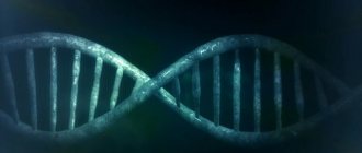
How does the liver work?
The liver is the largest gland in humans. It is located in the upper floor of the abdominal cavity on the right. It consists of two main lobes - right and left, which in turn are divided into 8 segments.
In an adult, it weighs about 1/40 of the total body weight (approximately 1.6 kg in men and 1.2 kg in women). The liver has a dual blood supply: approximately 80% of the blood entering the liver comes from the portal vein (venous blood collected from all unpaired abdominal organs), the rest (oxygenated arterial blood) comes from the hepatic artery. Having entered the portal of the liver, both vessels give rise to multiple branching smaller vessels. At the exit from the liver, 3-4 hepatic veins are formed, which flow into the inferior vena cava.
The main structural unit of the liver is the hepatic lobule, which consists of liver cells - hepatocytes. This cell is one of the most important biochemical laboratories of the body. The hepatocyte takes part in the metabolism of proteins, carbohydrates, fats, bile acids, and vitamins. Neutralizes toxic substances coming from the intestines, followed by the release of associated products into the intestinal lumen. An important function of the hepatocyte is the synthesis of bile, which is involved in digestion.
What is bile and why is it needed?
Bile is a secretion produced by hepatocytes. The color of bile ranges from light yellow to dark green. Bile contains bile pigments - bilirubin and biliverdin, cholesterol, fatty acids, lecithin, mucin.
Bile is the main participant in digestion. It neutralizes hydrochloric acid that enters the duodenum from the stomach, thereby creating favorable conditions for the action of pancreatic juice and parietal digestion in the jejunum.
Bile acids emulsify fats, allowing them to be absorbed in the intestines.
Bile has a bactericidal effect that prevents the proliferation of pathogenic microflora.
Causes of the disease
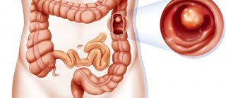
The most common cause of obstructive jaundice is complete or partial closure of the bile ducts.
However, other factors can also provoke the disease:
- inflammation of the gallbladder (cholecystitis);
- inflammation of the bile ducts (cholangitis);
- cholelithiasis;
- cysts in the bile duct area;
- hepatitis;
- liver cirrhosis, ascites;
- inflammation of the pancreas;
- tumor neoplasms in the area of the duodenum, liver, stomach;
- parasitosis;
- a sharp increase in hepatic lymph nodes;
- complication resulting from surgery in the biliary tract.
How are the bile ducts arranged and how do they work?
The bile ducts are a system of ducts that originate from hepatocytes and gradually gather into interlobular, segmental and right and left lobar ducts, which unite into the common hepatic duct.
One of the stages of bile removal is its accumulation in the reservoir - the gallbladder. The main function of the gallbladder is to concentrate bile and release it into the lumen of the duodenum during digestion. The volume of this organ is about 80 ml.
The gallbladder is located on the lower surface of the liver. It is a hollow muscular organ, externally covered with a serous membrane and internally lined with mucous membrane. The structure of the gallbladder is divided into the bottom, body and neck of the bladder, which passes into the cystic duct.
Prevention
Obstructive jaundice can affect any organism. However, there are a number of factors that can trigger its appearance. In this regard, it is recommended to follow simple preventive measures.
These include:
- timely detection and competent treatment of gallbladder diseases, as well as any malfunctions in the functioning of the hepatobiliary system;
- maintaining a healthy diet that excludes the consumption of fatty, fried, and excessively sweet foods;
- giving up alcohol;
- moderate physical activity;
- body weight regulation.
Why do gallstones form?
The exact cause of cholelithiasis has not yet been fully elucidated. Only a few factors are known that increase the likelihood of its occurrence.
- Genetic: family predisposition increases the risk of cholelithiasis by 4-5 times;
- Congenital anomalies of the biliary tract;
- Gender - the risk of developing gallstones in women is approximately 2-3 times higher, which is associated with the influence of estrogens on the ability to form gallstones;
- Age - the maximum frequency of clinical manifestations of cholelithiasis is recorded at the age of 40-69 years;
- Dietary (high-calorie diet, poor in plant fiber, as well as with an excess of simple carbohydrates, animal proteins, foods containing cholesterol);
- Use of medications (oral contraceptives, fibrates, ceftriaxone, somatostatin, nicotinic acid);
- Concomitant diseases and conditions (obesity - a tendency to form gallstones in persons with a body mass index of more than 30 is 3 times higher, type 2 diabetes mellitus, hypothyroidism, cirrhosis of the liver, etc.);
- Pregnancy - the risk of developing cholelithiasis increases during pregnancy, especially with repeated pregnancies (the likelihood of stone formation increases by 10-11 times). During pregnancy, bile heterogeneity, or the so-called biliary sludge, develops in 20-30% of patients, stones - in 5-12% of cases.
There is a so-called 5F rule that describes patients at greatest risk of developing stones:
1. Female (female);
2. Fat (overweight/obesity);
3. Forty (age 40 years or more);
4. Fertile (fertile women - that is, pregnant and giving birth);
5. Fair (light-skinned blondes).
How are gallstones formed?
The process of stone formation is quite well studied. The key links are:
- An increase in the lithogenicity of bile (the ability to form stones) means an increase in the content of cholesterol and bilirubin in it.
- Impaired contractile function of the gallbladder.
- Increased pressure in the bile duct system.
Mucin is a natural gel, constantly secreted by the wall of the gallbladder; when lithogenicity increases, it begins to “absorb” cholesterol crystals, thereby forming a “fossilization nucleus.” The formed suspension, with impaired contractile function, continues to attach salt crystals encrusted with calcium salts. Thus, gallstones can grow up to 5-7 mm per year.
How does gallstone disease occur?
The main factors influencing the course of the disease are the nature of nutrition and the size of the stones. Gallstone disease can occur both in the form of chronic stone carriage and in acute, and sometimes in complicated, form.
Where does pain come from with gallstone disease?
By themselves, gallstones do not cause any pain and can be an accidental finding during medical examination. When food enters the duodenum, the mechanoreceptors are irritated and cause contraction of the gallbladder. The stones wedge into the cervix, causing a prolonged spasm, which manifests itself as pain. When taking antispasmodic drugs, the calculus can “move” back from the cervix and the attack of hepatic colic is stopped. If the stone remains in the cervix, then inflammation begins to develop against the background of obstruction.
Chronic calculous cholecystitis is an asymptomatic stone carrier. The size of the stones can vary from 2 – 3 mm to 5 cm. This stage is characterized by an almost complete absence of clinical manifestations of the disease. Stones can remain in the lumen of the gallbladder for years without causing any symptoms. However, if there is an error, discomfort may occur in the right hypochondrium, which can be relieved independently or while taking antispasmodic drugs.
Hydrocele of the gallbladder - occurs against the background of obstruction of the neck of the gallbladder by a calculus, cutting off the latter from the entry of bile. The bladder is filled with mucus secreted by the mucous membrane and is not involved in digestion.
Acute calculous cholecystitis - occurs with prolonged obstruction of the neck of the gallbladder by a calculus. The pain syndrome persists against the background of ineffective conservative treatment. The patient is bothered by vomiting, which does not bring relief; pain can be felt behind the sternum - cholecystocardial syndrome, in the right shoulder blade. Body temperature rises and chills appear. Inflammatory changes begin to develop in the wall of the gallbladder, which progress from catarrhal, phlegmonous and gangrenous forms. Against the background of the latter, perforation of the gallbladder or abscess may develop.
Choledocholithiasis and acute biliary pancreatitis. Choledocholithiasis develops as a result of migration of biliary sludge or small stones from the gallbladder into the common bile duct. Once in the common bile duct, small stones freely descend to the sphincter of Oddi, which is a release mechanism - a circular muscle cuff. Sludge and stones “wedge” into the sphincter, causing its long-term spasm, causing a violation of the outflow of bile - obstructive jaundice. The patient's skin and sclera become a rich yellow color. This condition requires emergency decompression of the bile ducts - restoration of bile outflow.
An increase in pressure in the common bile duct promotes the reflux of bile into the main pancreatic duct and intraductal activation of pancreatic juice enzymes - a trigger for the development of acute biliary pancreatitis.
Mirizzi syndrome . A fairly rare complication of gallstone disease. Develops as a result of prolonged stone bearing. First, a stricture forms, then a bedsore between the gallbladder and the common bile duct.
Mirizzi syndrome presents significant difficulties for surgical treatment. This is due to the fact that in the human body there is no plastic material to close the defect of the common bile duct. Surgical treatment of Mirizzi syndrome is often accompanied by the formation of an anastomosis between the small intestine and the common bile duct. When a stone migrates into the small intestine, acute intestinal obstruction may develop, requiring emergency surgical treatment.
In the photographs, in the lumen of the resected section of the small intestine there is a calculus that caused intestinal obstruction.
Postcholecystectomy syndrome: diagnosis and treatment
Gallstone disease (GSD) is extremely common in civilized countries, including Russia. The incidence is especially high among women aged 35–40 years and older. GSD is characterized by the formation of gallstones in the biliary system, mainly in the gallbladder, and complications arising from the progression of the pathological process.
The main treatment for this disease is cholecystectomy. In 25% of patients who underwent cholecystectomy, abdominal pain and dyspeptic disorders that require treatment appear or persist after some time. Adequate therapy of patients depends on a correct understanding of the pathogenesis of clinical symptoms developing after cholecystectomy. The peculiarities of their formation are associated, on the one hand, with disturbances in cholesterol metabolism inherent in gallstone disease, and on the other, with the fact that the pathological process occurs in new anatomical and physiological conditions, i.e. in the absence of the gallbladder.
It is known that removal of the gallbladder for calculous cholecystitis does not relieve patients from metabolic disorders, including hepatocellular dyscholia, which persists after surgery. In most patients after cholecystectomy, lithogenic bile with a low cholate-cholesterol coefficient is determined. Loss of the physiological role of the gallbladder, namely the concentration of bile during the interdigestive period and its release into the duodenum during meals, is accompanied by a violation of the passage of bile into the intestines and indigestion. Changes in the chemical composition of bile and its chaotic entry into the duodenum disrupt the digestion and absorption of fat and other substances of lipid nature, reduce the bactericidal properties of duodenal contents, which leads to microbial contamination of the duodenum, weakened growth and functioning of normal intestinal microflora, disruption of the hepatic-intestinal circulation and decreased total pool of bile acids. Under the influence of microflora, bile acids undergo premature deconjugation, which is accompanied by damage to the mucous membrane of the duodenum, small and large intestines with the development of duodenitis, reflux gastritis, enteritis and colitis. Duodenitis is accompanied by duodenal dyskinesia and, first of all, duodenal hypertension with the development of duodenogastric reflux and reflux of contents into the common bile duct and pancreatic duct.
As a result, dyskinesia of the sphincter of the hepatopancreatic ampulla, bile and pancreatic ducts (sphincter of Oddi) is formed, and deconjugated bile acids cause the development of diarrhea. Thus, in patients with a removed gallbladder, the existing clinical manifestations may be associated with changes in the chemical composition of bile, impaired passage into the duodenum, dyskinesia of the sphincter of Oddi, as well as with excessive bacterial growth in the intestine, maldigestion and malabsorption syndromes, duodenitis and other pathological disorders in the digestive system. In addition, in a number of patients, organic obstructions to the outflow of bile associated with cholecystectomy may persist or develop again. Pathological conditions that are observed in patients after cholecystectomy can be divided into 3 groups.
- Functional disorders of the sphincter of Oddi - increased tone of the sphincter of the common bile duct or pancreatic duct or the common sphincter as a result of loss of a functioning gallbladder.
- Biliary hypertension with the presence of organic obstructions to the flow of bile, in the presence of which recurrent or residual choledocholithiasis is detected in 5–20% of patients, stenosis of the major duodenal papilla in 11–14%, strictures of the bile ducts and bile-ducting anastomoses in 6.5–20%, 0.1–1.9% have an excess stump of the cystic duct.
- Concomitant diseases that developed before or after surgery. The main ones are chronic pancreatitis, duodenal dyskinesia, irritable bowel syndrome, duodenitis, peptic ulcer, gastroesophageal reflux disease, hiatal hernia.
Currently, the term “postcholecystectomy syndrome” is used to designate only dysfunction of the sphincter of Oddi, caused by a violation of its contractile function and preventing the normal outflow of bile and pancreatic secretions into the duodenum in the absence of organic obstacles. It is proposed, instead of the previously accepted definitions “postcholecystectomy syndrome”, “biliary dyskinesia”, etc., to use the term “sphincter of Oddi dysfunction”.
Clinical manifestations of sphincter of Oddi dysfunction depend, first of all, on the involvement of one or another of its structures in the process. With isolated dysfunction of the common bile duct sphincter, biliary pain develops; with the predominant involvement of the pancreatic duct sphincter, pancreatic pain develops; and with pathology of the common sphincter, combined biliary-pancreatic pain develops. Dysfunction of the sphincter of Oddi is characterized by recurrent attacks of severe or moderate pain lasting 20 minutes or more, repeated for 3 months or more. With the biliary type, the pain is localized in the epigastrium or right hypochondrium with irradiation to the back and right scapula, with the pancreatic type - in the left hypochondrium with irradiation to the back, decreasing when bending forward, with the combined type - it has a girdling character. Pain can be combined with the following symptoms: a) onset after eating; b) appearance at night; c) nausea and/or vomiting.
However, assessment of clinical symptoms alone is not sufficient to exclude organic pathology of the biliary system. For this purpose, a number of screening and clarifying methods are used (table).
Non-invasive methods to suggest sphincter of Oddi dysfunction include blood levels of bilirubin, alkaline phosphatase, aminotransferases, amylase and lipase. Laboratory tests must be carried out during or no later than 6 hours after the end of a painful attack, as well as over time. A transient increase of 2 or more times in the level of liver or pancreatic enzymes during a period of at least 2 consecutive attacks of pain is important in confirming sphincter of Oddi dysfunction.
The provocative morphine-prostigmine test is of similar importance, when parenteral administration of drugs provokes the development of a painful attack, most often of a mixed biliary-pancreatic type in combination with an increase in the blood level of aminotransferases, alkaline phosphatase, and pancreatic enzymes. However, the diagnostic value of this test is limited by its low sensitivity and specificity.
During ultrasound, great importance is attached to the expansion of the common bile duct and main pancreatic duct, which indicates a violation of the flow of bile and pancreatic secretion at the level of the sphincter of Oddi. At the same time, in 3-4% of patients who have undergone cholecystectomy and have no symptoms, dilation of the common bile duct is noted. Of particular importance is ultrasound examination of the diameter of the common bile duct using fatty breakfasts, which stimulate the production of endogenous cholecystokinin and increase choleresis. After a test breakfast, the diameter of the common bile duct is measured every 15 minutes for 1 hour. An increase in its diameter by 2 mm or more compared to the initial one suggests the presence of incomplete obstruction of the common bile duct, both as a result of dysfunction of the sphincter of Oddi, and due to organic pathology of the biliary system.
To assess the condition of the pancreatic ducts, a test with the introduction of secretin at a dose of 1 mg/kg is used. Normally, after stimulation of pancreatic secretion by secretin, ultrasound shows an expansion of the pancreatic duct within 30 minutes, followed by its decrease to the initial level. If the duct remains dilated for more than 30 minutes, this indicates a violation of its patency.
With biliscintigraphy, the presence of dysfunction of the sphincter of Oddi is indicated by an increase in the transit time of the radiopharmaceutical from the porta hepatis to the duodenum, and it is proportional to the level of basal pressure of the sphincter of Oddi. At the same time, with dilated common bile duct, this study is of little information for assessing the function of the sphincter of Oddi.
Invasive methods for assessing the function of the sphincter of Oddi include an indirect method - endoscopic retrograde cholangiopancreatography (ERCP). Indirect signs of increased tone of the sphincter of Oddi are the diameter of the common bile duct more than 12 mm, the delay of contrast in the common bile duct for more than 45 minutes. Dysfunction of the sphincter of the main pancreatic duct is indicated by the expansion of the latter to more than 5 mm and a slowdown in the evacuation of contrast from its lumen. However, similar changes can be observed in the presence of organic pathology.
The most accurate method to confirm dysfunction of the sphincter of Oddi is manometry. In this case, it is possible to cannulate the common bile duct and Wirsung duct separately with manometry of their sphincters, which makes it possible to identify predominantly the biliary or pancreatic type of disorder, as well as to establish the etiology of recurrent pancreatitis in patients who have undergone cholecystectomy and papillotomy. Signs of sphincter of Oddi dysfunction during manometric examination are:
- increased basal pressure in the lumen of the sphincters;
- increased amplitude and frequency of phasic contractions (tachyodia);
- increased frequency of retrograde contractions;
- paradoxical response to the administration of cholecystokinin analogues.
Manometry of the sphincter of Oddi is not indicated for all patients. The choice of this study is based on an assessment of the severity of clinical manifestations and the effectiveness of conservative therapy.
Thus, we can assume that we are talking about dysfunction of the sphincter of Oddi in the following situations:
- the presence of pain in the epigastric region in patients who have undergone cholecystectomy, in cases where other causes that can explain their origin are not identified (concomitant diseases, structural changes in the biliary and pancreatic ducts);
- the presence of idiopathic recurrent pancreatitis;
- for recurrent biliary colic in patients with the presence of an unchanged gallbladder and common bile duct and with a normal composition of gallbladder bile.
Approximate formulations of diagnoses for patients who have undergone cholecystectomy:
Diagnosis: cholelithiasis, cholecystectomy in 1994 (for cases in which there are no clinical symptoms).
Diagnosis: dysfunction of the sphincter of Oddi of the biliary type. Gallstone disease, cholecystectomy in 1999
Diagnosis: chronic recurrent (obstructive) pancreatitis with exocrine and endocrine insufficiency in the acute phase.
Concomitant disease: cholelithiasis, cholecystectomy in 1994.
Diagnosis: irritable bowel syndrome with a predominance of constipation in the acute phase.
Concomitant diagnosis: cholelithiasis, cholecystectomy in 1994.
Management of patients who have undergone cholecystectomy. The goal of treatment: to restore the normal flow of bile and pancreatic secretions from the biliary and pancreatic ducts into the duodenum. Treatment objectives: normalize the chemical composition of bile; restore the patency of the sphincter of Oddi; restore the normal composition of intestinal microflora; normalize digestive processes and small intestinal motility to prevent duodenal hypertension.
To prevent the formation of bile sludge or stones in the extrahepatic biliary system, it is recommended:
- limiting (but not completely eliminating) the intake of foods containing cholesterol (fats of animal origin) and fatty acids (fats that have been heat-treated over 100°C - fried foods);
- regular 4-6 meals a day;
- slow weight loss. When using low-calorie diets (2110 kJ/day and below), during fasting, during shunt operations, additional administration of ursodeoxycholic acid at a dose of 10 mg/kg/day is necessary;
- adding dietary fiber contained in products of plant origin or food additives (bran, etc.) to the diet. At the same time, it is better to use vegetables, fruits, and herbs after heat treatment (boiled, baked). Bran can be used in the form of ready-made breakfasts (porridge, bread) and proprietary preparations;
- providing daily bowel movements.
Drug treatment for sphincter of Oddi dysfunction is aimed at relieving spasm of the smooth muscles of the latter. For this purpose, a number of drugs that have an antispasmodic effect are used. Nitrates: nitroglycerin is used for quick pain relief, nitrosorbitol is used for course treatment. The mechanism of action of nitrates is reduced to the formation of free radicals of nitric oxide (NO) in smooth muscles, which activate guamylate cyclase and increase the content of cGMP, which leads to their relaxation. However, pronounced cardiovascular effects, side effects and the development of tolerance make them unsuitable for long-term treatment of sphincter of Oddi dysfunction.
Anticholinergics block muscarinic receptors on the postsynaptic membranes of target organs. As a result, calcium channels are blocked, the entry of calcium ions into the cytoplasm of smooth muscle cells is stopped and, as a result, muscle spasm is relieved. Both non-selective (belladonna preparations, metacin, platiphylline, buscopan, etc.) and selective M-cholinergic blockers (gastrocepin, etc.) are used as antispasmodics. However, when taking drugs in this group, a fairly wide range of side effects can be observed: dry mouth, urinary retention, visual disturbances, increased intraocular pressure, tachycardia, constipation, drowsiness. The combination of rather low effectiveness with a wide range of side effects limits the use of drugs in this group for sphincter of Oddi dysfunction.
Blockers of slow calcium channels - phenylalkylamines (verapamil, gallopamil), 1,4-dihydroperidines (nifedipine, amlodipine) and benzothiazepines (diltiazem, etc.) close calcium (“slow”) channels of cell membranes, prevent the entry of calcium ions into the cytoplasm of smooth muscle cells and cause her relaxation. The drugs have numerous cardiovascular effects, primarily vasodilating, and therefore have not found widespread use in the treatment of sphincter of Oddi dysfunction. There are only a few publications on the use of nifedipine for sphincter of Oddi dysfunction, and the feasibility of their use requires further study.
Myotropic antispasmodics, the mechanism of action of which is the inhibition of phosphodiesterase, or the activation of adenylate cyclase, or the blockade of adenosine receptors, which leads to a decrease in the tone and motor activity of smooth muscles. Representatives of this group of drugs are drotaverine (no-shpa, no-shpa forte), bencyclane (halidor), dicycloverine (trigan-D), alverine (meteospasmil), etc.
The main disadvantages of drugs from all of the above groups are:
- significant differences in the effectiveness of treatment for sphincter of Oddi dysfunction in each specific case;
- lack of a selective effect on the sphincter of Oddi;
- the presence of undesirable effects caused by effects on the smooth muscles of blood vessels, the urinary system and all parts of the digestive tract.
In contrast, hymecromone (odeston) has a selective antispasmodic effect on the sphincter of Oddi and the sphincter of the gallbladder. In addition, the drug does not increase pressure in the biliary tract, does not affect the secretory function of the digestive glands and intestinal absorption processes.
In restoring the normal outflow of bile in the absence of a gallbladder, along with the patency of the sphincter of Oddi, the level of pressure in the duodenum is of great importance. If it exceeds the secretory pressure of bile and pancreatic juice, they will be deposited in the biliary and pancreatic ducts with corresponding consequences.
In this regard, resolution of duodenal hypertension is considered an indispensable condition for the management of patients with sphincter of Oddi dysfunction. When choosing treatment tactics, it should be taken into account that the main pathogenetic mechanism for the development of duodenal hypertension is the excess content of liquid and gas in the lumen of the duodenum as a result of fermentation and putrefactive processes caused by microbial contamination.
To decontaminate the duodenum, 1-2 seven-day courses of antibacterial therapy are carried out with a change of drug during the next course of treatment. The drugs of choice are: doxycycline 0.1 g 2 times a day, tetracycline 0.25 g 4 times a day, biseptol 960 mg 2 times a day, furazolidone 0.1 g 3 times a day, ersefuril 0.2 g 4 times a day day, ciprofloxacin 250 mg 2 times a day, metronidazole 0.25 g 4 times a day, Intetrix 1 capsule 4 times a day, less often - chloramphenicol 0.25 g 4 times a day.
The selection of drugs is usually carried out empirically. For severe symptoms of dyspepsia, 2 drugs are prescribed at once, one of which should act primarily on anaerobic microflora (for example, metronidazole). Along with taking intestinal antiseptics, in some cases prebiotics are prescribed: in the presence of diarrhea - Hilak Forte 60 drops 3 times a day for 1 week, then 30 drops 3 times a day for 2 weeks; in cases of predominance of constipation - lactulose 1-2 tablespoons 1 time per day until stool normalizes.
After completion of antibacterial therapy, probiotics (preparations containing normal strains of intestinal flora) are indicated. One of the well-known representatives of the group of probiotics is bifiform, which contains in one enteric-coated capsule, enterococci 107, bifidumbacteria 107 and a nutrient medium that promotes their growth in the small and large intestines. The drug is prescribed 1 capsule 2 times a day for 2 weeks. In combination with antibacterial agents, it is necessary to prescribe aluminum-containing buffer antacids (alugastrin, alumag, maalox, phosphalugel, smecta, etc.). Any of the drugs is taken 1 dose 1 hour after breakfast, lunch and dinner and before bedtime (cannot be combined with Hilak Forte). The duration of treatment is 5–7 days. The feasibility of using these drugs is due to their following abilities:
- bind organic acids, which leads to a decrease in the osmolarity of intestinal contents and cessation of fluid flow into the intestinal lumen;
- increase the intraduodenal pH level, which creates conditions for normal digestive processes;
- bind deconjugated bile acids, which reduces secretory diarrhea and their damaging effect on the mucous membrane;
- reduce the absorption of antibacterial drugs, which increases their concentration in the intestinal lumen and enhances the antibacterial effect, and also reduces the side effects of drugs.
The presence in most patients of relative enzyme deficiency as a result of the destruction of digestive enzymes by duodenal and small intestinal microflora, a decrease in intraduodenal pH levels, as well as disruption of the process of mixing them with food chyme is the rationale for the prescription of enzyme preparations. If you are prone to diarrhea, pancreatin preparations are prescribed: mezim forte, creon, licrease, pancitrate and others, 1 dose 2-3 times a day at the beginning of meals; if you are prone to constipation - combined products containing pancreatin, bile acids, hemicellulose: festal, enzistal, digestal, 1 tablet 2-3 times a day at the end of meals. The duration of treatment is individual and, as a rule, does not exceed 10 days.
We present indicative schemes for the management of patients with sphincter of Oddi dysfunction.
Diagnosis: dysfunction of the sphincter of Oddi of the biliary type. Gallstone disease, cholecystectomy in 1996
- Low-fat diet (40 g/day vegetable fats), excluding fried, spicy, sour foods.
- Gimecromon (odeston) 200 mg 3 times a day 30 minutes before meals for 7–14 days.
- Exclusion of drugs that have a choleretic and hydrocholeretic effect (bile acids, including in enzyme preparations, infusions and decoctions of choleretic herbs, synthetic choleretic drugs, gastric juice, etc.).
Diagnosis: dysfunction of the sphincter of Oddi of the biliary type. Gallstone disease, cholecystectomy in 1989. Concomitant diseases: chronic catarrhal, mainly distal duodenitis. Small intestinal bacterial overgrowth syndrome.
- Low-fat diet (40 g/day vegetable fats), excluding fried, spicy, sour foods.
- Gimecromon (odeston) 200–400 mg 3 times a day for 30 minutes. before meals - 1-2 weeks.
- Biseptol 480 2 tablets 2 times a day for 5–7 days, from days 6–8 doxycycline 0.1 g 2 times a day or ciprofloxacin 250 mg 2 times a day for 5 days, followed by bifiform 1 capsule 2 times a day for 2 weeks.
- Pancreatin 250–500 mg, or mezim forte, or creon, or lycrease, or pancitrate, 1 tablet or 1 capsule 3 times a day with meals - up to 2 weeks, then “on demand”.
- Alumag (Maalox protab, phosphalugel, etc.) 1-2 doses 3 times a day 1 hour after meals for 1 week.
Thus, timely and correct assessment of the clinical symptoms developing in patients after cholecystectomy makes it possible to select adequate therapy and, as a result, significantly improve the quality of life of patients with diseases of the hepatobiliary system.
For questions regarding literature, please contact the editor.
P. Ya. Grigoriev, Doctor of Medical Sciences, Professor, E. P. Yakovenko, Doctor of Medical Sciences, Professor N. A. Agafonova, Candidate of Medical Sciences, A. S. Pryanishnikova, I. P. Soluyanova, Candidate of Medical Sciences Sciences, Z. V. Bekuzarova, A. V. Yakovenko, Candidate of Medical Sciences, Russian State Medical University, Moscow
How to diagnose gallstone disease?
The gold standard for diagnosing gallstone disease is ultrasound examination of the abdominal cavity. This method allows you to identify stones in the gallbladder. Assess their size, shape and location. An important indicator is the thickness of the gallbladder wall. Normally, it does not exceed 2-3 mm. Thickening of the wall is an absolute sign of inflammatory changes - acute cholecystitis. An important criterion is the diameter of the common bile duct. If there are stones, its diameter increases from 3 – 6 to 20 mm.
Magnetic resonance imaging allows you to see the intra- and extrahepatic bile ducts, as well as the main pancreatic duct. This diagnostic method is used when there is a suspicion of the presence of stones in the common bile duct. This is necessary for treatment planning.
Multislice computed tomography is indicated for the development of acute biliary pancreatitis. This diagnostic method allows you to identify inflammatory changes in the pancreas and surrounding tissues.
Gastroduodenoscopy - this diagnostic method is especially valuable for complicated cholelithiasis, as it allows not only to evaluate the stomach, duodenum, but the flow of bile through the major duodenal papilla. If necessary, gastroduodenoscopy can easily be transformed from a diagnostic procedure into a therapeutic one. Using special instruments, you can “paint” the bile ducts and remove stones.
In the figure, the arrow indicates a stone in the common bile duct. Using a duodenoscope, a dissection of the sphincter of Oddi was performed in the duodenum. A radiopaque substance was introduced to allow visualization of the calculus. Using a special basket, the stone is captured and removed.
Diagnostics
The main and most informative diagnostic method for suspected duodenitis or gastric ulcer is esophagogastroduodenoscopy. This procedure refers to endoscopic examination methods, and allows in real time to assess the condition of the mucous membrane of the esophagus, all parts of the stomach and duodenum.
Additional diagnostic methods for suspected acute or chronic gastric duodenitis include:
- coprogram;
- duodenal intubation;
- blood chemistry;
- pH-metry of gastric juice;
- X-ray of the stomach and duodenum.
To carry out differential diagnosis and confirm the clinical diagnosis, when performing esophagogastroduodenoscopy, a biopsy of the duodenal mucosa can be performed, followed by histological examination of the biomaterial in the laboratory.
Treatment of gallstone disease
Treatment of gallstone disease is only surgical. Conservative treatment, taking choleretic drugs promotes the migration of stones from the gallbladder into the common bile duct and can cause acute biliary pancreatitis and obstructive jaundice.
Depending on the access and equipment used, cholecystectomy can be performed openly, laparoscopically through punctures in the anterior abdominal wall, and robotically using the DaVinci apparatus. Each method has its own indications and limitations.
Open cholecystectomy, this type of surgical intervention is indicated if there are contraindications for laparoscopic cholecystectomy. Such as severe cardiovascular or respiratory failure, abdominal adhesive disease, the presence of internal biliodigestive fistulas, Mirizzi syndrome.
Laparoscopic cholecystectomy is the gold standard in the treatment of gallstone disease. This method is low-traumatic and provides excellent visualization and functionality. Modern high-tech equipment reduces the risk of intraoperative complications.
One of the options for laparoscopic surgery is robotic cholecystectomy using the DaVinci device. Allows you to isolate extrahepatic bile ducts with pinpoint accuracy, thereby protecting the patient from a dangerous complication - intersection of the common bile duct.
Special categories of patients
Elderly and senile patients require special attention. Many concomitant diseases can cause refusal of planned surgical treatment in favor of surgical treatment “for health reasons” in other clinics. Our clinic has accumulated extensive experience in providing care to patients of the older age group. After preoperative evaluation and preparation, older patients can expect to undergo surgical intervention.
Patients suffering from diabetes with a high body mass index require increased attention. Against the background of diabetes mellitus, the body's reparative capabilities are significantly reduced, which, along with severe pain and prolonged bed rest, can lead to a number of undesirable complications.
In both groups of patients, laparoscopic cholecystectomy is the operation of choice, because minimally invasive access can significantly reduce surgical trauma and significantly speed up the process of patient activation. As a rule, you can get out of bed 3-4 hours after surgery. Thanks to early activation, the risk of developing cardiovascular complications and abdominal adhesive disease is significantly reduced. The use of special extended instruments allows laparoscopic surgery to be performed on patients with a body mass index of more than 40 kg/m2.
How is laparoscopic cholecystectomy performed?
The photographs show in detail the stages of laparoscopic cholecystectomy.
The picture shows the stage of mobilization of the neck of the gallbladder, its isolation from the adhesions with the duodenum.
In this image, using a laparoscopic instrument, the peritoneum in the area of the neck of the gallbladder is opened and the cystic duct is mobilized.
In this image, the cystic duct is highlighted and a clip is applied to it.
After applying two clips to the remaining part of the cystic duct and one to the outgoing part, it can be crossed.
After crossing the cystic duct, the cystic artery is isolated and clipped.
Now that the cystic artery has been crossed, all that remains is to separate the gallbladder from the bed and remove it from the abdominal cavity.
This image shows the separation of the gallbladder from its bed in the cervical region. Seemingly simple to perform, it requires very careful work to avoid liver damage.
The final stage of the operation. The gallbladder is separated from the bed and removed from the abdominal cavity.
What do gallbladder stones look like?
The photographs show removed gallbladders with extracted stones. The appearance of stones is varied. They differ in shape, size, appearance and structure.
Symptoms of the disease
The symptoms of obstructive jaundice are pronounced, so it is easy to suspect. However, they do not appear immediately, but over time, starting, as a rule, with inflammation of the biliary tract. In this regard, a person may feel:
- paroxysmal pain in the right hypochondrium;
- increase in body temperature to 38 degrees and above;
- general physical weakness, increased fatigue;
- sudden loss of appetite;
- almost permanent headache.
If nothing is done at this stage, the following signs of the disease will appear:
- yellowing of the patient's body skin;
- acquisition of a yellowish color by the mucous membranes of the body and sclera of the eyes, often with a greenish tint;
- obsessive skin itching;
- lightening of stool;
- darkening of urine;
- increased drowsiness, lack of strength.
Are you experiencing symptoms of obstructive jaundice?
Only a doctor can accurately diagnose the disease. Don't delay your consultation - call
Complications after laparoscopic cholecystectomy
The most common intraoperative complications are the intersection of the cystic artery and the common bile duct.
Bleeding from the transected cystic artery is stopped by clipping and does not cause technical difficulties for an experienced surgeon.
Damage to the common bile duct requires reconstructive intervention, which is associated with significant difficulties. In the long term, such a surgical injury can lead to a significant decrease in quality of life and disability.
Performing cholecystectomy using minimally invasive and high-tech equipment can significantly reduce the risks of intraoperative complications.
Diet
To get the maximum effect from the operation and not provoke a relapse, the patient must follow a diet before and after the intervention. It is also important to maintain a drinking regime: at least 2 liters of clean water are recommended per day. This rule allows you to quickly remove toxic bilirubin from the body and reduce its effect on other organs.
The diet includes:
- sweetened tea, compote, diluted glucose solution;
- vegetable soups;
- viscous porridge with milk;
- low-fat varieties of fish and meat, steamed or boiled;
- You can gradually introduce white bread (day-old or fresh dried), and butter in small quantities.
All dishes must be served at room temperature.
Postoperative period
In case of an uncomplicated course of the postoperative period, early activation of the patient is practiced, 3-4 hours after the operation. Eating is allowed on the second day. Discharge from the hospital occurs within 3-4 days, and depends on the individual threshold of pain sensitivity. Removal of sutures is not required, as skin wounds are sutured with cosmetic sutures and absorbable sutures.
A return to normal lifestyle and physical activity is possible within 10–14 days, after complete healing of skin wounds.
Appearance after laparoscopic cholecystectomy:
What questions can you ask the doctor at your appointment?
- Do I need surgery?
- Is it possible to dissolve stones?
- Why is it better not to dissolve stones?
- Is it possible to simply remove the stones and leave the bubble?
- How will my life change after cholecystectomy?
- Will there be dietary restrictions?
- Will it hurt after the surgery?
- What kind of anesthesia will it be?
- When can I eat fatty heavy foods and alcohol?
- Are dressings necessary after discharge from the hospital?
You can contact the clinic of coloproctology and minimally invasive surgery for diagnosis and treatment of gallstone disease. Thanks to modern operating rooms and highly qualified staff, the clinic provides all types of treatment for gallstone disease, including robotic surgeries.
Treatment under the compulsory medical insurance policy is possible.
Sign up for a consultation and receive quality treatment as soon as possible.
Surgical treatment
Initially, percutaneous surgical intervention is performed by applying an external fistula to the gallbladder area. This allows you to get a quick temporary effect, and in some cases this is enough.
If, despite all the measures taken and the patient’s compliance with clinical recommendations, obstructive jaundice does not go away, there is a need for transhepatic drainage of the ducts through the patient’s skin. The endoscopic method can also be used due to its low invasiveness.
In extreme cases, when the above methods are not suitable or have proven ineffective, the surgeon performs abdominal surgery. Drainage of the bile ducts is done from the outside in order to prevent bile from entering the abdominal area.