There are contraindications. Specialist consultation is required.
Manifestations of diseases Diagnostic methods Treatment methods
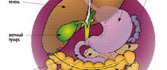
Bile, which is produced in the liver, enters the gallbladder and bile ducts through the hepatic duct. Part of the bile is immediately sent through the common channel to the duodenum. Sometimes there are malfunctions in this system, which can be immediately determined by the characteristic pain in the right hypochondrium. The nature of this pain is related to the type of disease. For example, if there are large stones in the excretory ducts, a person experiences stabbing pain.
Clinical manifestations and symptoms
Manifestations of the disease depend on the type of damage.
1. If bile flows freely into the abdominal cavity through the damaged bile duct
- Pain appears in the right hypochondrium and in the upper abdomen. The pain is constant, dull.
- There is a feeling of heaviness in the stomach.
- Then an infection sets in and the body temperature rises.
- Intoxication and bile peritonitis occur, requiring emergency surgical treatment.
2. If there is no leakage of bile into the abdominal cavity, then as a result of an obstruction (stricture), bile does not enter the duodenum
The pressure in the bile duct itself increases, as a result of which bile enters the blood through the vascular system of the liver and obstructive jaundice develops.
- Patients develop pain and increased body temperature.
- Yellowness of the skin, sclera and visible mucous membranes.
- If treatment for this complication is not started on time, the patient becomes lethargic and apathetic.
- General weakness, confusion and other severe disorders of the basic functions and systems of the body develop.
Complications of strictures and tumors of the bile ducts
- Mechanical jaundice.
- Biliary peritonitis.
- Abdominal abscesses are an accumulation of bile in certain areas of the abdominal cavity accompanied by infection.
- Cholangitis is inflammation of the bile ducts as a result of the action of complex negative factors.
Clinical forms of obstructive jaundice in choledocholithiasis
- Jaundice-pain: the main symptoms are pain, nausea, vomiting, jaundice, fever.
- Icteric-pancreatic: develops when a stone is strangulated in the large duodenal papilla or cicatricial stenosis of the large duodenal papilla. There are symptoms of obstructive jaundice and acute pancreatitis.
- Jaundice-cholecystitis: jaundice develops against the background of an attack of acute cholecystitis.
- Icteric-septic: this form is based on the development of obstructive purulent cholangitis, sometimes complicated by cholangiogenic liver abscesses and sepsis.
- Jaundice-painless: jaundice increases gradually, the patient’s condition suffers little, there is no indication in the anamnesis of a painful attack. Requires differential diagnosis with jaundice of tumor etiology.
Mechanical jaundice. Pancreas cancer
Return to section:
Department of X-ray surgical methods of diagnosis and treatment
Obstructive jaundice - what is it?
Rice. 1 - The anatomical structure of the biliary tract is normal |
First, let me remind you: bile is formed in the liver, after which it enters the duodenum through the bile duct system, where it helps digest food. The gallbladder is a kind of “reservoir” where bile “accumulates” between meals (Fig. 1).
So, any obstacle to the “movement” of bile into the intestine forms a pathological condition called “obstructive jaundice.” Bile begins to accumulate in the bile ducts and gallbladder, enlarging and expanding them. Excess bile is released into the blood, and the body removes it through other routes, including the kidneys and skin. Therefore, one of the initial signs of obstructive jaundice is an increase in bilirubin in the blood, a bile pigment that is the main component of bile. Since bile does not enter the intestines and does not color the stool, it becomes light, even gray. The urine becomes very dark due to increased secretion of bilirubin by the kidneys. The skin turns yellow, and due to an excess of bile salts in the blood, severe skin “itching” may begin.
Thus, “obstructive jaundice” is a pathological syndrome associated with a violation of the outflow of bile, which is externally manifested by yellowing of the sclera and skin, lightening of the stool and darkening of the urine, and can cause weakness, drowsiness, abdominal discomfort and itching. Quickly identifying the cause of this syndrome (that is, making a diagnosis) is the main task of the doctor in this situation.
Obstructive jaundice: causes.
| Rice. 2 The most common causes of obstructive jaundice. |
There are many diseases that cause disruption of the flow of bile.
The main ones are listed in Figure 2.
The method of treatment, its result, as well as the prognosis of the disease (i.e., the expected duration and quality of life with this disease and its "curability").
Unfortunately, in 40-67% of cases, the cause of obstructive jaundice is tumors, and they are benign only in 2-3% of cases.
The most common cause that doctors have to deal with is cancer of the head of the pancreas.
The tumor compresses the duct from the outside, disrupting the outflow of bile. (Fig. 3)
Obstructive jaundice in pancreatic cancer - treatment methods
| Rice. 3 Compression of the common bile duct due to a tumor of the head of the pancreas. |
It is impossible to consider all possible types of treatment for various causes of obstructive jaundice in one article. Therefore, I will dwell in more detail on treatment options for the most difficult category of patients - those with malignant tumors that cause compression of the bile ducts.
The only chance for patients with a malignant tumor causing obstructive jaundice is radical surgery (complete removal or resection of part of the organ), but this is possible in less than 30% of cases. This happens because the disease develops very slowly and begins to “manifest” already at an advanced stage.
In each case, the question of the possibility of complete tumor removal is decided individually; it depends on many factors: the extent of the process, the age of the patient, the presence of concomitant diseases, etc. These operations are considered one of the most difficult in modern abdominal surgery and are performed, as a rule, in specialized departments by experienced oncologist surgeons.
All other methods of treatment - radiation, chemotherapy (so-called dietary supplements, phyto- and homeopathic therapy, I do not consider “treatment” in principle) are ineffective and are aimed only at slowing down the growth of the tumor and improving the “quality of life” of the patient.
Why is obstructive jaundice dangerous?
As I already mentioned, the main parameter that assesses the severity of obstructive jaundice is the level of total bilirubin in the blood. All of the above treatment methods, including radical surgery, with rare exceptions, are possible when the level of total blood bilirubin is below 50-90 µmol/l (normal 3-17 µmol/l) due to the high risk of complications. However, visible jaundice of the sclera and skin occurs, as a rule, when the bilirubin level is above 100-120 µmol/l. At levels above 300-350 µmol/l, bilirubin begins to penetrate the blood-brain barrier, i.e. enter the brain and, with further growth, cause severe intoxication, even death.
According to the literature, in conditions of biliary tract obstruction and inflammation, surgical treatment is risky, accompanied by a large number of complications, and mortality reaches 10-34%, which is 4 times higher than in cases where obstructive jaundice can be eliminated before surgery.
Therefore, one of the first tasks in the treatment of obstructive jaundice is to reduce the level of bilirubin in the blood - to treat intoxication and prepare the patient for one or another type of specialized medical care (surgery, chemotherapy or radiation therapy).
Choosing a treatment method for obstructive jaundice
Conservative therapy (intravenous infusions of drugs) in patients with obstructive jaundice of tumor origin is rarely effective. of decompression of the bile ducts come to the fore - i.e. methods aimed at restoring the outflow of bile from the bile ducts into the digestive tract. Among the surgical methods for treating obstructive jaundice, three main areas can be distinguished:
decompression of the bile ducts – i.e. methods aimed at restoring the outflow of bile from the bile ducts into the digestive tract. Among the surgical methods for treating obstructive jaundice, three main areas can be distinguished:
- Endoscopic techniques for drainage and stenting of the bile ducts.
- Percutaneous transhepatic techniques under ultrasound and fluoroscopy control.
- Open surgery is the creation of an “anastomosis” (i.e., “connection”) between the bile ducts and the intestine, bypassing the tumor.
The latter method is used quite rarely today, as it is associated with a greater number of complications. It is used when it is technically impossible to perform the operation using the first two methods or when there are no specialists of the required profile in the hospital.
The choice between endoscopic (“retrograde”) (Fig. 4) or percutaneous transhepatic (“antegrade”) (Fig. 5) techniques, all other things being equal, largely depends on the specific situation.
| Rice. 4 “Retrograde” stenting of the common bile duct using an endoscope for the treatment of obstructive jaundice caused by compression of the common bile duct by a pancreatic tumor. | Rice. 5. “Antegrade” - percutaneous transhepatic drainage of the bile ducts for obstructive jaundice caused by Klatskin’s tumor |
Thus, for the technical feasibility of percutaneous puncture under ultrasound control, a necessary condition is the expansion of the intrahepatic bile ducts. At the same time, the use of endoscopic techniques in patients who have previously undergone surgery on the stomach or duodenum, as well as with obstructive jaundice caused by a tumor in the “gate” of the liver, is difficult and, at times, impossible.
In 70-80% of patients with obstructive jaundice, it is possible to use both methods of decompression, and then the choice largely depends on how common one or another method is in a particular hospital (technical equipment, experience of a particular specialist, on whom it largely depends percentage of successful interventions and number of complications).
Obstructive jaundice - surgical treatment in St. Petersburg
The State Budgetary Healthcare Institution “City Hospital No. 40” has implemented the ability to provide urgent and emergency medical care to patients with obstructive jaundice of any etiology using all of the listed methods. The availability of the latest equipment and experienced specialists allows us to provide timely, highly qualified medical care to this complex category of patients.
More information about minimally invasive methods of percutaneous transhepatic drainage and stenting of the bile ducts for relieving obstructive jaundice can be found in the second part of the article.
Percutaneous transhepatic drainage and stenting of the bile ducts for the treatment of obstructive jaundice.
As a specialist x-ray surgeon, I would like to dwell in more detail on the technique of percutaneous transhepatic decompression of the bile ducts for the malignant nature of obstructive jaundice.
Percutaneous transhepatic cholangiography – conditions for implementation, advantages and disadvantages of the method.
A necessary condition for performing percutaneous puncture is the expansion of the intrahepatic bile ducts to 3-5 mm. With obstructive jaundice of any etiology, this phenomenon is quite common; if the outflow of bile is disrupted, it begins to accumulate primarily in the ducts, gradually expanding them. If an obstacle (stone or tumor) does not completely compress the common bile duct, i.e. Some bile still flows into the intestine, but this process may take some time.
Fig.1. X-ray operating room.
Advantages of the method:
- Performed under local anesthesia (i.e. does not require general anesthesia)
- In experienced hands, the rate of successful drainage is 98-100% (which exceeds the technical success of endoscopic methods).
- Fewer complications (if the necessary equipment and experienced specialists are available).
Disadvantages of the method:
- It is performed under the control of fluoroscopy (although modern equipment makes it possible to reduce the radiation dose to minimal figures - less than during computed tomography).
- When installing external or external-internal cholangiodrainage, part of the bile flows into a special plastic container, which must be carried with you for 3 to 14 days, which worsens the patient’s quality of life.
In the hospital, patients with obstructive jaundice are admitted to the surgery/oncology departments. As a rule, operations aimed at decompressing the bile ducts are urgent - i.e. urgent enough to avoid complications associated with bilirubin intoxication, but at the same time not performed immediately upon admission of the patient. Usually doctors have 1-3 days to further examine the patient - to determine the cause of jaundice (stone, tumor, stricture), determine the level of bilirubin in the blood, and other tests that need to be taken into account when preparing for surgery.
The purpose of the operation, its risks and possible complications are explained to the patient, and voluntary informed consent to the procedure is signed. A light dinner is allowed the day before, and fasting on the day of intervention.
Percutaneous transhepatic drainage for pancreatic and bile duct cancer.
Rice. 2. Puncture of the bile ducts under ultrasound control on the right in the 8th intercostal space.
Percutaneous transhepatic cholangiodrainage (PTCHD) and stenting operations are performed in a specially equipped cath lab.
The intervention is performed under local anesthesia, usually 20-30 ml of a 1% lidocaine solution. In our hospital, an anesthesiologist-resuscitator is always present in the operating room, who, if necessary, provides intravenous anesthesia.
The puncture site is selected individually, depending on the anatomical structure and location of the obstacle. As a rule, access to the ducts of the right lobe of the liver is carried out from the 7-8 intercostal space along a line drawn perpendicular to the anterior angle of the axilla. Access to the ducts of the left lobe is from under the xiphoid process.
The correct choice of access has the greatest impact on the safety of the technique.
How is bile duct drainage surgery performed?
After treating the skin with an antiseptic solution and anesthesia, the skin at the puncture site is incised with a scalpel to facilitate insertion of the puncture needle. The needle itself has a diameter of less than 1 mm. Under ultrasound or fluoroscopy control, it is carried out to a depth of 5-10 cm until it enters the dilated bile duct.
Several milliliters of a non-ionic iodine-containing contrast agent (Omnipaque, Optireum) are injected through a needle. This is done to ensure that it enters the bile duct and not the liver vessels. A thin soft conductor with a diameter of up to 0.3 mm is inserted through the lumen of the needle, the needle is removed, and a thin plastic catheter (diameter less than 2 mm) is inserted along the installed conductor. Through it, 20-30 ml of contrast agent is injected - the so-called. Cholangiography.
| Rice. 3. Percutaneous transhepatic cholangiography. | Percutaneous transhepatic cholangiography. The following is determined: a) pronounced expansion of the intrahepatic bile ducts; b) complete block in the distal third of the common bile duct (compression of the head of the pancreas by the tumor) |
| Rice. 4. Cholangiography for obstructive jaundice caused by Klatskin tumor. | Cholangiography for obstructive jaundice caused by Klatskin tumor. A pronounced narrowing of the right (a) and left (b) lobar bile ducts is determined due to the germination of cholangiocarcinoma. Tight filling of the bile ducts allows you to accurately determine the level and degree of blockage of the bile ducts, the degree of their expansion, defects in their filling (large stones and intraluminal tumors are visible), as well as determine the tactics and method of further treatment - decompression of the bile ducts. |
| Rice. 5. Cholangiography for intrahepatic cholangiolithiasis | Cholangiography for intrahepatic cholangiolithiasis: a) multiple small concretions (stones) up to 2-3 mm in size inside the dilated bile ducts of the right lobe of the liver; b) benign (post-inflammatory) stricture of the terminal part of the common bile duct; c) entry of the contrast agent into the duodenum through the installed percutaneous transhepatic drainage. Bile obtained during primary puncture of the bile ducts is often taken for culture and determination of sensitivity to antibiotics. This greatly helps to combat such a common complication of obstructive jaundice as cholangitis - i.e. inflammation of the wall of the bile duct. |
Rice. 6 - Drainage tube for percutaneous transhepatic drainage of the bile ducts. | After determining the level of the block, the doctor, using catheters of various shapes and conductors of different stiffness, performs recanalization of the obstacle (the conductor is passed through the stricture or the common bile duct compressed from the outside into the small intestine). A plastic tube with a diameter of about 3 mm with a large number of holes is inserted into the intestine along the guide to restore the outflow of bile - drainage. It is positioned so that the drainage holes are located both before and after the obstacle. Thus, bile enters the drainage tube before the obstruction and exits the openings into the intestine after it. |
For the first 2-3 days, a plastic bag is connected to the outer end of the drainage (in the intercostal space). This allows you to eliminate excess bile located in the ducts and control (in time identify) possible complications, such as hemobilia - bleeding into the bile ducts.
If the obstacle cannot be passed, then drainage is left only for external outflow in order to reduce the level of bilirubin in the blood and its toxic effects. In such cases, the patient has to drink bile (along with juice or water), since with it the necessary liquid and microelements that are necessary for an already exhausted body are lost. After a few days, when the inflammation and swelling of the wall of the bile ducts passes, as a rule, a second attempt is made to pass the obstacle. After installing the drainage in the desired position, it is fixed to the skin with a suture, which reduces the risk of its displacement.
Treatment after a decrease in bilirubin levels. Caring for bile duct drainage.
Rice. 7. External-internal percutaneous transhepatic cholangiodrainage.
The success of drainage largely depends on the availability in the hospital of the entire range of instruments and the experience of the surgeon performing the intervention. In our department, the success of external-internal drainage with restoration of normal passage of bile into the intestine is 98-99%.
In the case where, after a decrease in the level of bilirubin in the blood, radical surgery is possible (i.e., completely eliminating the root cause of obstructive jaundice), the drainage is removed during or after this operation. In cases where the process is inoperable, the drainage is closed after a few days and remains with the patient on an ongoing basis. It must be washed - once a day, by injecting 20 ml of saline solution into the drainage. This is done in order to avoid its rapid “clogging” with bile salts or so-called “sludge” - thick stagnant bile. Patients are prescribed drugs that “thin” bile, such as Ursosan. Despite all these measures, the drainage has to be changed every 4-6 months. This happens quite quickly, since there is no need for repeated puncture of the bile ducts and the drainage channel has already been formed.
However, even the presence of a foreign body in a patient for a long time, even a thin plastic tube without a bag, causes psychological discomfort and reduces the quality of life. By itself, the drainage can shift and cause inflammation when food enters the bile ducts through its openings from the intestine; Bile may leak through the external drainage channel and stain clothing.
Stenting of the bile ducts for obstructive jaundice.
In order to avoid these complications, bile duct stenting surgery was developed in patients with inoperable malignant process (in some cases with other causes of obstructive jaundice). In fact, it is a logical continuation of the drainage operation, and, if possible, is performed on a stable patient with a satisfactory prognosis for survival.
Stenting of the bile ducts is usually performed 1-4 weeks after drainage surgery, after assessing the dynamics of the decrease in bilirubin levels and preparing the patient. It is performed through the same access - a thin conductor is inserted into the intestine through the existing drainage, after which the drainage tube is removed. A special balloon is inserted along this guide, which is positioned inside the stricture (benign or malignant), and opened for a minute for “plasticity” of the common bile duct - i.e. expanding it to allow insertion of a mesh metal structure – a stent – into it.
The diameter of the opened balloon is 6-8mm. The balloon is deflated and removed, and a stent is inserted along the same guidewire.
| Rice. 8. Bile duct stent | Rice. 9. Balloon catheter for “plasty” of common bile duct strictures. | Rice. 10. Balloon plasty of malignant common bile duct stricture before stent placement. A balloon with a diameter of 6 mm (a) is opened on a guide (b) in the area of the stricture. |
The size of the stent is determined in advance, according to the data of cholangiography. Most modern stents are coated with a special material (looks like fabric on the outside). Such stents are called “grafts” and have a much lower percentage of “tumor growth” through it - and therefore relapse of obstructive jaundice.
The stent (like the balloon) is rolled up on a special delivery system, which is quite thin and does not require additional expansion of the channel in which the drainage was previously located.
The stent is inserted and opened in such a way as to block the stricture, but not block the remaining bile ducts.
| Rice. 11. Stent-graft with polytetrafluoroethylene coating for stenting of bile ducts. | Rice. 12. Stent-graft installed in the common bile duct from the confluence of the lobar bile ducts (a) to the duodenum (b) for the treatment of obstructive jaundice caused by metastases of colon cancer to the porta hepatis. |
If necessary, intravenous anesthesia is added at the time of balloon and stent deployment. After installation of the stent, the patient is observed for several days in the surgical department, then, after making sure that there are no complications, he is discharged to continue treatment (chemotherapy, radiation therapy, photodynamic therapy) in a specialized institution or at the place of residence (symptomatic therapy).
Obstructive jaundice is the cost of an operation where drainage and stenting of the bile ducts are performed.
In most hospitals in St. Petersburg, minimally invasive operations are performed for a fee, because require quite expensive consumables and the presence of experienced specialists.
St. Petersburg State Budgetary Healthcare Institution “City Hospital No. 40” provides residents of St. Petersburg with the opportunity to perform such operations free of charge, using coupons for the provision of high-tech specialized medical care within the framework of the compulsory medical insurance program.
Doctor on X-ray endovascular methods of diagnosis and treatment, oncologist Popov V.V.
← Back
Causes of strictures
Most often, there are three groups of causes of the disease:
- traumatic - develop after surgical interventions: cholecystectomy, endoscopic manipulation, resection of the stomach or liver;
- inflammatory - they are caused by cicatricial changes in the walls of the ducts in various diseases (chronic pancreatitis, low duodenal ulcers, parasitic liver diseases, etc.);
- tumor - strictures that occur with cancer of the gallbladder or extrahepatic bile ducts.
Surgical relief of obstruction
If a person is diagnosed with pancreatic cancer, doctors install a stent in the narrowed area of the bile duct. First, specialists insert an endoscope into the esophagus, and then pass it into the stomach and at the very end reach the duodenum. Such actions help to study the blocking site for proper stent placement. Before taking an x-ray, a dye is injected into the patient's body, which will also ensure the correct placement of the device. If obstruction recurs, the medical device is replaced.
Classification of narrowing of the bile ducts
By etiology
- Post-traumatic scar strictures;
- Strictures of biliary anastomoses;
- Primary sclerosing cholangitis;
- Secondary inflammatory strictures;
- Tumor stenoses of the bile ducts 5.1. Bismuth-Corlette classification (for proximal bile duct cancer)
- Type I. Damage to the common hepatic duct
- Type II. Damage to the confluence of the hepatic ducts.
- Type IIIA. Damage to the right hepatic duct.
- Type IIIB. Lesion of the left hepatic duct.
- Type IV. Damage to both hepatic ducts.
- 5.2. Extrahepatic bile duct cancer
- Cancer of the distal common bile duct.
- Cancer of the large duodenal nipple.
According to the degree of narrowing of the ducts
- Complete strictures.
- Incomplete strictures.
- Limited strictures (up to 1 cm).
- Widespread strictures (1 - 3 cm).
- Subtotal strictures (more than 3 cm).
- Total damage to the extrahepatic bile ducts
According to the clinical course
- With external biliary fistula.
- With jaundice.
- With cholangitis.
- With liver and kidney failure.
- With biliary cirrhosis and portal hypertension.
Risk factors
Factors that may increase the risk of gallstones include:
- Female
- Age 40 and older
- Overweight or obesity
- Passive lifestyle
- Pregnancy
- High Fat Diet
- High Cholesterol Diet
- Low fiber diet
- Family history of gallstones
- Diabetes
- Having certain blood disorders, such as sickle cell disease or leukemia
- Very fast weight loss
- Taking medications that contain estrogen, such as oral contraceptives or hormone therapy.
- Liver disease
Treatment of strictures and tumors of the bile ducts
Currently, various types of treatment are used: from drainage to reconstructive operations. However, there is no systematic approach that takes into account both the main characteristics of ductal stenosis and the advantages and disadvantages of a particular surgical treatment technique for a particular patient. Today, an individual approach to each clinical situation is required, allowing the use of both modern minimally invasive techniques and time-tested reconstructive operations.
For non-tumor injuries of the bile ducts, the following types of surgical interventions are distinguished:
- drainage operations (bile diversion to the outside);
- reconstructive operations;
- reconstructive operations.
Drainage operations include: drainage of the common bile duct according to Vishnevsky, Kerta, Halsted, drainage of the proximal end of the crossed duct, percutaneous - transhepatic drainage.
In case of marginal injuries, preference is given to T-shaped drainage compared to other methods, since it has been proven that in choledochotomies completed with T-shaped drainage, neither gross deformations of the hepaticocholedochus nor its stricture at the site of the drainage were detected in the long term.
When the tumor affects the distal part of the common bile duct, preference is given to gastropancreatoduodenal resection.
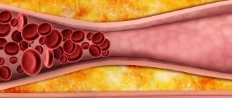
Types of gallstones
Types of gallstones that can form in the gallbladder include:
Cholesterol stones in the gall bladder. The most common type of gallstones, called cholesterol gallstones, are often yellow in color. These gallstones are composed primarily of undissolved cholesterol, but may contain other components.
Pigment stones in the gall bladder. These dark brown or black stones form when your bile contains too much bilirubin.
Treatment in the department of thoracoabdominal surgery and oncology of the Russian Scientific Center for Surgery
Treatment of tumors and strictures of the bile ducts in our department is carried out within the framework of the compulsory medical insurance , voluntary medical insurance, VMP , as well as on a commercial basis . Find out how to get treatment at the department of thoracoabdominal surgery and oncology of the Russian Scientific Center for Surgery.
We provide surgical treatment with highly qualified doctors , the best modern equipment and attentive staff. Read patient reviews here .
To schedule a consultation, call:
+7 (499) 248 13 91 +7 (903) 728 24 52 +7 (499) 248 15 55
Submit a request for a consultation by filling out the form on our website and attaching the necessary documents.