Protein in urine
– a serious disorder in the body. A healthy person does not have an increased amount of protein in the urine. If the concentration is no more than 0.03 g/l, as well as if there are traces in the physiological fluid, there is no need to sound the alarm, but if, after taking tests, the numbers exceed this norm, you should urgently consult a specialist. Before taking tests, do not use acetazolamide, colistin, aminoglycoside and other drugs.
Urologist
is a doctor who treats problems of the genitourinary system. In the modern world, especially with an urban lifestyle, many people suffer from such diseases. But urology has stepped far forward; it promptly recognizes the prerequisites for the disease, conducts a high-quality examination and prescribes adequate treatment, which quickly relieves a person of discomfort and inconvenience, returning him to a healthy life.
Causes of protein in urine
Often protein in urine
found in pregnant women. There is no need to worry too much, but it is better to consult a doctor. Typically, protein appears due to stress or hormonal imbalances, as well as due to an increase in the size of the uterus. In addition, protein is noticed after eating raw eggs or fresh dairy products.
You should not eat such foods before taking tests. In all other cases, the cause is a dysfunction of the kidneys and organs that accumulate and excrete urine:
- pathologies;
- cancers of the kidneys and urinary tract;
- concussion;
- epilepsy;
- consequence of stress;
- hypothermia.
Increased amounts of protein are also seen in athletes due to protein intake and heavy exercise. Follow the recommendations of doctors: you should go for an examination with a urologist at least once a year, undergo an ultrasound of the kidneys, do not have sex with casual partners, eat a balanced diet, lead an active lifestyle and avoid stress.
You can make an appointment with a urologist from our consultants by calling +7 (495) 125-49-50
Prices for urologist services Addresses of clinics Ultrasound of the prostate Paid urology Blood tests for PSA Call a urologist at home
References
- Nastausheva, T.L., Sitnikova, V.P., Shvyrev, A.P. and others. Proteinuria in children and adolescents: genesis, diagnostic algorithm, principles of therapy. Nephrology, 2011. - No. 2.
- Trukhan, D.I., Bagisheva, N.V., Goloshubina, V.V. and others. Differential diagnosis of urinary syndrome: proteinuria. International Journal of Applied and Documentary Research, 2016. - No. 12(7). — P. 1203-1207.
- Baranov, A.A. Clinical guidelines for providing medical care to children with nephrotic syndrome, 2015. - 11 p.
Why is protein in urine dangerous?
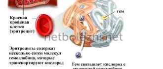
According to the physiological structure, the pattern of pathological deviation, indicating the appearance of protein in the urine, signals a reaction of its increased utilization from cells and tissues. This phenomenon is detected when the filtering ability of the kidney tissue membranes is impaired. Together with protein, red blood cells can be washed out of the bloodstream, leading to signs of anemia and a change from straw-colored urine to bloody.
Since the participation of protein structures in the functional activity of almost all areas of the body is vital, including the stabilization of protective parameters, allergenic and infectious resistance, ensuring hormonal balance, etc., their significant loss has negative consequences.
The lack of a sufficient level of protein in the bloodstream has a negative impact both on the functionality of individual structures of the internal space and on the activity of entire systems, leading to disruption of the homeostasis of the entire organism. This threatens to slow down all restoration functions in organs and systems, significantly delaying the healing process.
General urine analysis (with sediment microscopy)
IMPORTANT!
The information in this section cannot be used for self-diagnosis and self-treatment.
In case of pain or other exacerbation of the disease, diagnostic tests should be prescribed only by the attending physician. To make a diagnosis and properly prescribe treatment, you should contact your doctor. We remind you that independent interpretation of the results is unacceptable; the information below is for reference only.
General urine analysis (with sediment microscopy): indications for use, rules for preparing for the test, interpretation of the results and normal indicators. Indications for the purpose of the study
A general urine test refers to routine laboratory tests aimed primarily at screening for diseases of the urinary system, since pathological processes in the kidneys and urinary tract affect the properties of urine.
With this simple diagnostic test, infectious and inflammatory diseases such as glomerulonephritis
(inflammation of the renal glomeruli),
pyelonephritis
(inflammation of the renal pelvis),
cystitis
(inflammation of the bladder).
Microscopy of urine sediment allows one to suspect injury or infarction of the kidney, urolithiasis, some neoplasms, renal amyloidosis (a systemic disease in which a specific insoluble protein is deposited in the kidneys, which impairs the functioning of the organ).
In addition to diagnosing kidney and urinary tract diseases, the results of a general urine test with sediment microscopy can provide information about your general health.
Urine is formed as a result of ultrafiltration of blood plasma through the glomeruli of the kidneys. With the development of various diseases, pathological metabolic products enter the blood and are excreted from the body, including through the kidneys.
Preparation for the procedure
Preparation for a general urine test begins the day before the collection of biomaterial. Certain foods, the amount of fluid you drink, taking medications and dietary supplements, and intense physical activity can distort the results of the study.
The day before collecting urine, you should avoid foods that can affect the color of your urine: for example, beets and blueberries give your urine a reddish tint; consuming large amounts of carrots or carotene supplements may change the color of your urine to orange.
On the eve of urine collection, it is not recommended to drink alcohol, coffee, dietary supplements or strong tea. If possible, you should limit your intake of diuretics (diuretics). It is necessary to exclude serious physical activity, as well as visiting a bathhouse or sauna.
Women during menstruation are not recommended to donate urine for testing, since even a small amount of blood will significantly distort the test result.
You should warn your doctor about the medications you are taking, as well as about invasive examinations (for example, cystoscopy) on the eve of the test.
Method of collecting urine for general analysis
- It is necessary to prepare in advance a disposable sterile container for collecting urine (can be purchased at a pharmacy or taken from the INVITRO medical office).
- Before collecting urine, hygienic treatment of the external genitalia should be carried out, without using antibacterial and disinfectants.
For children, you need to adhere to the following rules: girls are washed from front to back (from the pubis to the tailbone) so that bacteria that populate the intestines do not enter the urinary tract. Only wash the skin with soap, since contact with mucous membranes causes irritation, dryness and itching. In boys, the glans penis is fused with the foreskin (physiological phimosis), so it is not recommended to forcefully open the glans penis, as this leads to trauma to the delicate tissue. You just need to slightly pull the skin and rinse with water, but it is unacceptable to direct a stream of water into the opening of the urethra. - For general analysis, as a rule, the first morning urine sample is collected. First, release a small amount of urine into the toilet, then, without interrupting urination, place a container and collect approximately 50 ml of urine. In this case, it is necessary to ensure that the container does not touch the skin and mucous membranes.
- After collecting urine, close the container tightly with a screw cap.
- Special urinals have been developed for newborns and infants. You should not use urine squeezed out of a diaper or diaper - the results will be unreliable, since the diaper is a kind of filter for microscopic elements of urine, which are counted during the study.
- When taking the test during the day, it is not recommended to drink large amounts of water, tea, coffee or diuretics to stimulate urination.
The turnaround time for a general urine test is usually 1 business day. What may affect the results
Factors that may distort the results of the study:
- Violation of hygiene procedures and urine collection techniques.
- Drinking large or small amounts of water.
- Consuming foods, medications, or dietary supplements that change the color of urine.
- Menstruation.
- High blood pressure.
- Intense physical and psycho-emotional stress on the eve of urine collection.
- Visiting a bathhouse, sauna, hypothermia.
- Carrying out invasive procedures on the urinary tract a week before the test.
take a general urine test
at the nearest INVITRO medical office. A list of offices where biomaterial is accepted for laboratory research is presented in the “Addresses” section.
Urine examination includes the study of physical and chemical properties, as well as microscopy of sediment.
Physical properties: quantity, color, odor, transparency, relative density (specific gravity), urine reaction (pH).
Chemical properties: determination of protein, glucose, ketone bodies, urobilinogen, bilirubin, hemoglobin, nitrites, leukocyte esterase.
Microscopy: identification of erythrocytes, leukocytes, cells of flat, transitional and renal epithelium, cylinders, crystals, mucus, bacteria, fungi.
Normal indicators
| Index | Result |
| Quantity | 50 ml |
| Color | Colorless, light yellow, straw yellow, yellow, amber yellow |
| Smell | Odorless or non-specific |
| Transparency | Transparent |
| Relative density of urine (specific gravity) | 1003-1035 |
| Urine reaction (pH) | 5.0-8.0 (in children under 1 month – 5.0-7.0) |
| Protein | > 0.140 g/l |
| Glucose | > 2.8 mmol/l |
| Ketone bodies | > 1 mmol/l |
| Urobilinogen | > 34 mmol/l |
| Bilirubin | Not detected |
| Hemoglobin | Not detected |
| Leukocyte esterase | Not detected |
| Nitrites | Not detected |
| Red blood cells | Up to 2 cells per field of view |
| Leukocytes | Up to 5 cells in field of view |
| Epithelium | Up to 5 squamous epithelial cells per field of view |
| Cylinders | Not detected |
| Crystals | Little or no detectable amounts of urates, calcium oxalates, amorphous phosphates |
| Slime | In small quantities |
| Bacteria | Not detected |
| Fungi | Not detected |
Interpretation of indicators
It should be remembered that a general urine test is a screening test, so its results can be used when prescribing other laboratory and instrumental examinations to clarify the diagnosis.
Urine color
depends on the concentration of substances dissolved in it and ranges from transparent to amber-yellow.
Under normal conditions, urine is colored by products of pigment metabolism (in particular, bilirubin): urochromes, urobilinoids and other substances. When the level of bilirubin in the blood increases, more of it enters the urine and gives it a rich brownish or even greenish-brown color. When erythrocytes (red blood cells), myoglobin (the main protein of muscle tissue) or hemoglobin (the protein contained in red blood cells) enter the urine, its color changes to brownish-red and takes on the appearance of “meat slop.” Taking vitamins and nitrofuran drugs can give urine a lemon-yellow to orange color. When there are a large number of leukocytes (white blood cells), the urine becomes milky in color (a condition called pyuria).
Transparency.
Under normal conditions, urine is clear. Its cloudiness can be caused by the presence of salts, crystals, cellular elements (erythrocytes, leukocytes).
Smell
. Normally, urine has a weak, non-specific odor. The appearance of an ammonia odor may be a sign of a bacterial infection; a peculiar fruity odor (“rotting apples”) appears with an increase in the concentration of ketone bodies (which most often indicates diabetes mellitus - a disorder of glucose metabolism).
Relative density of urine
, or specific gravity, is determined using a urometer. The relative density of urine gives an idea of the concentrating ability of the kidneys and the dilution function, which are reduced, like the relative density of urine, in renal failure.
Urine reaction (pH)
- a pH value that reflects the ability of the kidneys to maintain the acid-base balance of the body. The kidneys are involved in the excretion of hydrogen ions and bicarbonates, maintaining a constant blood pH. The pH value of urine is greatly influenced by diet, metabolic characteristics, infectious and inflammatory processes in the kidneys and urinary tract.
Protein
in urine is a significant marker in the diagnosis of diseases of the kidneys, urinary tract and cardiovascular system; it is also important in the diagnosis of gestosis, a severe complication of pregnancy. The appearance of protein in the urine is called proteinuria. Normally, urine does not contain protein because the kidney filter prevents the release of protein molecules from the blood into the urine. There are several causes of proteinuria.
- Prerenal causes: intense physical and psycho-emotional stress, heart failure, hypertension, fever, gestosis during pregnancy, nephroptosis, forced prolonged standing (often with hairdressers, surgeons, military personnel), injuries, disturbance of the protein composition of blood plasma.
- Renal causes: glomerular damage (glomerulonephritis and glomerulopathy), tubular damage, nephrosclerosis.
- Postrenal causes: infectious and inflammatory processes in the urinary tract, neoplasms.
Urine glucose.
The appearance of glucose in the urine depends on its concentration in the blood. Normally, glucose is completely reabsorbed from primary urine into the bloodstream if its concentration in the blood has not reached the “renal threshold” - 8.8-10.0 mmol/l. An increase in the level of glucose in the urine is possible due to a number of physiological reasons: errors in the diet (abuse of sweet foods, especially on the eve of collecting biomaterial), prolonged fasting, stress.
The appearance of glucose in the urine serves as a signal indicating pathology of the kidneys, endocrine system, side effects of drugs, poisoning, and complicated pregnancy.
Ketone bodies
are a non-specific indicator. The appearance of an increased amount of ketone bodies in the urine is the result of accelerated fat metabolism or decreased carbohydrate metabolism. Most often, an increase in their level is observed during fasting, fever, vomiting, alcohol intoxication and diabetes mellitus.
Urobilinogen
in urine increases in diseases of the intestines, liver, and hemolytic conditions (destruction of red blood cells).
Bilirubin
appears in the urine in case of liver pathologies, infectious diseases and pigment metabolism disorders.
Hemoglobin
is determined by a large number of red blood cells in the urine, with myositis, extensive injuries to muscle tissue, and thrombosis of muscle vessels.
Nitrites
detected in urine when pathogenic microflora is activated in the urinary system.
Increased red blood cell
observed in the following cases:
- Inflammatory diseases of the kidneys and urinary tract of infectious and non-infectious origin (glomerulonephritis, pyelonephritis, nephritis, cystitis, prostatitis, tuberculosis).
- Urolithiasis disease.
- Traumatic damage to the kidneys and urinary tract.
- Fever.
- Arterial hypertension with involvement of the renal vessels.
- Various blood coagulation disorders (hemophilia, thrombocytopenia, overdose of anticoagulants).
- Poisoning with benzene derivatives, aniline, snake venom, poisonous mushrooms, with intolerance to anticoagulant therapy.
- Tumor diseases of the genitourinary system.
The presence of leukocytes
in an amount of more than five cells in the field of view is noted in the following cases:
- Inflammatory diseases of the kidneys and urinary tract of infectious and non-infectious nature (glomerulonephritis, pyelonephritis, tubulointerstitial nephritis, cystitis, urethritis, tuberculosis).
- Urolithiasis disease.
- Renal transplant rejection.
- Systemic inflammatory diseases of non-infectious etiology (nephritis with systemic lupus erythematosus).
Epithelium
. There are cells of squamous, transitional and renal epithelium. A large amount of epithelium indicates increased desquamation of cells of the mucous membrane of the urinary tract when they are traumatized (by a stone, during an inflammatory process).
Cylinders
are formed in the tubules of the kidneys and allow us to determine the level of their damage. Most often found in glomerulonephritis.
Crystals
are detected in the sediment of salts at a certain pH of urine. Most often (though not always) found in patients with urolithiasis.
Slime
Normally, it can be found in urinary sediment in small quantities. An increase in mucus content may be associated both with an inflammatory process in the urinary tract and with errors made when collecting urine for research.
Bacteria and fungi
Normally they are not found in urinary sediment. Their presence indicates the presence of an infectious process in the kidneys and urinary tract or errors made during the collection of biomaterial for research.
If the general urine analysis indicators deviate from the norm, the following instrumental examinations and laboratory tests may be additionally prescribed:
- Comprehensive ultrasound of the urinary system (kidneys, ureters, bladder).
- Comprehensive ultrasound of the abdominal organs (liver, gall bladder, pancreas, spleen) for suspected diseases of the liver, gall bladder, etc.
- Study of biochemical blood test parameters (total protein, protein fractions, C-reactive protein, ALT, AST, LDH, GGT, total and direct bilirubin, cholesterol, creatinine, glucose, electrolytes: potassium, sodium, chlorine, calcium).
- Determination of fasting blood glucose, conducting an oral glucose tolerance test, studying the level of glycated hemoglobin for diagnosing diabetes mellitus.
- Urine culture for microflora and determination of sensitivity to antimicrobial drugs.
- Urine culture for microflora and determination of sensitivity to an extended range of antimicrobial drugs.
If the indicators of a general urine test change, you may need to consult a pediatrician or general practitioner, as well as a nephrologist.
Sources:
- Pediatric nephrology: Textbook / ed. P.V. Shumilova, E.K. Petrosyan, O.L. Chugunova. – M.: MEDpress-inform, 2021. – 616 p.: ill.
- Grebenev A.L. Propaedeutics of internal diseases: Textbook. – 5th ed., revised. and additional – M.: Medicine, 2001. – 592 p.: ill.
- Danilova L.A. Tests of blood, urine and other biological fluids at different age periods. – St. Petersburg: SpetsLit, 2014. – 111 p.
- .
IMPORTANT!
The information in this section cannot be used for self-diagnosis and self-treatment. In case of pain or other exacerbation of the disease, diagnostic tests should be prescribed only by the attending physician. To make a diagnosis and properly prescribe treatment, you should contact your doctor.
Symptoms of the disease
If a person has increased protein in the urine
, this indicates a urological disease. If you experience the following symptoms, you should contact a highly qualified specialist:
- fast fatiguability;
- bone pain;
- dizziness;
- fatigue, confusion and drowsiness;
- change in urine color - it becomes more whitish;
- chills and fever are symptoms that occur when there is a high level of protein.
Newborns also have increased protein, but don’t be alarmed, this is how it should be. After all, its absence is also wrong. Therefore, protein should be monitored for both children and adults and consult a doctor at the first symptoms.
How to treat high protein?
When protein is detected in the urine, this is not a disease, but only a sign of it. Therefore, before prescribing certain therapeutic measures, the doctor must discover the original cause of proteinuria. If the cause is diabetes, the doctor will treat diabetes. If the cause is kidney disease, the doctor will specify the disease (glomerulonephritis, pyelonephritis) and prescribe appropriate treatment.
The patient’s task is to seek medical help in a timely manner and not allow the pathological process to worsen its course.
An unambiguous positive addition to the successful treatment of proteinuria should be a balanced, nutritious diet, with the exclusion or limitation of salt, spicy seasonings, sugar, and alcohol. In no case should you completely exclude protein: the main thing is not to abuse it. Try to maintain a balance of carbohydrates, proteins and fats in your diet. Only a balanced diet will facilitate the functioning of the kidneys and allow for faster restoration of impaired functions.
Avoid hypothermia, injuries, stressful situations. Drink more clean water and herbal teas. Cranberry tea or fruit drink, which is consumed with honey throughout the day, has a particularly good effect on the urinary system.
Physiological limit of normal
With a healthy functional state of the body, in men and women, the quantitative content of protein in the urine reaches 0.14 g per liter of liquid, without indicating kidney dysfunction. If the value exceeds the threshold of 0.33 g/dm3, the development of a pathological deviation in the form of a disease is observed, the current indicator of which is proteinuria.
The pathology can be mild, moderate or severe. In the children's age category, the norm of proteins in the urine can be 0.036 g per liter; its increase to 0.1 g/dm3 diagnoses a moderate form of proteinuria. During gestation, the normalized threshold for protein in urine shifts to 0.03 g per liter. A more significant increase indicates the development of a pathological disorder in the urinary system or urine disposal.
An indicator of a shift in the protein normal limit upward is various pathologies, or a temporarily manifested deviation that is transient in nature. This form of proteinuria is observed during a fever or significant loss of water, stress, burns or prolonged hypothermia. In men, the presence of protein in the urine is observed during strenuous physical activity.
Correction
Conservative therapy
There are no independent methods for correcting protein loss in urine. It is necessary to treat the underlying disease. Short-term proteinuria resolves on its own and does not require any therapy. Orthostatic proteinuria in the vast majority of children disappears upon the onset of puberty, sometimes persisting until 18-20 years of age.
Patients with diabetes are prescribed a strict diet with limited foods high in easily digestible carbohydrates and animal fats. In case of interstitial nephritis provoked by taking nephrotoxic medications, their immediate withdrawal is necessary. Also, for various pathologies that cause proteinuria, the following medications are used:
- Insulin and hypoglycemic drugs.
For type 1 diabetes, daily injections of short- and long-acting insulin are required. For type 2 diabetes, hypoglycemic agents are prescribed - biguanides (metformin), sulfonylurea derivatives (glibenclamide), DPP-4 inhibitors (vildagliptin). - Antibacterial drugs.
For pyelonephritis, the drugs of choice are penicillin antibiotics (amoxicillin) and cephalosporins (ceftriaxone). For cystitis, fosfomycin trometamol is effective. - ACE inhibitors.
This group of drugs (lisinopril, perindopril) has a nephroprotective effect and is prescribed to all patients with nephrotic syndrome, especially to patients with diabetic nephropathy. - Glucocorticosteroids.
Hormonal agents (prednisolone, methylprednisolone) have anti-inflammatory and immunosuppressive effects. They are used in the treatment of glomerulonephritis and almost any rheumatological pathology. - Cytostatics.
Cytostatic drugs (azathioprine, cyclosporine) are used for severe forms of glomerulonephritis, necrotizing vasculitis, when steroid monotherapy is ineffective. - Chemotherapy.
Patients with confirmed paraproteinemia are shown courses of chemotherapy. Combinations of alkylating drugs (chlorambucil), nucleoside analogues (fludarabine) and monoclonal antibodies (rituximab) are prescribed. If the above agents are ineffective, thalidomide and bortezomib are used.
Surgery
For kidney cancer or polycystic disease, the main type of treatment is surgery (laparoscopic or open) - partial nephrectomy or total nephrectomy. Some patients with Waldenström's macroglobulinemia or multiple myeloma are candidates for hematopoietic stem cell transplantation.
Methods for laboratory detection of pathology
Proteinuria is diagnosed based on laboratory confirmation of the quantitative excess of the permissible threshold of protein present in the urine. The technique is based on its molecular weight, which is used to evaluate the filtering parameters of the kidney membranes. An overestimated value of the molecular weight of proteins signals serious damage to the renal tissue with a violation of its functional ability.
According to the laboratory conclusion of the presence of protein and leukocytes in the urine, an inflammatory process is diagnosed, and a combined increase in the concentration of proteins and the presence of red blood cells indicates traumatic damage to the tissues of the urinary tract. There is a significant variety of methods for quantitative and qualitative determination of protein in urine; the use of a specific one is determined by the doctor, depending on the individual parameters of the current pathology indicators.
Modern medicine makes it possible to treat diseases of the genitourinary system quickly and very efficiently. Clinics use many examination methods. Among them are the Bence-Jones method, and the method for determining protein breakdown products, and indicator paper, and the unified Brandberg-Roberts-Stolnikov method, as well as the Biuret method and photoelectrocolorimeter.
Classification of proteinuria
If normally there can be no more than 0.033 g/l of protein in the urine (and more often in the analysis they write “protein - negative”), then depending on its concentration, the following levels of proteinuria are distinguished:
- traces of protein in urine: 0.02-0.033 g/l;
- microalbuminuria: from 30 to 300 mg of protein is released per day;
- mild proteinuria: 300 mg – 1 g per day;
- moderate degree: 1-3 grams per day are lost in urine;
- severe proteinuria (when there is high protein in the urine): when more than 3.0 g/day is excreted.
The reasons for protein in the urine can be different - from physiological to indicating gross pathology. Proteinuria (that’s what this condition is called) develops with diseases of the urinary system, as well as structures located outside it. Depending on the level of damage, different types of proteinuria are distinguished:
- The tubular form is observed when the tubules are inflamed, which now stop catching and returning low molecular weight proteins.
- If the renal filter is damaged, proteinuria is called glomerular. “Large” – high molecular weight – proteins end up in the urine.
- If the first two types are caused by kidney pathology (renal), then with increased tissue breakdown or the appearance of pathological proteins in the blood, a prerenal form of proteinuria is observed.
- Postrenal proteinuria occurs when the urinary tract is damaged or inflamed. Then leukocytes and casts - protein carriers - will be released into the urine, as well as protein directly from decaying tissues.
Can there be protein in a child's urine?
The function of a child’s kidneys is to filter the contents of the bloodstream from toxic and unnecessary components, the molecular size of which is definitely small.
Such substances include:
- uric acid;
- urea;
- indican;
- ammonium salts;
- creatinine, etc.
At the same time, useful and necessary components of the blood, namely glucose and amino acids, are absorbed back through the membrane of the renal tubules at the primary stage of filtration of urine, which forms plasma, in the absence of proteins with high molecular weight. During a daily interval, about 50 dm3 of primary urine is transported through the kidneys of a newborn, but secondary fluid is released through the system, determining the components of diuresis per day.
In an adult, about 180 dm3 of fluid is utilized per day at the primary stage of filtration, while the total volume of diuresis per day averages two liters. In childhood, this figure depends on the general health of the child, his weight and surface area. If the child’s condition is good and the child is in full health, there is completely no protein in his urine, but even minor traces of up to 0.03 g per liter are not an indicator of the development of pathology.
Preparation
In order to reduce the likelihood of receiving false data, you need to properly prepare for the analysis. The day before collecting urine, you should completely exclude foods that can change the color of urine from your diet. You also need to minimize the amount of fatty, smoked and very sweet foods. It is also necessary to give up alcohol and medications, including vitamins and dietary supplements.
Only the morning average portion is suitable for analysis. It should be collected in sterile disposable containers. They can be bought at a pharmacy very cheaply. Special containers are always available in stock. Urine should not be stored for more than two hours, otherwise the reliability of the results will be reduced. It is important to exclude any foreign substances from entering the urine that will be examined.
Treatment and prevention
The primary task of any person is timely monitoring of his own well-being and the signs that the body signals about existing pathological problems. If they are detected, it is recommended to visit a urologist and undergo the necessary types of diagnostic confirmation of the well-being of the body.
This approach will help to establish the root cause of detecting protein in the urine and select the optimally effective method for rapid recovery, completely eliminating the current pathological problem. Against the background of diabetes mellitus with proteinuria, the doctor will recommend adjusting the diet; with an increased level of vascular pressure, constant monitoring, taking stabilizing drugs, as well as limiting the consumption of sugar, salt and protein-rich foods is necessary.
A conservative form of normalizing the protein threshold in urine includes bed rest, a certain diet and corrective drug therapy, namely corticosteroids, antirheumatic drugs, ACE inhibitors, cytostatics and others.
When diagnosing protein in the urine, indicating the development of an inflammatory process, the formation of solid conglomerates, or an existing congenital defect in the development of the kidneys, it is necessary to be systematically observed by a specialized specialist.
Pathological factors
There are also pathological reasons for detecting protein in the urine. Among them are:
- Intoxication of the body;
- Endocrine diseases: diabetes mellitus;
- Obesity of the third or fourth degrees;
- Acute inflammation of the appendix of the cecum;
- Hypertension of the second and third stages, when kidney damage is present;
- Severe hypersensitization of the body: Quincke's edema, anaphylactic shock and others;
- Systemic administration of certain groups of drugs: cytostatics, antibiotics and others;
- Infectious diseases that occur with fever: ARVI, influenza, pneumonia and others;
- Malignant diseases: leukemia, myeloma, bladder or kidney cancer;
- Diseases of a systemic nature: systemic lupus erythematosus, scleroderma, rheumatoid arthritis and others;
- Diseases of the urinary system: glomerulonephritis, urolithiasis, kidney injuries, pyelonephritis, inflammation of the prostate gland, specific kidney damage and others.
Protein in the urine in men most often appears due to inflammation of the prostate gland or urethra. In this case, you need to see a urologist.
As you can see, there are many reasons why protein appears in the urine. And since proteinuria is just a symptom of a particular disease, treatment will be selected individually for each patient.
Help from our clinic specialists
The main thing to do is to monitor changes in your body and detect problems in a timely manner. Then consult a doctor who will conduct a thorough objective examination,, if necessary, prescribe additional tests, and tell you about the causes in order to eliminate not only the symptoms, but also the core of the disease.
Multidisciplinary specialists in Moscow are always happy to provide the required range of specialized care for patients of any age category, including consultation with a urologist and laboratory testing of urine. The clinic has several branches geographically scattered throughout the city, which allows you to choose the most suitable location for your visit.
| Prices for urologist services in Moscow | |
| Initial appointment with a urologist | 900 rubles |
| Repeated appointment with the urologist | 700 rubles |
| Calling a urologist to your home | 2,800 rubles |
| Kidney ultrasound | 1,000 rubles |
| Urine protein test | 600 rubles |
This article does not constitute medical advice and should not serve as a substitute for consultation with a physician.