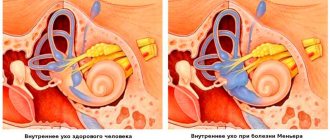
An important part of the inner ear is the labyrinth. It is located deep in the bone and is represented by interconnected fluid-filled channels. The labyrinth consists of the cochlea, which perceives sound vibrations and transforms them into electrical impulses, and the vestibular canals, which are responsible for the sense of balance. With labyrinthitis, hearing and/or vestibular function is impaired.
A dangerous complication of this disease is meningitis. Therefore, the symptoms of labyrinthitis are a reason to immediately contact an ENT doctor for treatment.
Causes
Often the cause of the disease remains unclear. It is believed that it can be caused by pathogenic viruses, such as influenza, or ARVI pathogens. Symptoms of labyrinthitis may be associated with infection with mycobacteria, streptococci, and meningococci.
Other possible causes of aseptic (microbial-free) inflammation of the inner ear:
- bruise, head injury, concussion;
- allergic reaction, for example, with hay fever;
- alcohol abuse;
- benign neoplasm of the middle ear (cholesteatoma);
- taking certain medications, such as aspirin or furosemide, in large doses.
Symptoms of labyrinthitis may resemble tumors of the base of the brain, strokes, or impaired blood supply to the inner ear due to atherosclerosis.
Microbial toxins or other damaging factors destroy the sensitive cells lining the labyrinthine channels from the inside, and the partitions between them. At the same time, the production of lymph located inside the cochlea increases, swelling develops, and serous inflammation occurs.
In the chronic course of the disease, the bone walls of the inner ear are destroyed, and the infection can easily penetrate the blood vessels and brain tissue. Sometimes such a process is limited to the bone shaft, and then labyrinthitis is called limited.
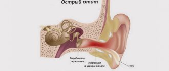
Acute otitis media of the middle ear: what causes inflammation?
The inflammatory process is always caused by pathogenic microorganisms, which means that the prerequisites for their activation must be present in the body. The causes of otitis media are:
- hypothermia;
- diseases caused by infection (flu, ARVI, measles);
- inflammatory processes of the ENT organs (the tympanic cavity is connected to the nasopharynx via the Eustachian tube, it is not surprising that the infection from the nasopharynx easily penetrates into the middle ear);
- improper nose blowing;
- hypertrophy of adenoid vegetations;
- rhinitis, sinusitis;
- allergic reactions;
- deviated nasal septum;
- foreign object in the ear;
- damage to the hearing organ.
Symptoms
The most common symptoms of labyrinthitis:
- dizziness, nausea and vomiting that do not bring relief;
- loss of balance, unexpected falls;
- moderate headache, ringing or rustling in the ear, hearing loss;
- nystagmus - movements of the eyeballs towards the lesion in the serous form or towards the healthy side - in the purulent version of the course.
These signs intensify when moving the head, turning and looking up. They may persist for several days or even weeks depending on the severity of the disease. Even after treatment for labyrinthitis has been started, symptoms may recur, so the patient should be careful when driving and working at heights for at least a week after recovery.
Cases requiring urgent consultation with a specialist and treatment of labyrinthitis:
- vomiting, making it difficult to eat, drink, or take medications;
- fever, ear pain, progressive hearing loss;
- severe headache or dizziness that lasts for several hours or more;
- double vision, speech impairment, weakness of facial or skeletal muscles;
- recent ear or head injury.
Cardiological pathology
5. Cardiac arrhythmia.
Dizziness is not always a sign of brain disease. It is often observed in patients with cardiac arrhythmia. The danger of cardiac arrhythmia lies in the fact that when the heart muscle works irregularly, the release of blood into the aorta and, consequently, into the arteries extending from it, carrying blood to the brain, decreases. Due to insufficient blood supply to the brain, a feeling of dizziness and lightheadedness occurs. What to do?
It is necessary to examine the heart. Sometimes cardiac arrhythmia is not visible with a regular ECG and it is necessary to monitor heart rate and blood pressure for a day (and sometimes more) to “catch” the arrhythmia. But with properly selected antiarrhythmic therapy (and sometimes after low-traumatic cardiac surgery), the patient begins to feel much better - performance increases, dizziness stops.
Kinds
Depending on the structural changes, the following types of pathology are distinguished:
- serous: fluid production in the labyrinthine channels increases, pressure increases; these changes are reversible if treatment for labyrinthitis is started in time;
- purulent: occurs with microbial damage, the fluid in the labyrinth becomes purulent, the disease is more severe, prone to chronicity;
- necrotic: characterized by the breakdown of the bone tissue of the inner ear with irreversible changes in hearing and vestibular function, most characteristic of scarlet fever.
Depending on the cause of the disease, the following variants of the disease are distinguished:
- bacterial - develops when an infection occurs from the middle ear with otitis (otogenic) or from the skull with meningitis (meningogenic); symptoms of labyrinthitis increase gradually, and if the infection gets directly from the skull, acute dizziness, nausea and other symptoms occur;
- hematogenous - caused by the entry of the pathogen or its toxins from the blood into the inner ear, found in measles, mumps (mumps);
- viral – characterized by a favorable prognosis, caused by influenza viruses, rubella, herpes, measles, hepatitis, Epstein-Barr virus;
- traumatic - occurs with fractures of the base of the skull, head injuries, as well as with unsuccessful operations on the middle ear.
The most common form of labyrinthitis is viral. This disease usually develops in adults aged 30–60 years and is rarely seen in children. In the group of patients under 2 years of age, purulent labyrinthitis of meningogenic origin is more common. Otogenic purulent labyrinthitis is observed in people of any age if they have cholesteatoma or otitis media. If the child’s illness is not caused by meningitis, then it has a milder serous nature and a favorable course.
Risk factors
Factors that increase the risk of developing labyrinthitis:
- decreased immunity;
- anatomical features of the structure of the ear cavity;
- frequent inflammatory processes in the middle ear.
There are 2 types of labyrinthitis - purulent and non-purulent. The first is a dangerous condition with serious consequences, the second is much easier.
And finally, like many other diseases, inflammatory processes in labyrinthitis can be acute or chronic.
Diagnostics
The success of treatment of labyrinthitis depends on its timely diagnosis. The doctor asks the patient about how long ago the symptoms appeared, their connection with head movements, medications taken, and concomitant diseases. Then a neurological and ENT examination, otoscopy are performed, and hearing acuity is assessed.
If necessary, additional research methods are prescribed - computer or magnetic resonance imaging of the skull.
The main task of diagnosis is to exclude a transient ischemic attack or stroke.
What can you see on magnetic resonance imaging of the ears?
What does a magnetic resonance imaging scan of the ears show:
- ear structure;
- anatomical features;
- foci of inflammation, tumors;
- condition of the vestibulocochlear (VIII pair of cranial nerves) and facial (VII pair) nerves;
- neighboring structures of the head - eyes, temporal bones, cerebellopontine angle, blood vessels.
MRI of the inner ear allows you to detect minor changes, the initial forms of diseases. Such information makes it possible to carry out timely treatment and stop the disease at the very beginning of its development.
MRI of the ear with contrast allows you to look at the inner ear in more detail and assess the extent of its damage. The method is based on the ability of the metal gadolinium, which is part of contrast agents, to give an enhanced signal from the tissues in which it accumulates. The inflammatory focus has a stronger blood supply than the healthy ear, so it is clearly visible. MRI of the ear with contrast clearly shows areas of insufficient blood flow and tumors.
Treatment
In mild forms of the disease, labyrinthitis can be treated on an outpatient basis. With severe manifestations, hospitalization is necessary. Before transportation, the patient is given medications to reduce dizziness. It is transported in a lying position on its side.
Home Remedies
In addition to the medications prescribed by your doctor, it is recommended to do the following at home:
- lie in the most comfortable position that relieves dizziness;
- limit the consumption of salt, sugar, coffee, chocolate and alcohol;
- no smoking;
- stay in a quiet room, avoid any stress and irritants.
Your doctor may recommend special exercises to speed up recovery:
- sit on the edge of the bed closer to the middle;
- turn your head to the right 45° and quickly lie on your left side, keeping your head turned so as to lie on the area behind your left ear;
- stay in this position for 30 seconds;
- then sit down and repeat the same exercise on the other side;
- repeat 6 – 10 times in 1 approach, do 3 approaches per day.
Folk recipes
Traditional medicine can be used in combination with conventional treatment after consultation with a doctor. Plants with anti-inflammatory effects are used: garlic, eucalyptus, string, yarrow. A collection is made from these herbs, a tablespoon of which is brewed daily in a glass of boiling water, then filtered and drunk throughout the day.
To relieve nausea and dizziness, you can use infusions of mint, lemon balm, and ginger.
Drug treatment
Depending on the causes of the disease, antibiotics, antihistamines, and glucocorticoids can be used. Dehydration and sedative therapy is indicated. During the recovery period, vitamins, restoratives, and physiotherapy may be prescribed.
Surgical intervention
Labyrinthitis is treated surgically for purulent and necrotic forms of the disease. The operation used is anthromastoidotomy - opening the cavity of the inner ear and cleansing it of pus and dead tissue.
Mental disorders
6. Panic attacks.
Anxiety and stress are common in our lives. Sometimes a person suffers from a seemingly hopeless situation, his “head is spinning” from the problems that have piled up, in a state of despair “the ground is disappearing from under his feet”... Finding himself in a crowd of people (in a large store, subway), he feels fear and anxiety (“everything flashes before his eyes”), on the plane he freezes from the horror of a possible plane crash... In a state of stress, and especially when overtired, some people experience anxiety attacks, which may be accompanied by a feeling of dizziness, which is purely psychogenic in nature. In this case, the help of a qualified psychoneurologist, psychotherapist, or psychologist can be effective. The combination of psychotherapy (both medicinal and non-medicinal) and breathing exercises helps to break the vicious circle of anxiety.
Possible complications and prognosis
If labyrinthitis is serous in nature and is not accompanied by complications, the prognosis for life and health is favorable, that is, the disease usually ends in complete recovery. The situation is different with the diffuse gnome or necrotic variant. Even despite full treatment, the patient often fails to preserve his hearing, and his vestibular functions remain impaired for life.
- With any variant of the disease, its acute manifestations (dizziness, nausea, vomiting) subside a few days after the start of treatment. The patient may experience positional vertigo for several weeks.
- The disease may have a relapsing course. The main complication of labyrinthitis is chronic decrease or complete loss of hearing, especially in children in whom the disease arose as a complication of bacterial meningitis. After meningitis, hearing loss occurs in 20% of children.
- To prevent the development of such a complication, after an intracranial infection, an examination by an audiologist or ENT doctor is recommended. With significant hearing loss after labyrinthitis, surgical assistance may be necessary - installation of cochlear implants. These devices are implanted in the ear and replace the lost function of the cochlea.
- Labyrinthitis can cause Meniere's disease, a chronic pathology of the vestibular apparatus.
- In severe cases, when bacteria enter the bloodstream, the disease can be complicated by meningitis or sepsis. In these cases, even the death of the disease is possible.
When to see a doctor?
You should make an appointment with your doctor if you have:
- Vertigo syndrome;
- fainting;
- slurred speech.
Contact your doctor immediately if you have:
- increased body temperature 38 C;
- convulsions;
- visual double vision;
- severe vomiting;
- paralysis.
It is important to consult a doctor in a timely manner to prescribe therapy. With timely treatment, the disease subsides within a week. In the case of a complicated course of the disease, its manifestations increase and have a pronounced character over 15–20 days. In advanced situations, the auditory nerve receptors are completely destroyed and hearing cannot be restored.
Prevention
Labyrinthitis can be prevented by the following measures:
- avoid ear and head injuries;
- promptly treat acute otitis media and other ENT diseases, as well as meningitis in children;
- Consult a doctor promptly if disturbing symptoms appear.
If you suspect a pathology of the organ of hearing and balance, we invite you for a consultation at the paid services department of NIKIO. The most experienced doctors work here and there is modern equipment for diagnosing and treating the disease. Early contact with a specialist will help preserve your hearing and health.
Contraindications for magnetic resonance imaging
Obtaining MRI images is possible due to the interaction of a high-power electromagnetic field with the structures of the body. If you place objects that have ferromagnetic properties in a magnetic field, they will react in a certain way. The special magnetic susceptibility of ferromagnets forms the basis of contraindications for MRI.
The examination is prohibited for people with implants containing a magnet and prostheses made of iron, cobalt, nickel, and steel. In a magnetic field, objects implanted into the body are displaced and cause pain and internal injuries. Implants can fail and distort the image.
MRI of the ear is not performed on patients who have inside the body:
- pacemaker;
- insulin pump;
- cochlear implant;
- hemostatic vascular clips;
- metal fragments after injury.
Modern prosthetics and implants can be made from MRI-compatible materials. If there are contraindications, it is necessary to study their characteristics to determine the feasibility of the procedure.
There are relative contraindications that, under certain conditions, allow examination. These include pregnancy in the first trimester, serious condition of the patient, age under 7 years, claustrophobia, mental illness, epilepsy.
MRI of the ear with contrast is a safe procedure, but it is contraindicated in people with severe renal failure and at any stage of pregnancy. Breastfeeding mothers can be administered a contrast agent, provided that feeding is stopped for 1-2 days - until the drug is completely eliminated from the body.
Classification
Meniere's disease should be distinguished from the syndrome of the same name.
Meniere's syndrome is a concomitant factor of a certain disease; BM represents an independent nosological unit. Meniere's disease, according to ICD-10, corresponds to class H81—vestibular function disorders, code H81.0.
Along the course, endolymphatic hydrops occurs:
- classic, when auditory and vestibular disorders appear simultaneously;
- if the balance is first disturbed - vestibular;
- in the cochlear form, auditory disorders primarily occur.
According to the severity, BM is classified into mild (short attacks with a break of at least a month), moderate (crises up to 6 hours) and severe (exacerbations once a day with loss of ability to work). There are also reversible and irreversible forms of the disease. If reversible, it is possible to restore the functions of the auditory analyzer.
