General information
Diaper dermatitis (syn. diaper dermatitis, diaper rash in newborns, diaper rash) in pediatric practice is one of the most common skin lesions in children of the first year of life. Diaper dermatitis was first described more than 100 years ago, and the term “diaper dermatitis” first appeared in the early 1960s, coinciding with the introduction of the production/use of disposable diapers. Diaper dermatitis (ND) is an inflammatory reaction of the baby's skin in the area of contact with the diaper/diaper, manifested in the form of irritation, redness, rashes or swelling of the skin. The photo below shows what diaper dermatitis looks like.
In the outpatient practice of a dermatologist, the incidence of the disease in the population of infants is on average 15%, debuting in the age interval of 3-12 weeks with a peak incidence at the age of 6-12 months. At the same time, the disease is more often diagnosed in girls. The incidence of diaper dermatitis among newborns who are breastfed is lower, which is due to the lower enzymatic activity of their feces/urine. Children with a tendency to allergic reactions, those receiving artificial feeding, and those who have undergone a long course of antibiotic therapy are more likely to suffer.
The disease occurs much less frequently after 3 years of age, which is due to the “maturation” of the protective functions of the skin and the child’s acquisition of hygienic skills. Diaper dermatitis in older children occurs mainly in those who need to wear diapers for a long time (with urinary/fecal incontinence).
The development of the disease is largely facilitated by the peculiarities of the skin in infants/young children, which functions in a state of unstable equilibrium and cannot fully perform a protective function. The main ones are:
- immaturity of the skin (fragility of the basement membrane, thinness/vulnerability of the epidermis, underdevelopment of the connective component of the dermis);
- reduced skin hydration (relatively low moisture content);
- high skin pH in skin folds;
- imperfect immune/thermoregulatory function;
- tendency to be easily injured.
In addition, reduced humoral/cellular immunity in the early periods of life and an insufficiently formed water-lipid mantle on the surface of the skin of newborns/increased alkalinity of the skin, in particular in intertrigenous areas, contribute to an increased susceptibility of the skin to infection by microorganisms that easily penetrate the damaged epidermal barrier. Also, the cause of prolonged inflammation, accompanied by excoriations , severe itching and the addition of a secondary infection, are metabolic disorders.
Diaper dermatitis in a newborn is provoked by a lack of skin breathing (greenhouse effect), wet diapers, irregular/improper hygiene procedures, and infrequent washing of diapers. The severity of the disease can vary widely: from local, mild irritation from diapers to deep/extensive infection of the skin.
Despite the good study of the ethology/pathogenesis of the rash in the diaper area, as well as the factors contributing to its development, the problem is still relevant and common in children of the first year of life.
Losterin for diaper dermatitis
In the complex treatment of diaper dermatitis in children, zinc-naphthalan paste “Losterin” is used. The paste is well suited for wounds and ulcerations with signs of weeping; it contains 2 main components:
- deresined naphthalan: resinous compounds have antiphlogistic, soothing, antipruritic and drying effects; have an immunocorrective effect, as a result of which they also have an anti-inflammatory and desensitizing effect;
- zinc oxide: has adsorbing, astringent, soothing and drying properties; relieves signs of inflammation and inhibits the proliferation of pathogenic microorganisms; improves normal cell proliferation and promotes healing of microcracks.
Zinc-naphthalan paste Losterin can be prescribed as part of a complex treatment of dermatitis, used as monotherapy during the rehabilitation period, and also used for the purpose of prevention and maintenance of results.
Pathogenesis
The pathogenesis of the disease is a cyclical process, at the beginning of which there are factors that have a damaging effect on the skin: physical (friction / high humidity), chemical (urea breakdown products / enzymes of feces and bacteria), and biological (microbial) factors. As a rule, the pathological process is triggered by an increase in skin moisture, which is caused by prolonged/frequent contact of the skin with wet diapers/diapers. This is accompanied by an increase in the coefficient of friction, which contributes to its mechanical damage.
Against this background, skin permeability increases and sensitivity to chemical and microbial damaging factors increases sharply, among which lipase / protease (stool enzymes) play a special role. Their adverse effects on the skin are caused by the loosening of all layers of the epidermis and connective tissue matrix and, as a consequence, an increase in the permeability of the dermis. The adverse effect of fecal enzymes increases significantly when they are combined with urine, from which ammonia urease , synthesized by fecal microbes . In turn, an increase in ammonia concentration increases skin pH and activates protease/lipase and increases the toxic effect on the skin, leading to accelerated destruction of the epidermal barrier
Insufficient/defective care of the baby's skin and the lack of air circulation under the diaper, creating a sealed environment, contributes to maceration of the skin and the rapid penetration of irritants and microorganisms into and through the epidermis. Factors contributing to the development of diaper dermatitis are schematically shown below.
Also, an increase in humidity and pH of the skin contributes to the increased reproduction on the surface of the skin of microorganisms of the genus Candida albicans, gram-positive/gram-negative flora (Proteus/Pseudomonas aeruginosa), that is, the inflammatory process “under the diaper” can be enhanced by an infectious component, which affects clinical manifestations and the degree of their severity .
Recipes for herbal decoctions:
- Take dry chamomile, string and calendula in equal proportions (10 grams each), add 250 ml of boiling water, let it brew for 30-35 minutes, pass the mixture through a sieve. The decoction is wiped over damaged skin and added to water when bathing.
- 2 tbsp. l. oak bark, brew 180 ml of boiling water, leave for 25-35 minutes, filter. The resulting solution is applied to the skin with a cotton pad or added to the bath when bathing.
Self-treatment of diaper dermatitis may become ineffective if the medicinal plant is incorrectly selected or the required concentration and ratio of ingredients are not observed.
Classification
There is no uniform classification. In practice, primary and secondary diaper dermatitis are distinguished. In turn, primary dermatitis is divided into:
- Uncomplicated , developing due to the individual characteristics of the constitutional development of the child’s skin, defects in care, as well as under the influence of various metabolic processes (for example, ammonia irritation).
- Complicated . It develops when bacterial (streptococcal, staphylococcal) flora is attached, infection with Candida albicans (candidal dermatitis) or viral infection (herpetic).
Depending on the predominance of provoking factors, several types of diaper dermatitis are clinically distinguished:
- Diaper dermatitis, formed as a result of mechanical action (friction) and damage to the baby’s skin by the diaper material. In this case, damage to the protruding surfaces of the skin adjacent to the diaper/diaper is typical. The folds of the skin are clean.
- Contact irritant diaper dermatitis. It is localized mainly in the anal area with the involvement of the skin of the inguinal/intergluteal folds, abdomen and thighs in the inflammatory process.
- Develops when there is a defect in care—prolonged contact of the child’s skin with urine/feces (as a result of bowel dysfunction).
- Intertriginous dermatitis (complicated diaper dermatitis), which develops mainly as a result of infection with Candida albicans (candidal diaper dermatitis).
Treatment methods
If there is irritation in the buttocks area caused by diaper dermatitis, it is necessary to ensure that the baby spends as much time as possible without underwear and diapers. It is important to maintain a comfortable temperature regime.
Medicines for treatment can be prescribed by a pediatrician. The most commonly used are: powders, creams and ointments for sedative purposes (“D-Panthenol”, “Bepanten”).
Causes
Diaper dermatitis is a polyetiological disease. A complex of directly irritating, provoking factors acting on specific background conditions of the body plays a significant role in the development of the disease. Among the main etiological factors it is customary to highlight:
- mechanical factors (high humidity/friction);
- chemical factors (bacterial/stool enzymes, urea breakdown products);
- infectious factors of bacterial (streptococcal, staphylococcal), fungal and viral nature.
Factors that provoke the disease include:
- Defects in the hygienic care of a child’s skin (improper treatment, refusal to bathe, infrequent diaper changes, etc.).
- Concomitant diseases (increased sensitivity to allergens , atopic / seborrheic dermatitis ; immunodeficiencies , diarrheal syndrome , etc.).
Predisposing (background) factors include constitutional/anatomical and physiological characteristics of organs and systems, including the skin, characteristic of young children (thin layer of the epidermis, insufficient connection between the epidermis and dermis, increased humidity and high vascularization of the skin, underdeveloped sweat glands), which determines the slight vulnerability of the skin and contributes to the development of the inflammatory process in it.
The reasons for the complicated course of diaper dermatitis (candidiasis) is the creation of a “greenhouse effect”, since diapers are poorly permeable to air, which increases the level of CO2 (carbon dioxide) and creates favorable conditions for the proliferation of fungi (candida/dermatophytes). When unfavorable factors appear (immunodeficiency states, taking antibiotics), fungi begin to actively multiply and synthesize proteases and hemolysins, causing manifestations of candidiasis. In most cases, fungi are an endogenous infection and, less commonly, infection occurs through household contact from a sick/healthy carrier.
Children at risk include:
- With a tendency to food allergies and other allergic diseases.
- With endocrine pathology.
- Mothers who are predisposed to allergic reactions.
- Exceeding normal body weight.
- With metabolic disorders.
Categories of patients at risk and danger
Diaper dermatitis occurs in 35-50% of infants. The most common irritations of the buttocks and genitals are:
- in girls;
- in children genetically predisposed to various types of allergies;
- in babies who are bottle-fed.
The danger from diaper dermatitis is minimal, however, treatment is necessary, since diaper rash causes significant discomfort and pain to the baby.
Symptoms
Manifestations of diaper dermatitis are characterized by varying degrees of symptom severity. At the initial stage, clinical symptoms are represented predominantly by acute inflammatory edematous confluent erythema , with a clearly defined edge localized in the area of contact between the skin and the diaper - in the inguinal/intergluteal folds, lower abdomen, genitals, buttocks area (irritation from diapers). Then the inflammatory process spreads to the skin of the thighs and overlying parts of the abdomen/back, taking on a more pronounced exudative character. Predominantly vesicular elements of the rash appear on the affected skin, and less commonly, a pustular rash . When the process becomes chronic, mild skin infiltration, peeling of varying severity, and erythema with a cyanotic tint appear.
Depending on the severity of the manifestations of the disease, mild, moderate and severe degrees of the disease are distinguished. In mild cases, the inflammatory process is predominantly localized around the natural openings in the perineum, the upper third of the thighs and buttocks. Characterized by mild hyperemia in the area of contact of the skin with the diaper and the presence of single small elements of a maculopapular rash.
The average degree of PD is characterized by pronounced infiltration in places of maximum damage to the skin, hyperemia , and a widespread papular rash .
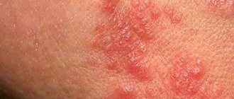
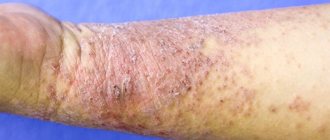
The transition to a severe form with the spread of inflammation over a larger area of the skin and the development of destructive changes in the form of pronounced skin maceration and erosion is typical for children with an unfavorable premorbid background. Characterized by the addition of a bacterial and fungal infection. Below is a photo of diaper dermatitis complicated by a fungal infection.
The rash is localized in the groin/buttock folds of skin and appears as well-demarcated bright red spots that are flaky at the edges. In a chronic course, it can manifest as granulomatous papules/nodules. The table below shows the grouped symptoms of diaper dermatitis depending on the degree of damage to the skin.
In severe cases of diaper dermatitis, the child’s general condition often suffers due to itching/burning in the affected area (sleeps poorly, often cries, is restless, and may have a decrease in appetite).
Causes of diaper dermatitis
The incidence of diaper dermatitis in infants (children under 12 months) is associated with the physiological structure of the skin. Immaturity of the epidermis, underdeveloped connective tissue structures of the dermis, unformed thermoregulation and immune response lead to skin damage and a decrease in protective function. From the photo you can see that the localization of the process is limited to the perineum area, the inner thighs, the anus and buttocks, namely in the places of greatest contact with the exogenous irritant.
Tests and diagnostics
The diagnosis is made based on the collection of anamnesis/complaints and physical examination of the child. It is extremely important to find out what is causing the rash, whether there is pain, restlessness/itching of the skin, especially during urination/defecation, the presence of diarrhea, how often diapers are changed, how the child’s skin is cared for (whether and what detergents, creams, powder), what kind of nutrition the child is on (breast or bottle feeding, whether the child took antibiotics, whether there are any concomitant diseases (gastroenteritis, atopic dermatitis, syndrome). Then the child is examined for the presence of irritations/damage to the skin in the diaper area, the nature of the rash is determined, affected area.
candidiasis diaper dermatitis is suspected, a scraping of the skin of the anogenital zone is performed with laboratory testing for the fungus. Differential diagnosis is carried out with candidiasis , contagious impetigo , psoriasis , seborrheic dermatitis .
Diaper dermatitis in children: how can we cope with it?
Both pediatricians and parents of young children consider diaper dermatitis one of the most common skin diseases. Almost every child experiences this condition to varying degrees of severity during the first months or years of life [1, 2]. Diaper dermatitis is diagnosed in 7–35% of infants, with peak prevalence at 9–12 months of age. life regardless of gender and race [3]. According to a study of the prevalence of skin lesions in the intensive care unit of the Children's Hospital of Philadelphia in 2008, diaper dermatitis was detected in 24% of inpatients [4]. This high frequency is largely due to the skin characteristics of young children.
Research in recent years has made it possible to obtain new information about the skin, explain its inflammatory response and justify the use of skin care products to prevent this condition. The skin as an organ is represented by the epidermis, the dermis itself and skin appendages. The skin is a dynamic system with the ongoing process of proliferation of keratinocytes in the basal layer of the epidermis, desquamation of corneocytes on the surface of the skin, and degradation of dense ligaments of corneodesmosomes [6]. In the apical layer of the epidermis, the stratum corneum, corneocytes are organized into an intercellular lipid matrix of laminar bodies consisting of cholesterol, ceramides and free fatty acids [7].
Details of infant skin morphology, such as surface structure, skin thickness, and cell size, have been visualized microscopically and calculated using computer analysis of microscopic images and skin impressions in silicone and adhesive tapes [8–10]. Video microscopy and in vivo confocal laser scanning microscopy make it possible to 3D image the topography of the skin and the internal structure of the dermal layers [9]. The results of these in vivo studies have expanded our understanding of children's and adult skin and articulated their differences.
The skin performs many different vital functions. Among them are physical and immunological protection from the damaging influences of external environmental elements (including microorganisms, ultraviolet radiation, humidity, extreme temperatures), sensory perception (pain, temperature, touch), temperature regulation, water and electrolyte homeostasis, gas exchange.
Understanding the physiology of normal healthy infant skin (the point of comparison) is important from a clinical perspective. Clinical studies on healthy infants, however, are ethically limited (in relation to the use of invasive methods), so they are few. New non-invasive technologies for measuring electrical capacitance, transdermal water loss, and absorption-desorption rates in infants and adults [10] have shown differences in skin barrier function, revealing dynamic changes in the level of natural hydration factor and water profile of the skin of infants, young children and adults [11] .
The structure, functions and constituent elements of infant skin differ from those of an adult organ - primarily in that during the first year of life and somewhat longer, a continuous maturation process continues in the skin. The epidermis of newborns is completely differentiated. In children of the first year of life and early age, corneocytes and granulocytic cells in the epidermis differ from those in adults in smaller size (by 20 and 10%, respectively) and in number [5]. The stratum corneum and epidermis of infant skin are 30 and 20% thinner than adults, respectively [5]. Microrelief lines are denser in children, while the depth of surface glyphs in children is similar to that in adults [5]. There is less melanin in the epidermis of children [12]. The dermal papillae, their density, size, and morphology are more homogeneous in children than in adults [5]. In children, there is no clear boundary between the papillary and reticular layers of the dermis [5]. In the components of the epidermis in infants, the concentration of natural moisturizing factor is lower than in adults [10]. It is important that children have higher skin pH values than adults, with the highest pH levels in newborns [13]. The level of surface skin lipids and sebum in children is lower than in adults [14], and the water content in the stratum corneum in infants is higher [11]. The density of collagen fibers in the dermis of children is lower than that of adults [5].
The listed features of the structure and composition of the skin determine the uniqueness of its function in children. Newborns have drier skin than adults, but during the first month of life the picture completely changes. In children 3–24 months. life, there is increased skin hydration compared to adults. The barrier function and moisture of the skin are also related to the intercellular lipid matrix, a natural moisturizing factor and the passage of water molecules through the stratum corneum. The rate of water absorption and desorption in the skin is higher in children than in adults, as is transepidermal water loss [10].
In addition to structural and functional changes, the composition of the skin microflora forms and changes in the first year of life. The predominant microbes on infant skin are Firmicutes (mainly Staphylococci), followed by Actinobacteria, Proteobacteria and Bacteroidetes. In adults, the phylum Proteobacteria dominates, followed by Actinobacteria and Firmicutes [15]. How to apply these research data is currently not entirely clear. Early microbial colonization may influence the development of skin immune function. The barrier function is vital for survival, preventing pathogens from entering the body. If the skin barrier is damaged, bacterial factors gain access to epidermal keratinocytes and can induce a protective immune response. Keratinocytes produce antimicrobial peptides. In the absence of these proteins, pathogenic microorganisms can invade the skin, disrupt the commercial/pathogen balance, or lead to infection.
The morphological characteristics of infant skin determine its easy vulnerability. Increased humidity, insufficient function of the sweat glands, and abundant vascularization of the skin can contribute to the rapid development of certain pathological conditions, such as irritant contact dermatitis and inflammatory processes.
Diaper dermatitis is common in young children. Diaper rash affects the lower abdomen and back, buttocks, perineum, and inner thighs. Wearing diapers causes an increase in moisture in the skin and an increase in pH (alkalinization). If high humidity persists for a long time, softening and loss of integrity of the skin (maceration) may occur, making it more susceptible to friction from the surface of the diaper. It also increases the risk of further skin damage and problems caused by exposure to other irritants, particularly feces containing proteases and lipases and ammonium in the urine. Other factors that can worsen the condition and worsen the rash include repeated washing and cleansing of the skin, inadequate skin care, infections, antibiotics, diarrhea and bowel and urinary tract problems [16].
Diaper dermatitis manifests itself as a rash in the diaper area (when diagnosing, other causes of rash in this area should be excluded) (Table 1). It is also necessary to take into account and evaluate the following factors (Table 2):
1. Hygiene skills (for example, how often to wash a child, change a diaper). Poor hygiene and prolonged contact with urine/feces predispose to irritant dermatitis.
2. Type of diapers used - disposable or reusable cotton.
3. Use of irritant products for washing and cleansing the skin, such as soaps, detergents, wet wipes with fragrance/alcohol.
4. Skin trauma: for example, diaper friction, excessive washing with vigorous cleansing.
5. Recent use of antibiotics, which predisposes to Candida colonization and diarrhea.
With diaper dermatitis, there is usually redness of the skin in the area covered by the diaper (buttocks, perineum, pubic area, upper thighs), with no redness in the deepest skin folds. The skin may have a slight shine, mild flaking, and hypopigmentation. The rash usually does not cause other symptoms until it becomes severe or painful. Mild diaper dermatitis involves a faint, pink rash covering less than 10% of the diaper area, with or without occasional scattered papules, slight sheen, and dryness. A mild course, as a rule, does not cause concern for the child and can usually be stopped with simple care measures. Moderate/severe course involves moderate to severe hyperemia, occupying an area of more than 10% of the skin under the diaper, with or without the presence of papules, spots, swelling or ulceration. With this degree of severity, irritability of children is noted, secondary infection with Candida albicans is determined, and often stomatitis. The erythema expands to areas outside of contact with feces, the diaper, and develops into confluent lesions with distinct edges with papules, pustules, involving the skin folds of the area of skin in contact with the diaper. Associated injuries and stomatitis are typical [17]. Signs of a bacterial infection include redness with exudate, blisters, small blisters, and pustules. If the rash in the diaper area is severe or does not disappear despite treatment, or there is doubt about the diagnosis, you should consult a dermatologist and conduct additional examination.
Relieving diaper dermatitis requires good child care, ongoing skin assessment, and parent education. Caring for the diaper area includes:
1) reducing the interval between changing diapers;
2) changing the diaper after soiling;
3) use of modern diapers;
4) cleansing the perineal area - by washing with warm water and emollient or wiping with wet wipes without alcohol and fragrances;
5) use of a water-repellent emollient or barrier agent after each diaper change to reduce irritation from contact with urine and feces;
6) air baths after washing [18].
In most cases, diaper dermatitis can be successfully treated with the above measures. If this is not enough, in moderate cases, topical corticosteroids and topical antifungals may be prescribed, applied to the skin under a barrier drug.
If candidal lesions are suspected, nystatin, clotrimazole or ketoconazole, miconazole are most often used. To reduce inflammation, if dermatitis persists, weak steroids, hydrocortisone, are prescribed [19]. Basically, 0.5–1% hydrocotisone is used, applied until the skin is cleared of the rash, but not longer than 1 week. Damaged infant perineal skin in a warm, humid environment has enhanced percutaneous absorption of drugs. In the USA, medium and strong steroids are used, often in combination with antifungal components (nystatin - triamcinolone, clotrimazole - betamethasone dipropionate) [19]. However, due to possible side effects for mild and moderate dermatitis in pediatric practice, their use should be avoided [20]. If dermatitis is complicated by a secondary infection (bacterial), then topical antibacterial drugs are used [21]. When choosing treatment tactics for dermatitis, you should be guided by scientifically based recommendations and the results of clinical studies.
Diapers. There are different types of diapers available today. The development of superabsorbent polymers has made it possible to produce new thin materials with a gel layer, which are currently used in diapers. New products are characterized by an increased rate of capture and absorption of liquid, high moisture capacity, which makes the skin surface drier. Diapers are created for babies of different ages, taking into account physiological characteristics. But at present, clinical studies do not provide sufficient evidence that certain types of diapers can be used to prevent diaper dermatitis [22].
The use of soaps and lotions is not recommended for cleansing the skin. Their use changes the pH of the skin, leading to greater dryness and damage [23]. A recent study found that using alcohol- and fragrance-free wet wipes was as effective at cleansing and hydrating the skin without damaging the skin as bathing in water using a cloth skin cleanser [24].
Barrier products create a protective layer between the skin and feces. The ideal barrier product should be one that has proven effectiveness in children and works similarly to the skin's natural function, forming a long-lasting barrier to maintain optimal moisture levels. It should not contain unnecessary ingredients, irritants, toxic ingredients or ingredients with undocumented safety [18]. However, there is insufficient evidence that barrier cream is more effective in the treatment of uncomplicated diaper dermatitis than simple water-repellent ointments (vaseline, paraffin) [16]. Water-repellent emollients or barrier preparations are applied at every diaper change. They form a protective lipid film on the surface of the skin and thereby protect against irritation upon contact with feces. Most products contain zinc oxide, petrolatum, or both. Under this layer, damaged skin is protected from irritation, healing occurs, and there is no excess skin moisture [25].
Barrier creams also use other ingredients, such as cod liver oil, aloe, dimethicone, and dexpanthenol [26]. In some European countries, a 2% aqueous solution of eosin is used to treat diaper dermatitis [27]. The absence or deficiency of barrier creams is associated with relapses of diaper dermatitis [28]. Barrier preparations may contain antiseptics and fragrances. Zinc preparations and castor oil may contain parts of peanut (groundnut) oil. Children with allergies to nuts and soy should not use such products. Ointments and creams containing vitamin A, calendula, honey, tea tree, recommended for the prevention and treatment of diaper dermatitis, do not have a sufficient evidence base of effectiveness. More research is needed.
To heal the skin and restore its protective properties, rapid regression of clinical manifestations of diaper dermatitis, positively proven topical dermato-reparative drugs, in particular Bepanten ointment, should be prescribed. The active principle of these drugs is dexpanthenol (provitamin B5). Pantothenic acid is a vital component of normal epithelial function. It is a component of coenzyme A, a cofactor for various reactions in the metabolism of carbohydrates, fatty acids, proteins, gluconeogenesis, steroid hormones, and porphyrins.
Dexpanthenol has been well studied. By the end of the twentieth century. Many works have been published that have proven its effectiveness in dermatology. A review of German researchers [29] summarized data from 56 publications. The use of dexpanthenol is based on its good penetration and high local concentration in the skin when used in the appropriate form, in particular as a water-in-oil emulsion. Topically, it acts as a humectant, improving hydration of the stratum corneum, reducing transepidermal moisture loss, maintaining skin softness and elasticity.
In vitro and in vivo studies revealed the effect of dexpanthenol on the activation of fibroblast proliferation, and accelerated reepithelialization during healing of the wound surface was proven. The anti-inflammatory effect of dexpanthenol was demonstrated in an experimental model of ultraviolet-induced erythema. Beneficial effects of dexpanthenol have been revealed in patients with skin grafts, in the treatment of scars, burn injuries, and various dermatoses. The most significant effects of dexpanthenol were stimulation of epithelialization, granulation and reduction of itching.
Double-blind, placebo-controlled studies assessed the effectiveness of dexpanthenol in wound healing. A decrease in erythema and more elastic and durable tissue regeneration have been shown in the treatment of epidermal wounds with dexpanthenol emulsion compared to treatment without its use. Monitoring of transepidermal water loss showed a significant acceleration of epidermal regeneration as a result of dexpanthenol therapy compared to placebo. In an experiment on a model of irritation, in the group that used dexpanthenol cream before irritation, significantly less damage to the stratum corneum barrier was detected compared to that in the group that did not use this drug. Skin care with dexpanthenol significantly improved symptoms of skin irritation such as dry skin, roughness, flaking, itching, erythema, erosions/cracks after 3-4 weeks. Usually, dexpanthenol preparations are well tolerated, the risks of irritation or hypersensitivity are minimal [29].
Of interest is a prospective observational study to evaluate the effectiveness of self-administered ointments containing dexpanthenol in treating irritated skin [30]. The study was conducted in Germany by pharmacists at a network of 392 pharmacies and included 1,886 patients with symptoms of irritated skin, including non-acute atopic eczema, as well as other conditions with skin xerosis and a damaged skin barrier. Efficacy assessment was carried out on the 7-10th day after treatment with calculation of the therapy effectiveness index / economic benefit. As a result, 91.5% of patients noted the benefits of therapy, 94.7% directly indicated the success of treatment. All symptoms of irritated skin (xerosis, erythema, desquamation) decreased significantly. There were no differences in responses depending on age, gender, or skin diseases. A high index of therapeutic effectiveness/economic benefit has been shown in the treatment of irritated skin with dexpanthenol ointment.
In the last decade, the volume of data on the protective effect of dexpanthenol has expanded. Thus, in a double-blind, placebo-controlled study, its effectiveness in protecting against irritation [31] and in the treatment of diaper dermatitis in infants was proven [26]. A multicenter study demonstrated the effectiveness of 5% dexpanthenol and zinc oxide ointment in the treatment of irritant diaper dermatitis in diarrheal syndrome in children [32].
Dexpanthenol is registered in Russia in the pharmacological group of regenerants and reparants. The results obtained in a number of clinical studies led by N. A. Korovina et al. (2004), V. A. Revyakina (2004), Yu. S. Akoeva et al. (2005), indicate the high clinical effectiveness and tolerability of Bepanten ointment (5% dexpanthenol) in the treatment of children with diaper dermatitis. It was found that in most cases, already on days 2–3 of therapy, there was a decrease or disappearance of clinical manifestations on the skin. It should be especially emphasized that among the subjects studied there were not only infants, but also newborns (both full-term and premature). At the same time, the positive effect of Bepanten was noted when used both in newborns and older children [33].
We present a clinical case of diaper dermatitis.
Girl E. Age 1 month. 5 days. During a routine examination by a pediatrician at the age of 1 month. Mom reported to the doctor about a rash in the diaper area on the 2nd day. Hereditary history is aggravated on the maternal side: my grandfather has allergic rhinitis and asthma during the grass flowering season, and my older sister has allergic rhinitis during the tree flowering season. The girl is the second desired child in a prosperous family; from the 2nd successful pregnancy with mild nasopharyngitis suffered without the use of drugs at the 6th month, timely physiological birth at 40 weeks. She was born with a weight of 3940 g, height 53 cm, chest circumference 35 cm, head circumference 35 cm. Apgar score - 8 points. She screamed immediately. They put me to the breast in the delivery room. In the early neonatal period, toxic erythema was noted. Vaccinated according to the calendar without complications. Psychomotor development by age. Breastfed girl. In the first month I gained 950 g of weight.
At the age of 14 days there was a single episode of skin hyperemia in the diaper area. Mom explained this by saying that she had not calculated her urine output that day and was late in changing the diaper. The hyperemia went away within a day when the diaper was changed; no doctor's consultation was required. The mother bathed the child with baby soap and used baby cream. After this episode, I began using body foam when bathing. The 2nd episode of rash at one month of age was associated with the fact that the child’s stools became up to 7 times more frequent with a sour smell, which caused skin irritation. Mom assumed that this was a response to the excessive amount of fried potatoes she had eaten the day before. The stool was restored after 1 day. The girl's behavior, sleep, and appetite have not changed. At the pediatrician's examination, the child is calm. The child's general condition does not suffer.
Objectively: locally on the skin of the perineum, pubic area, upper third of the inner thighs, buttocks against the background of moderate hyperemia, papular elements. Deep folds are not involved. There is no itching.
In a general blood test at the age of 1 month: hemoglobin - 132 g/l, erythrocytes - 4.0 × 10¹², color index - 0.9; leukocytes - 8.2×10⁹; eosinophils - 2, band neutrophils - 2, segmented neutrophils - 18, lymphocytes - 74, monocytes - 4, ESR - 4 mm.
In the coprogram: consistency - mushy, color - yellow, smell - fecal, mild, pH - slightly acidic, mucus was detected; no blood or undigested food was found. Microscopic examination of stool: mucus - moderate, leukocytes - 0-5 in the mucus, no other ingredients were detected.
Diaper dermatitis was diagnosed. Advice is given on rational nutrition for a nursing mother and keeping a food diary. For skin care, Bepanten ointment is recommended after each diaper change. Examination after 5 days showed a positive effect with the restoration of pink, clear skin and satisfactory condition.
In this case, diaper dermatitis occurred in a child with a high risk of developing allergic diseases. Early onset in the first month of life indicates the vulnerability of the skin. Episodes of its damage can serve as a trigger for the development of inflammation, including allergic inflammation. Restoring the integrity and function of the epidermal barrier is protective against the development of chronic pathology. Long-term use of Bepanten ointment to preserve the integrity of the skin in this case is justified.
Thus, it is necessary to emphasize that new data on the morphology and function of the skin in different periods of its maturity at different ages make it possible to reveal the mechanisms of its damage and create the necessary skin care products. Diaper dermatitis is a common inflammatory skin reaction in infants. The diaper, covering the skin surface, creates conditions for external environmental influences that contribute to skin irritation, which begins with damage to the skin barrier caused by components of urine and feces. Maintaining the integrity of the skin barrier with protective creams along with frequent diaper changes is an effective preventative strategy.
Prevention
Prevention of the disease comes down to a number of rules for caring for a newborn, which parents must follow:
- Adequate thermal conditions, preventing the child’s skin from drying out/wetting due to overheating.
- Careful selection of diapers (breathable disposable ones, matching the size and gender of the child).
- Do not wear diapers all the time (the baby should not be in diapers for more than 3 hours).
- Take air baths between changing diapers (from 5-10 minutes and up to 30 minutes at the age of about a year).
- Regular bathing/washing the child, ironing diapers/clothes.
- When choosing wet wipes, avoid products containing fragrances that can cause allergic contact dermatitis.
- Washing/wiping the skin with special wet wipes at each diaper change and drying it thoroughly.
- Apply protective creams to the skin every time you change diapers.
- Wash baby diapers only with special products adapted for children.
- Thorough hand washing/antiseptic treatment before any contact with the newborn's skin.
- If children are prone to food allergies, the child should be on a diet. When breastfeeding, exclude allergenic foods from the mother's diet.
Currently, for the prevention of PD, the so-called “A-E standard” has been developed, which indicates the basic principles of caring for a child’s skin (table below).
Treatment of diaper dermatitis
The primary principle in the treatment of diaper dermatitis in infants is proper care and hygiene procedures. With mild to moderate severity, parents can independently prevent dermatosis.
It is recommended to wear disposable diapers, which are changed after each act of defecation and urination; in infants aged 1 to 4 months, the frequency of replacement is 8 or more times. Afterwards, the anogenital area is washed under running warm running water with or without hypoallergenic liquid soap. Use an ironed diaper or towel to blot away any remaining moisture.
To eliminate inflammation, use soft cotton swabs to gently wipe the affected areas of the perineum with a decoction of medicinal herbs. It is impossible to get rid of plaque and crusts by friction. Bathing in a decoction of chamomile, string and calendula is allowed. Air baths of 10-30 minutes a day are required.
Treating the skin before putting on a diaper. If it becomes wet, dry it; if it becomes crusty and dry, moisturize it. Medications:
- Washing with antiseptic solutions: Furacillin solution; decoctions of medicinal plants: chamomile, string, oak bark, calendula, oats.
- Combined powders containing trace elements, minerals, talc and zinc oxide.
- Wound healing agents: Dexpanthenol ointment, Bepanten cream, D-panthenol ointment; Zinc Oxide cream and paste, Desitin cream, Sudocrem; Drapolene cream.
In some cases, the doctor may prescribe more active anti-inflammatory drugs for a short period - hormonal creams, antibacterial or antifungal drugs.
Sometimes antihistamines are prescribed to children with a history of allergies (atopic dermatitis, urticaria, etc.) in order to relieve severe swelling and inflammation. Treatment with the selection of a personal dosage is performed by the treating specialist.
Treatment of severe diaper dermatitis in newborns is carried out in a hospital setting.
Consequences and complications
After the first signs of film dermatitis appear, in the absence of timely treatment, adequate care of the child’s skin and failure to eliminate provoking factors, rapid progression of the disease and deterioration of the skin condition are observed. In almost 15% of children, a severe form develops within 1-2 days, and they have a tendency to relapse in cases of diarrhea / in the presence of the slightest errors in care. Severe and complicated forms of it are more common in children with signs of lymphatism and transient failure of cellular immunity .
Quite serious complications of diaper dermatitis include the development of a purulent-inflammatory process in the form of impetigo , abscesses , infiltrates, often accompanied by symptoms of intoxication, fever, disturbances in appetite, stool patterns, sleep, and malnutrition . At the same time, the most common pathogen is fungal flora and Staphylococcus aureus. With dominant candidal inflammation, a pronounced, rapidly progressing clinical picture is observed, forming extensive affected areas in the form of papules and vesicles in the genital area, inguinal folds, thighs, buttocks, and abdomen.
Symptoms of diaper dermatitis
The main symptom complex is skin hyperemia, the appearance of dryness, peeling and crusts, after a while weeping occurs and vesicles filled with serous fluid are formed. In advanced cases, the contents of the vesicles become purulent - pustules, and tissue swelling appears. Microcracks form, which quickly turn into ulcers and ulcers.
The child’s general well-being changes, painful sensations appear, he is often capricious, appetite decreases, and sleep is disturbed. Body temperature remains within the normal age range, however, when microbial flora joins, hyperthermia may occur.
It is classified according to the severity and characteristics of the course:
- mild: moderate hyperemia, the skin is pink or pale pink, crusts are possible, eliminated by drying and treatment with special solutions;
- medium: tissue swelling with infiltration is noted, skin color is from bright pink to burgundy, pustules are characteristic;
- severe: the skin is stretched, shiny, red or burgundy in color, pronounced weeping, crusts, ulcerations that tend to merge, deep painful cracks.
The lack of pathogenetic therapy leads to abscess formation, severe intoxication, and sepsis is possible. The condition is life-threatening for the baby.
List of sources
- Koval G.S. Prevention and treatment of diaper dermatitis // Issues of modern pediatrics, 2004, vol. 3, no. 5, p. 60-64.
- Geppe N.A., Belousova N.A. Diaper dermatitis. Attending doctor. 2004. No. 1. pp. 24-28.
- Suvorova K. N., Kasikhina E. I., Grishko T. N., Basse F. B. Modern features of the course of dermatoses in children of the first year of life. I Moscow Forum “Dermatovenereology and cosmetology: synthesis of science and practice.” Abstracts of reports. Moscow, 2011. 31 p.
- Studenikin V. M. Caring for children’s skin: more gentle, even more gentle // Pharmaceutical Bulletin. 2007. No. 40. pp. 16–17.
- Nechaeva O. S. Diaper dermatitis: modern etiopathogenetic aspects and approaches to prevention // Clinical dermatology and venereology. 2009; 3:77–79.
Diaper dermatitis in the photo
Photo 1. Diaper dermatitis
Photo 2. Diaper dermatitis
Photo 3. Diaper dermatitis
Photo 4. Diaper dermatitis