Increasingly, during gynecological examinations during pregnancy, doctors say that the sizes of the female pelvis and the fetus do not correspond to each other. This interferes with the normal course of labor.
Often this situation is so dangerous that the woman in labor is offered a caesarean section to avoid unwanted consequences.
What is a narrow pelvis during pregnancy and how can it harm the baby?
The essence of the concept
The pelvic bones are a dense ring through which the baby's head will have to pass during birth. The problem is that this bone formation is practically inextensible. Only a slight discrepancy is possible (only half a centimeter) due to the fact that the symphysis (cartilage) softens slightly before childbirth.
At its core, the pelvis is motionless. And if the circumference of the child’s skull is larger than this bone ring, gynecologists are forced to diagnose this anatomical feature of the female skeleton and recommend a cesarean section. What could be the reason for such an unusual pathology?
According to statistics. Recently, the frequency of diagnosis of narrow pelvis has dropped compared to previous years. It is only 7%.
Causes
Most women who were diagnosed with a narrow pelvis during pregnancy believe that this is an individual feature of the structure of their skeleton, with which they were born. In fact, in 90% of cases this problem turns out to be acquired.
The main causes of a narrow pelvis include:
- health problems in childhood: rickets, poor nutrition, excessive stress provoke deviations in physical development;
- injuries in the pelvic area: bone fractures lead to serious deformation and reduction in size; tumors in this area: osteomas narrow the gap between the bones;
- hormonal disorders leading to hyperandrogenism, which is characterized by broad shoulders and a masculine narrow pelvis;
- acceleration of girls during adolescence, which leads to a transversely narrowed pelvis;
- bone infections: tuberculosis, osteomyelitis, which destroy bone tissue and lead to pelvic deformities; orthopedic diseases (for example, scoliosis).
The same phenomenon is said to occur if the fetus is too large and risks not passing into the pelvic ring, even if it is of normal size.
The parameters for which pelvis is considered narrow for childbirth have long been developed in gynecology, so the doctor will answer this question after appropriate measurements and examinations. Depending on the type of pathology, a decision will be made on how the baby will be born - by caesarean section or naturally.
What's the secret? If previously a narrow pelvis was mainly an anatomical feature of the female skeleton, today women in labor have to face this problem due to the fact that large children are being born more often.
Anatomical
Gynecologists diagnose an anatomically narrow pelvis when there is a narrowing of the bones, which is a deviation from the average norm. It is not always an indication for a cesarean section, because the fetus may refuse to be small and pass freely through the birth canal without injury. This type of pathology has its own special classification.
By type of narrowing:
- Evenly tapered.
- Flat.
- Transversely tapered.
By degree of narrowing (Litzman classification):
If a woman is diagnosed with a narrow pelvis of the 1st degree during pregnancy, she is allowed to give birth on her own. However, the young mother and the team of doctors must be prepared for various complications of labor. In such cases, the surgeon and anesthesiologist are usually notified to be on the safe side. Their intervention may be needed at any moment.
The situation is a little more complicated when a woman is diagnosed with a narrow pelvis of the 2nd degree during pregnancy: natural childbirth is allowed, but under certain conditions. Most often, you are allowed to give birth on your own if the pregnancy is premature and the fetus is not too large.
Natural childbirth is not possible. If a narrow pelvis of the 3rd degree is diagnosed, this is a medical indication for cesarean section. The woman is hospitalized in advance (2 weeks before the cherished date), assigning her bed rest and absolute rest.
If during pregnancy it turns out that the expectant mother has a narrow pelvis of the 4th degree, her child can only be born by caesarean section.
Clinical
If a woman in labor is of normal size, but on the eve of birth it turns out that the fetus is too large and will not be able to pass through the pelvic ring without injury, they speak of a clinically narrow pelvis. However, in subsequent pregnancies, if the child turns out to be smaller, such a diagnosis will not be made. So if there are no other indications for a cesarean section, the birth will take place naturally.
Clinically, a narrow pelvis is diagnosed only during the last months of pregnancy or even immediately before childbirth, and its classification in obstetrics has not been developed. The most common causes of a clinically narrow pelvis:
- incorrect insertion of the head;
- large fruit size;
- hydrocephalus;
- various malformations of the child;
- incorrect presentation.
All these phenomena can be clarified immediately before the birth itself or already during its process. The decision must be made very quickly; the diagnosis of a clinically narrow pelvis is based on specific obstetric signs and symptoms. In this case, an emergency caesarean section is performed.
Regardless of its type, a narrow pelvis in obstetrics is regarded as a serious complication that can lead to dangerous consequences if handled incorrectly.
An experienced, professional doctor, at the first suspicion of this feature of the female skeleton, takes appropriate measures and controls the size of the pelvic bones throughout pregnancy so that no unforeseen situation arises during the birth of the baby. How is this pathology diagnosed?
For reference. Hydrocephalus is a dangerous and common disease, hydrocephalus in a baby, which is characterized by the enormous size of its head. There is no way it will pass through the pelvic ring.
Anomalies of the second stage of labor
If hypotonic dysfunction of the uterus occurs in the second stage of labor, weakness of pushing or inadequacy of voluntary expulsive forces is diagnosed. These complications usually manifest as slowing or delayed descent of the fetal head. Although deceleration of head descent is defined as a decrease in the rate of descent of <1 cm/h in primiparous women and <2 cm/h in multiparous women, this criterion does not have much clinical significance.
Stopping the descent of the head requires an urgent further examination of the contractile activity of the uterus, the condition of the mother and fetus, and the relationship between the fetal head and the mother’s pelvis. In the absence of fetal-pelvic disproportion, labor stimulation with oxytocin is possible, but if there are no contraindications, the method of choice will be surgical vaginal delivery (obstetric forceps, vacuum extraction). If operative vaginal delivery is not possible, a cesarean section is performed.
Weakness of pushing may be a consequence of epidural anesthesia, which reduces the reflex resistance of the pelvic floor muscles and the patient’s ability to voluntarily contract the muscles of the anterior abdominal wall. Treatment in this case can be surgical (obstetric forceps, vacuum extraction or caesarean section).
In order to prevent birth anomalies, perinatal morbidity, mortality and reduce the frequency of cesarean sections, Driscoll (Dublin, England) proposed the tactics of the so-called active management of labor in first-time mothers. The active labor management protocol consists of the following criteria:
1) strict criteria for patient selection (gestational age, normal pelvic and fetal dimensions, insertion of the fetal head, etc.);
2) early amniotomy;
3) hourly vaginal (cervical) examination;
4) administration of oxytocin when the rate of cervical dilatation in the active phase is <1 cm/h;
5) the use of high doses of oxytocin to achieve effective labor;
6) waiting no more than 12 hours in the first stage of labor and no more than 2 hours in the second stage of labor, in case of ineffective labor - delivery by cesarean section.
Diagnostics
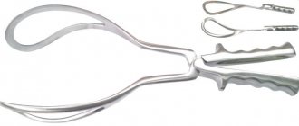
Many business and most active mothers try to find out on their own how to determine whether the pelvis is narrow for childbirth, and whether they can give birth themselves at certain sizes. In fact, this cannot be done either at home or “by eye”. Diagnosis is possible only in a hospital; it is carried out exclusively by a professional doctor using a specific obstetric instrument called a pelvisometer. With its help, the following dimensions are determined:
- the interspinous distance is measured between the anterior iliac (connecting the pelvis to the spine) spines (processes), normally it should be more than 25 cm;
- the gap between the most distant points of the iliac bones is normally more than 28 cm;
- the distance between the trochanters (greater) of the femurs, the desired norm is more than 30 cm;
- the true conjugate is measured during a vaginal examination, this is the distance between the pubic joint and the highest point (promontory) of the sacrum; it is considered normal when the obstetrician cannot reach this point;
- external conjugate - the gap between the suprasacral fossa, which is located in the lumbosacral region, and the upper corner of the pubic symphysis, a certain norm - more than 20 cm;
- Michaelis's diamond above the coccyx, in the area of the sacrum, the boundaries of which are normally clearly visible, all sides are symmetrical: transverse are 10 cm, vertical - 11 cm;
- The Solovyov index allows you to estimate the thickness of the bones, which can also interfere with normal childbirth - this is the circumference of the wrist, the maximum norm is no more than 14 cm. To clarify the parameters, in rare cases, radiography is performed, but it can harm the fetus.
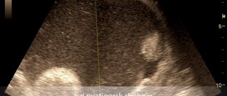
An ultrasound examination can also help assess the size of the narrow pelvis during pregnancy. In clinical cases where this data cannot be obtained in advance, obstetricians are guided by specific signs and symptoms.
Through the pages of history. S. A. Michaelis is a German gynecologist of the 19th century, whose name is the famous sacral rhombus, which determines whether a woman can give birth on her own or not.
Publications in the media
An anatomically narrow pelvis is a pelvis in which at least one dimension is shortened by 1.5–2 cm compared to normal. A functionally narrow pelvis (clinically narrow pelvis) is a pelvis that impedes the course of labor due to the disproportion between the fetal head and the woman’s pelvis. Etiology: infantilism, delayed development of the body, rickets, tuberculosis of bones and joints, fractures of the pelvic bones, deformities of the pelvis, spine, lower extremities, acceleration, etc. Classification According to structural features • Gynecoid pelvis - a normal pelvis of the female type •• Android pelvis - a female pelvis of a male or funnel-shaped type •• Anthropoid pelvis - a pelvis with an elongated anteroposterior size and a shortened transverse diameter •• Platypeloid pelvis - a flat, narrow pelvis. According to the shape of the narrowing • Generally uniformly narrowed pelvis - all dimensions are reduced by the same amount, usually by 1.5–2 cm • Transversely narrowed pelvis - reduction in transverse dimensions with a normal (or increased) size of the true conjugate • Flat pelvis - shortening of direct dimensions with a normal value of transverse and oblique sizes •• Simple flat pelvis - all direct dimensions are shortened •• Flat scorachitic pelvis - shortening only the direct size of the inlet (true conjugate). Signs: change in the shape of the sacrum and other pelvic bones, a decrease in the true conjugate, a kidney-shaped shape of the entrance to the pelvis, the size of the entrance to the pelvis is smaller than the size of the exit • Generally narrowed flat pelvis - a decrease in all sizes, but the straight lines are shortened more than all others • An oblique (asymmetrical) pelvis occurs after history of rickets, dislocation of the hip joint, scoliosis. The cause is depression of the acetabulum on one side • Lordotic pelvis is observed with lordosis in the lumbosacral region; the direct size of the inlet to the pelvis is reduced, the pelvis is anatomically narrow • Funnel-shaped pelvis is a pelvis in which the inlet dimensions are normal, and the outlet dimensions are narrowed in the transverse or transverse and anteroposterior directions. The sacrum is elongated, the pubic arch is narrow • Kyphotic pelvis - kyphosis of the spine causes deformation of the pelvis: an increase in the true conjugate, a decrease in the transverse size of the pelvic outlet, the pubic angle is acute, the pelvic cavity is funnel-shaped • Spondylolistic pelvis - an anatomically narrow pelvis with a reduced direct size of the inlet due to slipping of the fifth lumbar vertebra from the base of the sacrum • Osteomalactic pelvis - a pelvis deformed as a result of osteomalacia; pressure from internal organs on the pelvis and lateral pressure from the heads of the femurs lead to deformation of the pelvic aperture; the aperture is either triangular or shaped like a stylized heart, while the pubis takes on a beak-like shape • Pelvis narrowed by exostoses and bone tumors. By degree of narrowing •• I degree. The true conjugate is 9–11 cm. In most cases, childbirth occurs without complications •• II degree. The true conjugate is 7.5–9 cm. Natural delivery is possible, but complications often occur •• III degree. The true conjugate is 6.5–7.5 cm. Delivery of a full-term fetus through natural means is impossible. For vaginal delivery, fetal destruction surgery is indicated •• IV degree. The true conjugate is less than 6.5 cm. Vaginal delivery is impossible even with the use of fetal destruction surgery. Diagnostics • History: infantilism, past illnesses and injuries, obstetric history • Objective examination: general examination, height 150 cm and below, assessment of Michaelis rhombus, spinal curvature, joint mobility; a saggy belly in multiparous women and a pointed belly in primiparous women. In addition to the generally accepted external pelviometry, it is necessary to measure the distance between the anterosuperior and posterosuperior spines of one side (14–15 cm), and the height of the symphysis pubis (4–5 cm). To clarify the thickness of the bones, the circumference of the wrist joint is measured - the Solovyov index (14–15 cm). Vaginal examination: relief of the inner surface of the pelvis, true conjugate. The course of pregnancy • Due to the high position of the uterine fundus, pregnant women complain of shortness of breath, palpitations, fatigue • In the second half of pregnancy, gestosis often occurs • Premature discharge of amniotic fluid often occurs, because the fetal head is mobile, therefore, the amniotic fluid is not divided into anterior and posterior. Incorrect position of the fetus and breech presentation are often observed. The course of labor . With I and II degrees of pelvic narrowing, the course of labor depends on many factors, for example, on the size of the fetal head, intensity of labor, fetal presentation, etc. With III and IV degrees of pelvic narrowing, a cesarean section is indicated. Complications during childbirth • Early rupture of amniotic fluid with prolapse of the umbilical cord loop or fetal arm • Abnormalities of labor • Asynclitic insertion, extensor presentation of the head • Overdistension of the lower segment of the uterus and uterine rupture • Compression of the soft tissues of the birth canal between the pelvic bones and the fetal head with the formation of necrosis and fistulas • Impaired uteroplacental circulation • Bleeding in the placenta and postpartum periods • Complications from the fetus: fetal hypoxia, cerebral hemorrhage, cephalohematoma, depressions and cracks of the skull bones, fractures of the clavicle and limbs • Death of the mother and fetus.
Mechanism of birth • Generally uniformly narrowed pelvis •• Maximum flexion of the head, the small fontanelle is located on the center line of the pelvis •• The sagittal suture of the fetal head corresponds to the oblique size of the entrance to the pelvis, accordingly, the large transverse size of the head also passes through the oblique size •• The area of the suboccipital fossa cannot approach symphysis, so the head moves toward the perineum at birth, and deep ruptures often occur. • Transversely narrowed pelvis. When the sagittal suture corresponds to the direct size of the pelvis, the occiput of the fetus is facing the symphysis, and the head is small, strong flexion of the head occurs, and labor proceeds as in an anterior occipital presentation. When the back of the fetal head is turned posteriorly, the head can be rotated 180°, so labor takes place in the anterior or posterior view. With a transversely narrowed pelvis, there is often a high erect position of the head, leading to complications and requiring a cesarean section. • Flat scorachitic pelvis •• Prolonged high standing of the head, the sagittal suture corresponds to the transverse size of the pelvis •• Slight extension of the head, as a result of which the head passes through the true conjugate (smallest size) with a small transverse size •• Asynclitic insertion of the head. Anterior asynclitism is usually observed: the posterior parietal bone rests on the promontory and lingers in this place, and the anterior one gradually descends into the pelvic cavity. After a strong configuration, the posterior parietal bone slides off the promontory and the asynclitism disappears. • Simple flat pelvis. Often there is no internal rotation of the head, because the direct dimensions of the pelvis are reduced; the sagittal suture corresponds to the transverse size of the pelvis (low transverse position of the head). If spontaneous rotation of the head does not occur, surgical delivery is necessary.
Management of labor • For III and IV degrees of narrowing, a cesarean section is indicated • Childbirth is expectant; if signs of discrepancy between the sizes of the pelvis and head appear or if complications develop, surgical intervention is indicated • Signs of compliance of the head and pelvis •• Zangemeister's sign - measure the degree of elevation of the anterior surface of the head above the symphysis. The external conjugate is measured with a pelvis, then the rear button of the pelvis is not moved, and the anterior button (located on the symphysis) is placed on the protruding point of the anterior surface of the head. The distance from the head to the suprasacral fossa should be 3–4 cm less than the external conjugate. With the same value, the discrepancy is small, the prognosis of labor is doubtful •• Vasten's sign. After the water breaks and the head is inserted, one palm is placed on the surface of the symphysis, the other on the area of the presenting head. When the sizes of the mother's pelvis and the fetal head correspond, the anterior surface of the head is located below the plane of the symphysis (Vasten's sign is negative). If the anterior surface of the head is flush with the symphysis (flush Vasten's sign), there is a slight size discrepancy. If there is a discrepancy between the sizes of the mother's pelvis and the fetal head, the anterior surface of the head is located above the plane of the symphysis (Vasten's sign is positive) •• Ultrasound • Careful monitoring of the condition of the fetus • Prevention of complications, their detection and timely treatment.
ICD-10 • O33 Medical care for the mother with established or suspected discrepancy between the sizes of the pelvis and the fetus
Signs of a clinically narrow pelvis
Immediately before birth, if a woman in labor exhibits signs of a clinically narrow pelvis, a cesarean section is recommended. These symptoms include the following pathologies and complications:
- the baby’s head does not press against the pelvic bones upon entry;
- the biomechanism of childbirth is disrupted;
- amniotic fluid is discharged untimely;
- contraction of the uterus is disrupted: weakening of its activity, incoordination, premature appearance of attempts;
- the cervix has already fully opened, and the advancement of the fetus has not yet begun;
- the head remains in the pelvic plane for too long;
- protracted labor;
- deformation of the head, birth tumor, hematomas, fetal hypoxia;
- problems with the bladder: pressure, urinary retention, blood in the urine;
- threat of uterine rupture.
If a woman has a clinically narrow pelvis and a large fetus due to at least one of these signs, the team of doctors in 98% of cases performs an emergency cesarean section to avoid death or injury to the fetus during its movement through the birth canal. This is the only correct way out of this situation, medically completely justified and recommended.
Of course, such a birth with a narrow pelvis is much more difficult than with an anatomical one, since you can prepare for the latter in advance.
On a note. Intrauterine hypoxia is oxygen starvation of the child, which can be fatal if the fetus is not removed in time.
What complications can there be for the mother?
A narrow pelvis often leads to a risk of cervical tumors. If the head remains in one place for a long time, contractions become painful and sharp, which leads to severe stretching or rupture of the uterus.
The uterus also gets very tired during a long labor. To return to its normal size, the uterus continues to contract after childbirth. At this time, all injured vessels are tightened. If uterine contractions stop, there is a risk of postpartum hemorrhage.
Signs of an anatomically narrow pelvis
The main sign of an anatomically narrow pelvis is the discrepancy between its size and the standards indicated above. But there are such impatient young mothers who cannot wait for laboratory measurements and want to know in advance whether they are predisposed to such a diagnosis. There are such signs, and they usually include:
- short arms (hand length - no more than 16 cm);
- short fingers: thumb length - no more than 6 cm, middle finger - no more than 8;
- small foot size: less than 36;
- small height: no more than 150 cm;
- curvature of the spine, limbs, lameness, orthopedic diseases;
- pelvic injuries;
- complications during previous births;
- irregular menstrual cycle;
- androgenic (male type) physique.
However, do not think that if one of the listed features applies to you, this means that you have an anatomically narrow pelvis.
These are indicative signs that are observed in 98% of women who were diagnosed with this during pregnancy. You just need to keep these facts in mind in order to prepare in advance for all possible consequences.
And there is no need to be afraid of them: an anatomically narrow pelvis has a huge advantage over a clinical one: it allows you to prepare for childbirth in advance.
Sometimes it happens. Often, small women turn out to be much tougher than those who have more impressive sizes in terms of childbirth. They give birth to even large babies on their own.
Successful outcome: every chance
Newborns with a narrow pelvis are at high risk for various complications. They need constant monitoring, as they often suffer from cerebrovascular accidents and injuries to the cervical spine. They are more likely to experience asphyxia (insufficient oxygen supply) at birth. But the capabilities of modern medicine make it possible to quickly make adjustments to the birth process so that after 9 months of waiting the woman becomes a mother.
Expert: Irina Isaeva, obstetrician-gynecologist Author: Elena Nersesyan-Brytkova
The material uses photographs belonging to shutterstock.com